MRI Safety Policies 1
|
|
|
- Christina Ramsey
- 5 years ago
- Views:
Transcription
1 MRI Safety Policies 1
2 YALE-NEW HAVEN HOSPITAL DEPT. OF DIAGNOSTIC RADIOLOGY POLICY AND PROCEDURE MANUAL SUBJECT: CONTACT INFORMATION MRI Safety Manual YNHH Fitkin MRI 789 Howard Ave, New Haven CT Smilow MRI 20 York Street, New Haven CT Smilow MR Breast Center 20 York Street, New Haven CT Smilow MR OR 20 York Street, New Haven CT YNHH Pediatric MRI 20 York Street, New Haven CT Shoreline MRI 111 Goose Lane, Guilford CT Temple St MRI 60 Temple Street, New Haven CT ST Raphael Campus MRI 1450 Chapel Street, New Haven CT North Haven MRI 6 Devine Street North Haven, CT Park Ave MRI 5525 Park ave Trumbull, CT
3 YALE-NEW HAVEN HOSPITAL DEPT. OF DIAGNOSTIC RADIOLOGY POLICY AND PROCEDURE MANUAL MRI Safety Manual Reviewed Date SUBJECT: INDEX Last Revision Policy No. GENERAL SAFETY INFORMATION YALE NEW HAVEN MRI CONTACT INFORMATION...2 INDEX...3 ACRONYMS... 5 INTRODUCTION...6. GENERAL POLICY...7. SAFETY TERMINOLOGY...8 ACR ZONES (SITE ACCESS RESTRICTION)...11 EAR PLUGS AND OTHER TIME VARYING MAGNETIC FIELD RELATED ISSUES...17 TATTOO..19 FIREFIGHTERS, POLICE AND SECURITY CONSIDERATIONS...20 CRYOGEN RELATED ISSUES...26 MRI SCREENING PERSONNEL FAMILY/PREGNANT COMPANIONS..29 GUN SHOT WOUNDS 92 OUTPATIENTS INPATIENTS UNRESPONSIVE PATIENTS OXYGEN CYLINDERS...38 PENILE IMPLANT FOREIGN BODY ORBITS..40 CARDIAC IMPLANTATIONS: STENTS, VALVES, LEADS, PACEMAKERS. 41 VASCULAR COILS, FILTERS, STENTS...42 ANEURYSM CLIPS...43 BRAIN STIMULATORS...44 OTHER NERVE STIMULATORS, IMPLANTABLE PUMPS ORTHODONTIIC DEVICES...47 TRANSDERMAL MEDICATION PATCHES AND WOUND DERSSINGS EAR IMPLANT INFORMATION..50 MISCELLANOUS IMPLANTS. 52 MRI PREGNANCY RELATED ISSUES PREGNANCY EMPLOYEES, PATIENTS, COMPANIONS AND CONTRAST ISSUES... 57/58 PRE PROCEDURE PREGNANCY TESTING...58 ADMINISTRATION CONTRAST WHILE BREAST FEEDING MRI CONTRAST RELATED ISSUES CONTRAST AGENTS...60 EGFR SCREENING GAD QUICK REFERENCE SHEET...66 DOTAREM DOSING INFORMATION...88 PREMEDICATION
4 PHYSICIAN CONTRAST COVERAGE.. 67 MRI SCHEDULING RELATED AND OTHER POLICIES ED AND STAT EXAMS DEFINITON OF LIFE THREATENING HYPER ACUTE STROKE.. 71 OVERNIGHT CASES MAGNET ROOM CLEANNG.73 NURSING RELATED AND OTHER POICIES LATEX...74 MONITORING OF PATIENTS...75 CODE PROCEDURE/ CODE CART LOCATION...77 RN RESPONSIBILITIES...80 FERROUS OBJECT IN MR SCAN ROOM MRI SAFETY OFFICER RESPONSIBILITIES...83 MRI MEDICAL DIRECTOR RESPONSIBILITIES.84 WANDING 85 EAR PLUG POLICY EXEMPTION. 87 DOTAREM DOSING INFORMATION...88 GUN SHOT WOUNDS..91 PROGRAMMABLE SHUNTS...93 Appendix A Contrast Doses, Company Contact info Appendix B Bayer Short Form Adverse Reaction form Appendix C MRI Safety Website Information Appendix D Screening Forms Appendix E Techniques to Lower SAR and B1RMS List of Images 1 ASTM Approved Labeling 2 YNHH Stop Sign 3 ACR Zones 4 Zone 4 signage 5 Ferromagnetic wand 4
5 YALE-NEW HAVEN HOSPITAL DEPT. OF DIAGNOSTIC RADIOLOGY POLICY AND PROCEDURE MANUAL MRI Safety Manual Reviewed Date SUBJECT: ACRONIYMS Last Revision Policy No. Acronyms 0.5 mt 5 Gauss Line ACR American College of Radiology AED Automated Emergency Defibrillator ASTM American Society for Testing and Materials ARRT American Registry of Radiologic Technologists CPR Cardiopulmonary Resuscitation DB Decibel FDA Food and Drug Administration GBCA Gadolinium based contrast agent MR Magnetic Resonance MRI Magnetic Resonance Imaging RF Radio Frequency RMS Root Mean Square SAR Specific Absorption Rate SMS staples/superficial metallic sutures SOP Standard Operating Procedure TVMF Time Varying Magnetic Fields T Tesla W/kg Watt/kilogram 5
6 YALE-NEW HAVEN HOSPITAL DEPT. OF DIAGNOSTIC RADIOLOGY POLICY AND PROCEDURE MANUAL MRI Safety Manual Reviewed Date SUBJECT: INTRODUCTION Last Revision Policy No. Magnetic Resonance Imaging is an ever changing, evolving technology. There are potential risks in the MR environment, not only for the patient but also for the accompanying family members, attending health care professionals and others who find themselves only occasionally or rarely in the magnetic fields of MR scanners, such as security, housekeeping personnel, firefighters, police, etc. This manual has been developed to help guide the MR staff regarding these issues. It is the intent of the Yale New Haven Hospital safety manual to: Protect and educate all patients, direct and ancillary personal about the possible risks, associated with the MR Suite including but not limited to static, time-varying magnetic fields and RF pulses. To be in compliance with the most up to date MR safety information provided by the Joint Commission and the ACR Prove helpful as the field of MRI continues to evolve and mature, providing MR services that are among the most powerful, yet safest, of all diagnostic procedures to be developed in the history of modern medicine. 6
7 YALE-NEW HAVEN HOSPITAL DEPT. OF DIAGNOSTIC RADIOLOGY POLICY AND PROCEDURE MANUAL MRI Safety Manual Reviewed Date Last Revision SUBJECT: GENERAL POLICY Policy No. 1.All clinical and research MR sites, irrespective of magnet format or field strength, including installations for diagnostic, research, interventional, and/or surgical applications, should maintain MR safety policies 2. These policies and procedures will be regularly reviewed by the MR Safety Officer and the Medical Director to account for the significant changes in the MR center environment. This will take into account ACR, Joint Commission and international standards. 3. The responsibility for implementation and maintenance of these policies and procedures belong to the Medical Director of the YNHH MRI Centers. 4. Annually, all MR personnel will review safety within the MR environment 5. Provide all non-mr staff, patients and their families with appropriate materials (e.g., guidelines, brochure, and poster) that explain the potential for accidents and adverse events in the MRI environment. 6. Provide Access to all updates safety policies to all MR staff online and/or an updated hard copy in every MR area. 7. MR safety incidents or "near incidents" that occur in the MRI center are to be reported to the Manager of the center, the MR safety officer, and the Medical Director in a timely manner, an Event Report (RL solutions) should be documented by the technologist via the intranet and to the FDA Maude website if any equipment was involved 7
8 YALE-NEW HAVEN HOSPITAL DEPT. OF DIAGNOSTIC RADIOLOGY POLICY AND PROCEDURE MANUAL MRI Safety Manual Reviewed Date Last Revision SUBJECT: SAFETY TERMINOLOGY Policy No. The MR task group of the American Society for Testing and Materials (ASTM) International has developed a set of MR safety terms. This terminology is NOT being applied retrospectively to implants and devices that previously received FDA approved labeling using the terms "MR safe" or "MR compatible". This applies to those objects tested prior to December Particularly with regard to nonclinical and incidental equipment, current products marketed with ill-defined terminology such as nonmagnetic, or the outdated classifications described above ( MR compatible ), should NOT be presumed to conform to a particular current ASTM classification. To go along with the new terminology, the ASTM introduced corresponding icons consistent with international standards for colors and shapes of safety signs. They are intended for use on items that may be brought into or near the MRI environment as well as in product labeling. 8
9 MR SAFE - is an item that poses no known hazards in all MRI environments. Using the new terminology, "MR Safe" items include non-conducting, nonmetallic, non-magnetic items such as a plastic Petri dish. The "MR Safe" icon consists of the of the letters "MR" in green in a white square with a green border - or - the letters "MR" in white in a green square. MR CONDITIONAL - is an item that has been demonstrated to pose no known hazards in a specified MR environment as long as specified conditions of use are met. The "MR Conditional" icon consists of the letter "MR" in black inside a yellow triangle with a black border. The item labeling must include the results of testing and the specific conditions of use sufficient to characterize the behavior of the item in the MRI environment. MR UNSAFE - is an item that is known to pose hazards in all MRI environments. MR Unsafe items include magnetic items such as a pair of ferromagnetic scissors. The "MR Unsafe" icon consists of the letters "MR" in black in a white field inside a red circle with a diagonal red band. Safety in MRI Not Evaluated- For devices that have historically not provided any information about MRI safety 9

10 NEW ASTM Approved Labeling, No other Labeling is acceptable Image 1 10
11 YALE-NEW HAVEN HOSPITAL DEPT. OF DIAGNOSTIC RADIOLOGY POLICY AND PROCEDURE MANUAL MRI Safety Manual Reviewed Date Last Revision Policy No. SUBJECT: ACR ZONES : Site Access Restrictions The ACR established the 4 zone concept as defined in the ACR Guidance Document for Safe MR Practices: The four zone concept provides for progressive restrictions in access to the MRI scanner. All MRI Suites are marked with Zone signs Zone I: General public freely accessible to the public. This area is typically outside the MR environment. Zone II: Limited Access: This is the Zone located between the public uncontrolled Zone 1 and the strictly controlled Zone 3. This area has limited access - available to patients, family members and hospital personnel who have been safety trained or safety screened by Level 2 MR personnel. It is in Zone II that the answers to MR screening questions, patient histories, medical insurance questions, etc. are typically obtained. 11
 may be accessed.")
12 Image 2 Stop sign entering Zone III Zone III: The MR scanner (Zone 4) itself is located adjacent to this space. Zone III can be defined as regions from which potentially hazardous energies (related to the MR imaging process) may be accessed. Zone III regions should be physically restricted from general public access by, for example, key locks, passkey locking systems, or any other reliable, physically restricting method. Only MR personnel shall be provided free access, such as the access keys or passkeys, to Zone III. Patients, family members, or hospital staff that has undergone safety screening or safety training will be allowed access to this area only when accompanied by appropriate MR personnel. Zone IV: Is the room housing the MR scanner itself. Zone IV should also be demarcated and clearly marked as being potentially hazardous due to the presence of very strong magnetic fields. Zone IV, by definition, will always be located within Zone III as it is the MR magnet and its associated magnetic field which generates the existence of Zone III. Only patients and family members, or hospital staff accompanied by Level 2 MR personnel who have undergone safety screening or safety training will be admitted to this Zone. 12

13 Image 3 the magnet is always on outside Zone IV Non-MR Personnel should be accompanied by, or under the immediate supervision of and visual contact with, one specifically identified MR person for the entirety of their duration within Zone III and a level 2 MR person in Zone IV restricted regions. SITE ACCESS RESTRICTIONS: MRI Center, Fitkin Basement The MRI outpatient center is located at 789 Howard Avenue. Stretcher/wheelchair bound patients will be transferred to MR safe equipment in the prep hold area located adjacent to MR 5 and MR 1. The amount of additional hospital staff for any procedural MRI s will be restricted to 3 people per service; this will help alleviate overcrowding and potential safety incidents. 13
14 Smilow (Level 2), MRI Suite The Smilow MRI Suite is located at 20 Park Street, second floor of the Smilow Cancer Hospital. Stretcher and wheelchair bound patients will be transferred to MR safe equipment in the prep hold area. The amount of additional hospital staff for any procedural MRI s will be restricted to 3 people per service; this will help alleviate overcrowding and potential safety incidents in the suite Smilow (Level 1) Breast Center MRI Suite The Breast Center MRI Suite is located at 20 Park Street; first floor of the Smilow Cancer Hospital is divided into four zones. All stretcher bound patients will attempt to be scheduled on the second floor MRI suite. Wheelchair bound patients will be transferred to MR safe equipment in the changing area. North Haven, MRI Suite The North Haven MRI suite located at 6 Devine Street in North Haven is divided into four zones. Stretcher and wheel chair bound patients will be transferred to MR safe equipment in the transfer area. Pediatric Suite West Pavilion: The Pediatric MRI Suite is located on the second floor of the YNHH Children s Hospital. Due to limited space in the suite, stretcher bound patients will be transferred to MR safe stretchers in the MRI intake room. The amount of additional hospital staff for procedural 14
15 MRI will be restricted to 3 people per service; this will help alleviate overcrowding and potential safety incidents. For the safety of patients, families and staff, the detachable table at this location must be used. Temple Street, MRI Suite The YNHH Temple Street MRI is located in New Haven CT. The address is 60 Temple Street New Haven. The detachable table will be used to transfer wheelchair or stretcher patients from MR safe area to MR scan room. Shoreline Medical Center Guildford, CT MRI Suite The YNHH Temple Street MRI suite located at 111 Goose Lane Guilford CT. The amount of additional hospital staff for procedural MRI will be restricted to 3 people per service; this will help alleviate overcrowding and potential safety incidents. The detachable table will be used to transfer wheelchair or stretcher patients from MR safe area to MR scan room. St Raphael, MRI Center The St Raphael s Campus MRI center located 1450 Chapel Street New Haven CT. Due to limited space in the suite, stretcher bound and wheelchair patients will be transferred to MR safe equipment in the nursing area. The amount of additional hospital staff for procedural MRI will be restricted to 3 people per service; this will help alleviate overcrowding and potential safety incidents. 15

16 Image 4 STANDARD ACR ZONE 16
17 YALE-NEW HAVEN HOSPITAL DEPT. OF DIAGNOSTIC RADIOLOGY POLICY AND PROCEDURE MANUAL MRI Safety Manual Reviewed Date Last Revision Policy No. SUBJECT: EAR PLUGS and other TIME VARYING GRADIENT MAGNETIC FIELD-RELATED ISSUES The time varying magnetic fields in MRI produce auditory, induced voltage and thermal issues that we should be aware of. Auditory Considerations: Patients ALWAYS need to have ear plugs as well as their family members who accompany them into the scan room. Hearing loss can occur in those that don t wear the earplugs during the scan. The earplugs we supply are Latex Free and have an acceptable NNR rating. In pediatric patients, sometimes the earplugs are not a perfect fit but in addition, MRI staff will place towels by their ears and tape the earplugs in place. Induced Voltage Considerations: Implanted wires pose a possibility of creating a current along the wire inside the MRI Patients with implanted wires in anatomically and/or functionally sensitive area should be considered at a higher risk. The decision to perform imaging and/or limit the rate of magnetic field change and strength of the magnetic field should be reviewed by the radiologist supervising the case. Thermal Considerations: SAR-Specific Absorption Rate is defined as the RF power absorbed per unit of mass of an object. It is measured in watts per kilogram (W/kg). The SAR describes the potential 17
18 for heating of the patient s tissues due to application of the RF necessary to produce the MR signal. Technologists will monitor SAR levels introduced to the patient, and will stay within appropriate levels. Electric currents can be created during MR imaging which could cause burns to the patient. All electrical connections such as surface coil leads, monitoring devices, etc., must be physically checked by the scanning technologist before beginning the scan to ensure the integrity of the thermal and electrical insulation. All unnecessary or unused electrically conductive materials external to the patient should be completely removed from the MR system before scanning starts. For electrically conductive material, wires, leads, implants, etc., that are required to remain in the bore of the magnet with the patient during imaging, Pads, etc. should be placed between the patient and the electrically conductive material during imaging to keep the electrical conductor from directly contacting the patient. Pads can also be placed between the conductive material and the wall of the magnet if the body coil is being used, no loops should be created. Care is needed to ensure that the patients' tissues do not directly come into contact with the inner bore if the scanner during the imaging process. Pads should be placed between the patient and the magnet walls. It is also important to ensure that the patients' own tissues do not form large conductive loops. Therefore, care should be taken to ensure that the patients' arms and legs not be positioned in such a way as to form a loop within the bore of the magnet. 18
19 There have been rare reports of thermal injuries/ burns associated with clothing that contained electrically conductive materials, such as metallic threads and silver impregnated clothing. As such, all patients remove their own clothing and instead change into provided gowns. Tattoos should be discussed. In certain circumstances RF heating may cause a burn around the tattooed area. Patients will be instructed to contact the technologist during the exam if any sensations are felt. A cold compress/ice pack may be placed over the area to minimize RF heating at the patient s request. All unconscious/unresponsive patients should have attached leads insulated from their skin during scanning. It is important to follow established product MR Conditional labeling and safety guidelines carefully and precisely, applying them to and only to the static magnetic field strengths at which they had been tested. MR scanning at either stronger and/or weaker magnetic field strengths than those tested may result in significant heating where none had been observed at the tested field strength(s). The patient should immediately report any burning/or discomfort to the MR technologist, the scan should be stopped and the situation accessed. A RL solution should be documented by the technologist via the Intranet and to the FDA Maude website if any equipment was involved 19
20 YALE-NEW HAVEN HOSPITAL DEPT. OF DIAGNOSTIC RADIOLOGY POLICY AND PROCEDURE MANUAL MRI Safety Manual Reviewed Date 11/2016 Last Revision 11/2016 SUBJECT: FIREFIGHTERS, POLICE AND SECURITY CONSIDERATIONS Policy No. A.) All MR sites should arrange to prospectively educate their local fire marshals, police and security personnel about the potential hazards of responding to emergencies in the MR suite. B.)It should be stressed that even in the presence of a fire or other emergency the magnetic field may be present and fully operational. Free access to Zone IV by firefighters and other non-mri personnel with air tanks, axes, crowbars, guns, etc. can prove to be catastrophic or even lethal. Helium is not flammable and does not pose a fire hazard directly; however, the liquid oxygen that can result from the super cooled air might well increase the fire hazard in this area. If there are appropriately trained MRI personnel available during the emergency who are able to keep the emergency responders from the magnet room and the five gauss line, then quenching the magnet should not be a requirement. As part of the Zone III and IV restrictions, all MR sites must have clearly marked, readily accessible MR Conditional or MR Safe fire extinguishing equipment physically stored within Zone III or IV. C.) If the fire or emergency is in the magnet room, and the emergency response personnel and their equipment must enter the room, a decision to quench the magnet should be made to protect the health and lives of the emergency responders. Should a quench be performed, appropriately designated MRI personnel still need to ensure that 20
21 all non MRI personnel continue to be restricted from the magnet room until the designated MRI personnel have verified that the static field is either no longer detectable or at least sufficiently attenuated so as to no longer present a potential hazard (see Cryogen related policy) In the event of a fire at any YNHH MRI suites and please follow the following procedure: R.A.C.E. RESPONSE PROTOCOL: R.A.C.E. Stands for RESCUE, ALARM, CONFINE AND EXTINGUISH. RESCUE: Injured visitors, employees or staff must rapidly be rescued from the immediate area of the fire/smoke origin. ALARM: At the sight of flames or smoke, immediately activate a Fire Alarm Pull Station. CONFINE: Fire, Smoke and Toxic combustion products must be confined to the area of fire origin as much as possible. Close the door to the room of fire origin as soon as any rescue is accomplished. EXTINGUISH: If at all possible, staff should make one attempt to extinguish the fire with a hand-held fire extinguisher. They are to be used only after Rescue, Alarm and Confine have been completed. A fire extinguisher is not a replacement for activating the fire alarm system. Any fire that a fire extinguisher has been used on is to be reported to the Governing Police Department and/or Fire Marshal. To Use a fire Extinguisher use the PASS Protocol P.A.S.S. FIRE EXTINGUISHER PROTOCAL: P.A.S.S.: method is used for the proper operation of a hand held fire extinguisher. 21

22 P.A.S.S.: Stands for Pull, Aim, Squeeze, Sweep. PULL: Pull the safety ring/pin at the top of the fire extinguisher. AIM: Aim the discharge nozzle at the base of the fire. SQUEEZE: Squeeze the handle of the fire extinguisher together to discharge the agent. SWEEP: Sweep the agent side to side at the base of the fire. Whenever using a fire extinguisher the following is to be remembered. Maintain a clear exit Keep your back to that clear exit. If at anytime you feel as if you are in danger evacuate the area and close the door behind you. YNHH MRI CENTERS GENERAL EVACUATION PLAN IN CASE FIRE OR SMOKE 1. Stay calm. 2. Always sound the building fire alarm immediately. If the alarm fails to operate, warn other occupants by knocking on doors and shouting warnings. 3. Call your centers Emergency number. (Temple ST 911, YNHH 155, ST Raphael s=155, Guilford 911, North Haven 911, Yale University Office of the Fire (203) ) from a safely located telephone. Give as much information as possible to the dispatcher. Do not assume that someone else has already notified them. They will immediately notify the Fire Department and 22
23 dispatch officers to the scene. Do not hang up until told to do so by the dispatcher. 4. Before opening the door, feel it with the back of your hand. If it is not hot skip to STEP 5. If it is hot, do the following Seal cracks around the door with towels, tape, bed clothing or similar items to keep out the smoke. Shout for help. Call your centers Emergency number. (Temple ST 911, YNHH 155, ST Raphael s=155, Guilford 911, North Haven 911 Yale University Office of the Fire (203) ) and tell them that you are unable to get out of your room. They will be in contact with officers at the fire. Remain calm until firefighters reach you from the hallway or window. Their first duty upon arriving at a fire is to search for persons trapped in the burning building. 5. If you are able to leave your room, do so immediately and: Take your key with you in case you are forced to return. Close all doors behind you as you exit. This will lessen the spread of smoke and damage. Go to the nearest exit or stairway. Do not use an elevator. If smoke, heat or fire blocks your exit, go to an alternate exit. If all exits from a floor are blocked go back to your room and follow the procedures described above in step 4 6. If smoke is present, keep low to the floor. Take short breaths to avoid inhaling any more smoke than necessary. 7. Leave the building immediately. When the Police and/or Firefighters arrive, direct them to the fire. Non-essential staff should follow exit signs and or the directions of Police and Firefighters. Do not re-enter the building for any reason until the Fire Department has declared it safe. FIRE ALARM ACTIVATION PROCEDURE 23
24 In the event of a fire alarm activation alarm or other emergency, visitors, employees and non-essential staff are to evacuate the building using the nearest stairway or exit. Essential staff and patients undergoing procedures should be aware of their surroundings and be vigilant in checking for smoke or fire. An immediate evacuation order may be issued by Governing Fire, Security, Police Department, or a representative from the Fire Marshal s Office. If an evacuation order is issued, it must be followed immediately. Elevators are not to be used during a fire alarm or smoke/fire condition. During a fire alarm evacuation, non-essential staff should assist ambulatory patients in evacuating the building by following the exit signs which lead to the building exits. Once outside, everyone should move away from the exit discharge doors of the building and to avoid congregating close to the building where they may hamper emergency operations. Do not block the exits or fire department access to the building. The onsite administrator or business manager of each department will be responsible for the management of the evacuation process. They are to have a list of all employees in their department with them and are to advise the fire department of any employee that is missing. If the onsite administrator or business manager is not available, they are to have designated someone who is to manage the evacuation process for their department or group. Any disabled individual who cannot evacuate using the outside means of egress are to be moved to the other side of the smoke barrier doors and wait for the fire department to remove them safely from the building. The buddy system is to be used for these employees. YNHH staff is to notify the fire department when they reach the street that there may be disabled individuals still in the building and where they are located. The fire alarm system is not to be silenced or reset without the permission of the fire department. Occupants are not to reenter the building until the "All Clear" is given by the fire department. MRI EMERGENCY PROCEDURES 24
25 When a call has been placed for a fire or police emergency in the MRI Center, MR technologists at all scanners should immediately stop scanning and remove patients from the scanner room. The MR safety officer should be called and informed of the emergent situation so that they can be on site prior to the arrival of emergency personnel. The MR safety officer and a designated MR staff member should monitor the scanner room doors to prevent free access by emergency personnel. (NOTE: Even in the event of a fire or other emergency the magnetic fields are likely to be present and fully operational.) In the case of a fire that is not in the scanner room, quenching the magnet should not become necessary. If the fire is in a location that fire fighters and there equipment (oxygen, canisters, crowbars, axes, defibrillators, ETC.) need to enter the scanner room, a decision to quench the magnet may become necessary to protect the health and lives of the emergency personnel. If a quench is performed the MR safety officer and MR technologists need to ensure that all emergency personnel are restricted from the scanner room until the static magnetic field is no longer present. FIRE PREVENTION PLAN: Accumulations of flammable or combustible waste material are not to be left in the building. Computer rooms are not to be used for storage. All combustible waste materials are to be removed each day. Smoking is not allowed anywhere in the building. Corridors and stairs are not to be used for storage or equipment areas since they become an obstruction during an emergency. Storage and equipment can cause a fire if it is energized equipment, or contribute fuel to a fire, which will fill the corridor with smoke and toxic fumes. The building has a fire alarm system with consists of smoke detectors and pull stations and a fire sprinkler system. The building has also been provided with emergency lighting and exit signs to locate the exit stairwells. Hand held fire extinguishers have been provided throughout the building. Questions regarding Yale University building be directed to the Yale University Office of the Fire (203)
26 YALE-NEW HAVEN HOSPITAL DEPT. OF DIAGNOSTIC RADIOLOGY POLICY AND PROCEDURE MANUAL Reviewed Date Last Revision Policy No. MRI Safety Manual SUBJECT: CRYOGEN RELATED ISSUES In the event of a system quench it is imperative that all personnel/patients be evacuated from the MR scan room as quickly and as safely feasible. Stop all scanning and open the scan room door immediately. If the door to the scan room is closed the pressure may build up making it impossible to open the door. In this event, it may become necessary to break the glass window to allow the gasses to escape and the pressure to lessen so that the scan room door may be opened. The access to the scan room should be immediately restricted to all individuals until the arrival of the MR equipment service personnel. Do not rely upon the oxygen sensors in the room to warn of low oxygen levels in the room. This technology is now considered by industry experts not to be sufficiently reliable to allow for continued operations during situations of power outage, etc. It is especially important to ensure that all police and fire response personnel are restricted from entering the MR scan room with their equipment (axes, air tanks, guns, etc.) until it can be confirmed that the magnetic field has been successfully dissipated, as there may still be considerable static magnetic field present despite a quench or partial quench of the magnet. MR Safety Officer and MR Medical Director need to be informed immediately 26
27 YALE-NEW HAVEN HOSPITAL DEPT. OF DIAGNOSTIC RADIOLOGY POLICY AND PROCEDURE MANUAL MRI Safety Manual Reviewed Date Last Revision SUBJECT: MRI SAFETY SCREENING: PERSONNEL, FAMILY MEMBERS Policy No. MRI Personnel Screening: All MR personnel are to undergo an MR Screening process as part of their employment interview to ensure their own safety in the MR environment. At this time it is the employees' responsibility to fully disclose any trauma, procedure or surgery that they have experienced or undergone in which ferromagnetic objects or devices may have become introduced within them or on them. This will permit an appropriate screening to be performed upon the employee to determine the safety of introducing them to the MR environment. Personnel Definitions: Non-MR Personnel Patients, visitors or facility staff who do not meet the criteria of level 1 or level 2 MR personnel will be referred to as non-mr personnel. Specifically, non-mr personnel will be the terminology used to refer to any individual or group who has not within the previous 12 months undergone the designated formal training in MR safety issues defined by the MR safety director of that installation. 27
28 Level 1 MR Personnel Individuals who have passed minimal safety educational efforts to ensure their own safety as they work within Zone III will be referred to as level 1 MR personnel (e.g., MRI department office staff and patient aides.) Level 1 MR personnel are not permitted to directly admit, or be designated responsible for, non-mr personnel in Zone IV. Level 2 MR Personnel Individuals who have been more extensively trained and educated in the broader aspects of MR safety issues, including, issues related to the potential for thermal loading or burns and direct neuromuscular excitation from rapidly changing gradients, will be referred to as level 2 MR personnel (e.g., MRI technologists, radiologists, radiology department nursing staff.) MRI Personnel and Non MRI Personnel: All individuals working in Zones III and/or IV of the MR environment should be documented to have completed the MR safety program consisting of at least one MR safety lecture a year approved by the medical director. These educational efforts should be documented and reviewed annually. Personnel MR Training: All employees must be safety training before entering the suite. Specific training will be up to the medical director and continually accessed to meet department needs. At many YNHH facilities training includes filling out an employee MRI screening 28
29 sheet, watching a designated MR safety video and speaking with a level 2 MR employee.. Family Members and Non MR Personnel: All family members and companions entering Zones III and/or Zone IV will be required to complete an MRI safety form. Any positive responses and possible contraindications to these areas will be discussed with the MR technologist. If the technologist cannot resolve the issues the radiologist will be consulted. In addition to the screening form, pregnant family members who want to go into zone IV will also be given information about pregnancy and MRI. It is their choice to stay in the room during the MRI. 3&Command=Core.Download All non MR Personnel and family members who are going into Zone IV - the scanner room - will remove all metal objects and personal belongings, and will be wanded by a ferrous metal detector in Zone II before given permission to enter Zone III and Zone IV.. Device/Object Screening: MRI staff members should pay particular attention to the stretchers and beds of inpatient and remove all oxygen tanks and any other potential hazards. All portable metallic or partially metallic devices that are on or external to the patient (e.g., oxygen cylinders) are to be positively identified in writing as non- 29
30 ferromagnetic and either MR safe or MR compatible prior to admitting them into Zone III. As part of the Zone III site restriction and equipment testing and clearing responsibilities, all sites should have ready access to a strong handheld magnet (1000-Gauss) and/or a handheld ferromagnetic detection device. This will enable the site to test external, and even some superficial internal devices or implants for the presence of grossly detectable ferromagnetic attractive forces. If external devices/objects are demonstrated to be ferromagnetic and non MR safe/conditional, they may still, under specific circumstances, be brought into Zone IV if deemed by MR personnel to be necessary and appropriate for the care of the patient (ex. arterial line, catheter bag with clip). These devices/objects must be appropriately secured at all times. The safe utilization of these devices is the responsibility of MR personnel to ensure that they do not inadvertently become introduced too close to the MRI scanner and become a hazardous projectile or no longer accurately function. Never assume MR safety information about any device if it s not clearly documented in writing and following ASTM testing standards. If a devices MR compatibility/safety status is unknown it should not be permitted in the magnet field. A prior MR examination with an implanted device at any given static magnetic field strength (stronger or weaker) is not in and of itself sufficient 30
31 evidence, and will not be relied upon to determine the MR safety/compatibility for that device 31
32 YALE-NEW HAVEN HOSPITAL DEPT. OF DIAGNOSTIC RADIOLOGY POLICY AND PROCEDURE MANUAL MRI Safety Manual Reviewed Date Last Revision SUBJECT: MRI SCREENING PROCESS FOR GENERAL OUT PATIENTS Policy No. Out patients will be checked in by the receptionist as the front desk of the MRI department. Two Patient identification markers will be used. (Example Full Name, DOB, Address). They will be given a wrist band and be handed a safety sheet to fill out. Anyone accompanying the patient will also fill out a safety sheet. The MR personnel (level 1 or 2) will review the safety sheet to ensure that there are no positive responses. Any positive responses will be discussed between the patient and a level 2 staff member to confirm that the patient has understood the safety form and understands any risks that are involved with the MRI procedure. If the patient is getting contrast two additional steps should be taken before scheduling the patient. The patients egfr and pregnancy status (if applicable) should be known. The patient will be instructed to remove any metal objects and secure their belongings in the lockers. All patients will be requested to change into hospital attire. After the patient has changed, the patient and any accompanying companions or facility staff members will have a final check for metallic objects, pregnancy screening and any contraindications to the MRI procedure with Level 2 MR staff and using a ferromagnetic wand. 32

33 Having safely undergone a prior MR examination with an implanted device at any given static magnetic field strength (stronger or weaker) is not in and of itself sufficient evidence, and will not be relied upon to determine the MR safety/compatibility for that device Figure 5 Ferromagnetic wand yid=1623&command=core.download patient screening form yid=1625&command=core.download companion screening form 33
34 YALE-NEW HAVEN HOSPITAL DEPT. OF DIAGNOSTIC RADIOLOGY POLICY AND PROCEDURE MANUAL MRI Safety Manual Reviewed Date Last Revision Policy No. SUBJECT: MRI SCREENING INPATIENTS MRI Screening Process for all Inpatients Inpatient safety needs to be reviewed every time a patient has an MRI. They will be screened utilizing the inpatient screening form located in Epic. The form will be filled out on the floor with the patient. The patient needs to be alert and orientated x3 to be able to fill out their own screening sheet. If the patient is unable to complete the form (i.e.due to altered mental status) a nurse, physician, and/or family member may complete the form on the patient s behalf. (Unresponsive patients with no family members please see page 36) If the patient is getting contrast two additional steps should be taken before scheduling the patient. The patients egfr and pregnancy status (if applicable) should be known Inpatients will have their safety form reviewed for any contraindications to the MR procedure by the MR personnel before an appointment will be scheduled. Inpatients will not be scheduled without a complete and signed safety form. After these steps the patient will be scheduled. The patient will be put into the transport system. The inpatient transporter will transport the patient to the designated Zone II area, where staff will inform the technologists of the patient arrival 34
35 The patient will be changed into appropriate attire as needed, any unsafe medical equipment shall be removed. If metallic objects or personal belongings have come with the patient they will be secured in a locker.. If the patient is on oxygen they will be put on walled oxygen. The patient will be transferred to MR conditional equipment Before entering Zone III the patient will be wanded with a ferromagnetic detector and have a final recheck of all safety concerns with level 2 MR staff During Transport to Zone IV, the patient is disconnected from the ventilator and is manually ventilated with room air into the MRI scanner room. Any accompanying companions or facility staff members will also be wanded and safety screened by MR staff for any ferromagnetic objects or safety concerns Having safely undergone a prior MR examination with an implanted device at any given static magnetic field strength (stronger or weaker) is not in and of itself sufficient evidence, and will not be relied upon to determine the MR safety/compatibility for that device yid=1623&command=core.download patient screening form yid=1625&command=core.download companion screening form 35
36 YALE-NEW HAVEN HOSPITAL DEPT. OF DIAGNOSTIC RADIOLOGY POLICY AND PROCEDURE MANUAL Reviewed Date Last Revision SUBJECT: Unresponsive Patients in MRI Policy No. MRI Safety Manual I. Policy Screening of patients for whom an MR examination is deemed necessary but who are unconscious, unresponsive, not AOx3 or for other reasons unable to provide their own reliable histories regarding prior surgery or exposure to metallic foreign objects, and for whom such histories cannot be reliably obtained from others: The patient should be physically examined for evidence of possible surgery or trauma involving metal, and these areas should be subject to plain-film radiography (if recently obtained imaging of such areas is not already available). The patient should undergo plain film radiography of the chest and skull or orbits to exclude potentially dangerous metallic foreign objects/devices. Final determination of whether or not to scan any given patient will be made by the supervising radiologist. II. Procedure If a patient is unconscious, unresponsive or for other reasons unable to provide their own reliable histories, the responsible health care professional or supervising radiologist will order plain-film radiographs of the chest and skull or orbits. A verbal order to MRI technologist is acceptable. The ordering health care professional will examine the patient for any additional areas of scars or deformities that might be anatomically indicative of an implant, or foreign body. This examination will be documented in Epic (progress notes) before the patient will be scheduled for their MRI exam. If any areas of concern are found, they can be imaged with plain film radiography based on the ordering physician and/or the radiologist s request. A radiologist from the service (i.e. Neuro, MSK, Body, Peds) that will interpret the ordered MRI examination will determine whether there are any contraindications for the MRI. The same service will interpret and dictate the plain-film radiographs obtained for MRI screening. 36
37 Prior to the patient being scheduled, the supervising radiologist will sign a safety sheet that approves the patient for an MRI study. The sheet will be scanned into the chart via EPIC. Alternatively, the radiologist may review the safety sheet with the MRI technologist and provide verbal approval, but such approval must be documented and scanned into the patient s chart After the patient has been cleared by the radiologist, the standard inpatient process will continue. 37
38 YALE-NEW HAVEN HOSPITAL DEPT. OF DIAGNOSTIC RADIOLOGY POLICY AND PROCEDURE MANUAL MRI Safety Manual Reviewed Date SUBJECT: Last Revision OXYGEN CYLINDERS Policy No. Under no circumstances will standard ferromagnetic oxygen tanks be brought into Zones III or Zone IV. Hospital staff transporting inpatients with standard ferrous oxygen cylinders will exchange the O2 from tank to wall O2. All ferromagnetic oxygen cylinders will be stored in a holder MRI compatible oxygen cylinders are silver with a light green top. These tanks will be stored in a Zone II holder MRI compatible oxygen cylinders are allowed in Zone III only NEVER Zone IV The "Oxytote" oxygen cylinders are not MRI safe. They indeed have ferromagnetic components. They are to be treated as any other ferrous oxygen cylinder and will be exchanged for our traditional aluminum cylinders as outlined above. While the patient waits for their exam or wants to be transported back to their room the wall mounted oxygen will be used. When the patient enters Zone IV - the scan room - the patient will be hooked up to wall mounted oxygen. MRI compatible oxygen cylinders will not be filled with the general population of the ferromagnetic cylinders in the hospital. The aluminum MR compatible cylinders will be sent out to be filled separately at the request of the MRI center. No oxygen cylinders of any kind will be allowed in Zone IV - the scan room 38
39 YALE-NEW HAVEN HOSPITAL DEPT. OF DIAGNOSTIC RADIOLOGY POLICY AND PROCEDURE MANUAL MRI Safety Manual Reviewed Date SUBJECT: Penile Implants Last Revision Policy No. There are 2 penile Implants that are considered unsafe for MRI. They are the Omniphase by Dacomed, which was discontinued and replaced with the Duraphase by Dacomed. The Duraphase model was discontinued in YNHH all penile implants implanted after 1997 are considered safe on our 1.5 and 3T systems. 39
40 YALE-NEW HAVEN HOSPITAL DEPT. OF DIAGNOSTIC RADIOLOGY POLICY AND PROCEDURE MANUAL MRI Safety Manual Reviewed Date Last Revision SUBJECT: Foreign Body in Orbits Policy No. I. Policy All patients who have a history of orbit trauma by a potential ferromagnetic foreign body for which they sought medical attention are to have their orbits cleared by either plain X-ray orbit films (2 views) or by a radiologist s review and assessment of contiguous cut prior CT or MR images (obtained since the suspected traumatic event) if available. -ACR white paper 2013 II. Procedure At YNHH a patient will have orbital x-rays for foreign body if the following criteria are met: They have sought out medical attention in the past for a foreign body in the orbit. No available CT or MRI for a radiologist to verify they are cleared. If a patient arrives for an MRI and needs orbital x-rays, it is appropriate for the technologist to take a verbal or written order from the radiology attending physician. There is no need to obtain an order from the patient s referring physician. Ordering & Scheduling Foreign Body X-ray of Orbits: Please use Order entry in Epic. From Tech Worklist: From tech worklist Select the patient, right click and select Order Entry Change Order Mode to cosign required OR per protocol:no cosign required Under New Order select XR EYE Foreign Body as the exam Click the Provider Button Change the Ordering DR to Radiology Referring MED Physician Click Sign Order 40
41 YALE-NEW HAVEN HOSPITAL DEPT. OF DIAGNOSTIC RADIOLOGY POLICY AND PROCEDURE MANUAL MRI Safety Manual Reviewed Date Last Revision SUBJECT: CARDIAC IMPLANTATIONS: VALVES, STENTS, LEADS, PACEMAKERS Policy No. CARDIAC VALVES All cardiac valves are considered immediately safe on 1.5T and 3T. CORONARY ARTERY STENTS All coronary stents are considered immediately safe on 1.5T and 3T CARDIAC LEADS Abandoned Epicardial Leads are considered safe on 1.5 and 3T. Abandoned intracardiac pacing or ICD leads should be brought to an attending radiologist s attention for further evaluation. If it is unclear if there are abandoned leads a recent chest xray should be reviewed or a new chert xray ordered if needed. Any MR scan where the intracardiac leads are in the RF field is not recommended due to potential of significant heating and conduction and should only be done after attending radiologist approval after risk/benefit discussion. Informed patient consent is required. Scans where the retained leads are not within the RF field for the ordered MRI are at very low risk for heating. These require attending radiologist approval but do not require informed consent from the patient. Note, abandoned/retained intracardiac leads may be present when the patient still has a working pacemaker/icd. These abandoned leads can also effect leads of a MRI conditional pacemaker and should be handled in similar fashion. CARDIAC PACEMAKER/ICD Patients with cardiac pacemakers or ICDs may be able to undergo an MRI at YNHH Main Campus. Devices and models have specific processes that must be followed before a MR imaging. For specific information contact the MRI department. IMPLANTED CARDIAC MONITORS CARDIAC LOOP RECORDERS Please check miscellaneous implants for for information on Revel Loop recorders 41
42 YALE-NEW HAVEN HOSPITAL DEPT. OF DIAGNOSTIC RADIOLOGY POLICY AND PROCEDURE MANUAL MRI Safety Manual Reviewed Date Last Revision Policy No. SUBJECT: VASCULAR STENTS, FILTERS, AND COILS (NON CARDIAC) There is no waiting period for coils, stents, or filters on a 1.5 or 3T device. All devices are able to be scanned on a 3T magnet with proper documentation of the implanted device. To be imaged on 3T specific information (i.e., manufacturer or model) must be known and documents. The technologist will check the manufacturer s recommendations. Implant must be tested with ASTM standards. Some exceptions are listed below. EMBOLIZATION COILS Embolization coils implanted at YNHH Main campus are considered safe from the onset on 1.5T and are safe on a 3T. STENTS All biliary, urethral and coronary stents can be immediately scanned on a 1.5T and 3T. All other stents are immediately safe on a 1.5T and with proper documentation 3T FILTERS All filters are immediately safe on a 1.5T and 3T. 42
43 YALE-NEW HAVEN HOSPITAL DEPT. OF DIAGNOSTIC RADIOLOGY POLICY AND PROCEDURE MANUAL MRI Safety Manual Reviewed Date 11/2016 SUBJECT: ANEURYSM CLIPS Last Revision 11/2016 Policy No. In the event that it is unclear whether a patient does or does not have an aneurysm clip in place, plain films should be obtained If the patient is identified to have an aneurysm clip the type of aneurysm clip must be documented. All documentation must be in writing, phone or verbal histories are not permitted. Having safely undergone a prior MR examination with an aneurysm clip at any given static magnetic field strength is not in and of itself sufficient evidence, and will not be relied upon to determine the MR safety/compatibility or that aneurysm clip. All Aneurysm clips surgically placed here at YNHH Main Campus after and including 1986 are MR Conditional and can be scanned on a 3T. (Sugita aneurysm Clip, Yasargil Phynox aneurysm clip (FE), Yasargil Titanium aneurysm clip (FT)) Other aneurysm clips may acceptable on a 3T as long as guidelines are met. To image an aneurysm clip on 3T, specific information (i.e., manufacturer, type or model, and material) about the aneurysm clip must be known and documented. The technologist will check the implant against Dr Shellocks The List located at for confirmation, or the company s site if ASTM guidelines for testing have been followed. If the type of aneurysm clip cannot be identified and documented the MRI will not be done. If the artifact is great, moving the patient to a 1.5 might be considered if possible 43
44 YALE-NEW HAVEN HOSPITAL DEPT. OF DIAGNOSTIC RADIOLOGY POLICY AND PROCEDURE MANUAL MRI Safety Manual Reviewed Date 7/ Last Revision 7/ SUBJECT: BRAIN STIMULATORS Policy No. The lead of a stimulator may heat and cause injury during an MRI scans using the body coil. This may occur even when a part of the body remote from the head or neck is scanned. Magnetic and RF fields produced by MRI may change the pulse generator settings or activate the device. Always identify the device and follow its specific instructions. LIVA NOVA (formally CYBERONICS) VAGAL NERVE STIMULATOR The vagal nerve stimulator must be turned off by qualified personnel prior to starting the exam. The patient should have it turned back on after the exam. MRI areas and parameters are restricted based on VNS model and implant location. SCHEDULING LIVA NOVA (CYBERONICS) VNS Before the procedure starts, identify the make/model and confirm it is eligible for the exam ordered. Contact the adult or pediatric epilepsy fellow. The Continuous Auditory and Visual EKG Department (C.A.V.E ) can provide current adult or pediatric epilepsy fellow pager numbers. PLEASE CONTACT AS EARLY AS POSSIBLE TO CONFIRM AVAILABILITY 44
45 DEEP BRAIN STIMULATORS Depending on the Brand some DBS systems can be imaged in a MRI Medtronic Deep Brain Stimulators, imaging areas and parameters are restricted based on model. o/documents/documents/contrib_ pdf SCHEDULING MEDTRONIC DBS Before the procedure starts, identify the make/model and confirm it is eligible for the exam ordered and follow the appropriate guidelines For the head only T/R approved models, please schedule them on a 1.5 and use the transmit receive coil head only imaging. (no scan time limits) SAR as low as possible for useable clinical imaging. For the whole body DBS models, an eligibility sheet must be filled out by the DBS physician and scanned into EPIC before the procedure can start. B1 RMS under 2mT for all sequences and total scanning time >30 mins in a 90 min period. A qualified person should be made aware of the time and location of the scan to coordinate the turning on/off of the Medtronic DBS. DBS Rep PLEASE CONTACT AS EARLY AS POSSIBLE TO CONFIRM AVAILABILITY NEUROPACE RNS therapy The RNS therapy system by Neuropace is currently contraindicated for MRI. Any imaging would be on a case by case basis, after a risk verse benefit decision by the attending radiologist the ordering physician and the patient. This should be documented in EPIC. 45
46 YALE-NEW HAVEN HOSPITAL DEPT. OF DIAGNOSTIC RADIOLOGY POLICY AND PROCEDURE MANUAL MRI Safety Manual Reviewed Date Last Revision Policy No. SUBJECT: OTHER SPINAL STIMULATORS, IMPLANTABLE PUMPS (BACLOFEN, INSULIN) Many implantable devices can be imaged safely under specific conditions. All devices should be researched through the manufacturer, referencing the make model and serial number of the patient s device. The manufacturer s current recommendations should always be followed. Medtronics Manual Resource: INFO Technical Support Common Device List: MEDTRONIC INTERSTIM bladder control CHECK THE MODEL AND SERIAL NUMBER ONLY SOME 3023 ARE MR Conditional need SERIAL NUMBER The patient should have their own remote control to turn on and off the device before and after the MRI. MRI Imaging only allowed in transmit receive head coil, 1.5T Only. MEDTRONIC Local Bladder Stimulator REP Medtronic Synchomed II baclofen pain pump allowed on a 1.5T and 3T needs to be checked by a device programmer after the MRI (melanie.s.grippe@medtronic.com (203) or the patient s device programmer.) 8626,8627 Medtronic Synchomed EL baclofen pain pump allowed on a 1.5T and 3T. Pump settings need to be known before MRI starts and needs to be checked by a device programmer after the MRI (melanie.s.grippe@medtronic.com (203) or the patient s device programmer.) MEDTRONIC Technical Support
47 YALE-NEW HAVEN HOSPITAL DEPT. OF DIAGNOSTIC RADIOLOGY POLICY AND PROCEDURE MANUAL MRI Safety Manual Reviewed Date Last Revision SUBJECT: Orthodontic Dental Devices Policy No. Orthodontic Appliances Policy Most dental braces/orthodontic hardware is non-ferromagnetic, but some exhibit measurable deflection in a strong magnetic field, and others include magnetic components. Patients may experience vibrations. Loosening is possible if the dental implant is not firmly bonded or ligated. Artifacts from metal components may interfere with assessment of certain parts of the brain or cervical spine, especially at 3T. MRI PROCESS Prior to appointment During the pre-appointment call, the MRI tech aid 1 will alert patients with orthodontic appliances that they may feel slight vibrations during MRI. The patient will be advised that they are not required to remove fixed appliances (including wires) prior to any spine/chest/abdominal/pelvic/extremity MRI. However any components that are easily detachable should be removed before the exam, and loose components should be tightened or secured prior to the exam. Patients undergoing MRI of the brain should be informed that their MRI images will be monitored for significant MRI artifact, which may necessitate a repeat exam following removal of orthodontic appliance. Outpatients who require general anesthesia (intubation) to undergo MRI are required to have orthodontic appliances removed prior to exam. Patients who are intubated for other reasons (eg. from the ED) do not require hardware removal. Patients undergoing MRI with conscious/moderate sedation do not require orthdontic hardware removal. Immediately prior to exam Confirm with patient that their orthodontic appliance is not loose, does not employ magnets, and is not otherwise MRI conditional or unsafe. 47
48 During MRI If there is interfering artifact, the images should be reviewed by the supervising radiologist to ensure the scan is diagnostic. If necessary, sequences may be repeated after swap of phase and frequency encoding directions to move artifact away from area of interest and/or moving patient to a 1.5 Tesla scanner, if available. If the radiologist determines that the artifact is serious enough to request removing the wires from the braces, the patient will reschedule after removal. In truly emergent (hospitalized or ED patients) cases where the scan must be repeated for patient care, the referring service (not MRI or radiologist) should contact pediatric or adult dentistry services via page operator to assist with hardware removal prior to attempting re-scan Recommended script for tech aid for a patient that has orthodontic hardware/braces that is undergoing a brain MRI: Almost all dental appliances such as braces decrease the MRI scan quality, but the test is usually still diagnostic. We do not recommend that you have them removed unless you need general anesthesia to complete the MRI, but if they make it difficult for the radiologist to evaluate certain parts of the brain, we may ask that you return for a repeat MRI after having them removed. Any parts that are easily detachable should be removed before the exam, and any that are loose should be tightened or secured prior to the exam. Recommended script for tech aid for a patient that has orthodontic appliance that is undergoing MRI of any body part besides MRI brain: Your braces/dental hardware, including wires, should not affect the quality of your MRI and do not need to be removed, but you may feel some vibration or mild tugging during the test. Any parts that are easily detachable should be removed before the exam, and any that are loose should be tightened or secured prior to the exam. 48
49 YALE-NEW HAVEN HOSPITAL DEPT. OF DIAGNOSTIC RADIOLOGY POLICY AND PROCEDURE MANUAL MRI Safety Manual Reviewed Date Last Revision Policy No. SUBJECT: Trans-Dermal Medication Patches and Wound Dressings Wound Dressings and Trans-Dermal Medication Patches There are thousands of medical dressings and trans-dermal medication patches that are available and have never been tested for MRI safety. Some wound dressings may contain silver, and some Trans dermal medication patches have a metallic backing. Even clear medication patches could contain metallic particles that are invisible to the naked eye. Exposing these dressings or patches to excessive (RF) can increase their temperature. Increased temperature of metallic particles could cause burns to the patient. Also an increase temperature of a trans dermal patch could in theory alter the dosage given by the transdermal patch. Transdermal Medication Patches Patients at YNHH will remove all transdermal drug delivery patches that are in the radio frequency (RF) field before a MR exam. After the exam the patient will be given the original patch back and should contact their prescribing physician for a replacement if needed. Wound Dressings that do not contain metallic components or medication Wounds dressings that don t contain silver or any medication have no MRI restrictions. Wounds Dressings that possibly contain metallic components or medication Wound dressings that contain or possibly contain silver /medication are evaluated on an individual basis before having an MRI by the MRI team and the Radiologist Some of the questions to consider are Is the dressing in the RF field? Will the chemical makeup of the dressing (i.e. does it contain significant amounts of silver) degrade image quality a significant percent? Can the dressing be removed easily before the exam? How urgent is the MRI? Can the exam be shortened? 49

50 50

51 51

52 MISCELLANOUS IMPLANTS Device **Comments, Guidelines ** Bivona Trach yes* yes* If you are imaging the Brain Cspine Tspine, or Chest area these should be replaced with a shiley due to the massive artifact they produce. Imagine in the Brain, Cspine, Tspine or chest with a Bivona trach present is typically not diagnostic. Device **Comments, Guidelines** Tissue Expander including Breast Expander Some Models yes Some Models yes MENTOR SPECTRUM Post Operatively Adjustable Saline Breast Implant YES, MENTOR BECKER Expander/Breast Implant*Yes, and Smooth Tissue Expanders Yes All CONTOUR PROFILE Breast Tissue Expanders /CPX Tissue Expanders (product codes 354 6XXX, 354 7XXX, 354 8XXX, 354 9XXX) NO Device **Comments Guidelines** Cardiac Loop Recorder Implantable Cardiac Monitor Yes Yes See Revel Devices or other brand names page 55 Device **Comments, Guidelines ** Cochlear Implants Some models yes Some models yes Some types of cochlear implants employ an internal magnet used in conjunction with an external magnet to align and retain a radio frequency transmitter coil. Other types of cochlear implants are electronically activated. Follow manufacturers FDA approved guidelines. For these devices Cochlear USA surgical hotline 6am-6pm 52
 is a device used in neurosurgery that relieves raised intracranial pressure and monitors CSF fluid levels.")

53 Device Comments, Guidelines External Ventricular Drain Yes Yes An external ventricular drain (EVD) is a device used in neurosurgery that relieves raised intracranial pressure and monitors CSF fluid levels. Both are safe to use on either the 1.5 or 3.0T. This device does not cause artifacts. Device **Comments Guidelines** Intra osseous Vas acces NO NO Should be removed before MRI EZ-10 is a brand paramedic but these in in the field sometimes if rapid access is necessary Device Comments, Guidelines Linux Reflux Some models yes No Patients who have the newer LINX device implanted June 2015 and later can undergo a 1.5T MRI. These patients should have a blue implant card. Device Magnimplant Magnatract Sternum Comments, Guidelines No No Magnimplant and Magnatract in a combined system to correct for pectus excavatum or sunken chest Deformity, in pediatric patients. 53

54 Device **Comments Guidelines** Paraguard Yes Yes ALL copper IUD, Paraguards can be imaged on 1.5,3T Device Comments, Guidelines Perifix Polyamide Catheter Yes Yes This Catheter is used in epidural procedures. It is made of polyamide nylon and tungeston powder. It has been tested for use on 1.5 and 3 Tesla magnets. All Perfix catheters, gauges and length and tip configurations are considered safe and these strengths Device **Comments Guidelines** Reveal XT 9529 DX 9528 Yes Yes 6 week waiting period, Its best practice to send the data collected on the device before the MRI. If an appointment was never set up it is not necessary to delay care. The technologist should continue with the MRI. For more information contact (800) ask for the representative in the area Check the most up to date Specific MRI instructions on the site below or call INFO Device **Comments Guidelines** Reveal LINQ Yes Yes NO waiting period, Its best practice to send the data collected on the device before the MRI. Patients are able do this themselves if they have the MyCareLink Patient Monitor. If the patient doesn t have it, there is no need to delay care. The technologist should continue with the MRI. For more information contact (800) ask for the representative in the area Check the most up to date Specific MRI instructions on the site below or call INFO 54
55 Device **Comments Guidelines** Scleral Buckle AFTER WANDING AFTER WANDING Tantalum Clips used in scleral buckle surgery are acceptable on 1.5 and 3T Please Wand the orbital area with the ferromagnetic wand. Device **Comments Guidelines** Swanz-Ganz Catheters Some models yes Some models yes Some brands are safe for 1.5 and 3T Check the manufacturers FDA approved guidelines Ex. Edwards Life Sciences model numbers Pedi catheter 040F4, 040HF4, 015F4, 015HF4 flow directed catheter 111F7, 114F7, 115F7, 123F6 are SAFE on 1.5 and 3T Device **Comments Guidelines** Temperature Foley Catheters Some models yes Some models yes Some brands are conditional for 1.5 and 3T Check the updated manufacturers FDA approved guidelines Ex. All Bard Temp Foleys are OK on 1.5 and 3T. they should be ran straight down the center or the table no loops wires and disconnected from any temp monitoring devices Device **Comments Guidelines** X-stop Vertebrae implant Yes Yes May cause increased artifact 55
56 Miscellaneous Surgical Objects safe on 1.5 and 3T Burr hole reservoir Vicrly Raney Clip Pexy Clip Neuro Hemo Clip Weck Clip Neuro Ligating Clip Surgi Clip Resolution Clip Gastro Duodeno or Jejuno tube procedures TIPPS Procedures Retention disc Retention ring Screw Implant Plate Implant Omhya Reservoir Surgicel Surgiflo DuraGen Duraguard Durepair Burr hole cover Prolene PICC placement Quinton Hickman Skin Staples Catheters Rickmans Reservoir Zenith AAA Endovascular Graft 56
57 YALE-NEW HAVEN HOSPITAL DEPT. OF DIAGNOSTIC RADIOLOGY POLICY AND PROCEDURE MANUAL MRI Safety Manual Reviewed Date Last Revision Policy No. SUBJECT: PREGNANT MRI HEALTHCARE PROVIDERS AND PREGNANT PATIENTS a.) Pregnant Health Care Employees: All pregnant health care practitioners are permitted to work in and around the MR environment throughout all stages of their pregnancy. Although permitted to work in and around the MR environment, pregnant healthcare practitioners are requested not to remain within the MR scanner room during actual data acquisition. b.) Pregnant Patients 1.5 and 3T Pregnant patients may undergo MR scans at any stage of their pregnancy if the referring physician and the supervising radiologist determine that the risk-benefit ratio to the patient warrants that the study be performed. 1.) Pregnant patients may be allowed to have an MRI exam on a 3 Tesla magnet, if the area being imaged is a brain or extremity. Pelvis, abdomen or spine work should be on a ) The technologist is required to give the patient an information sheet about MRI s and pregnancy. This should be documented. mand=core.download 3.) MR contrast agents should generally NOT be administered to pregnant patients, although exceptions may be made at the radiologist discretion. 57
58 c.)possible Pregnant Patients and NON Contrast Patients will not be given a urine hcg test unless getting contrast. In the event the patient is scheduled for a non-contrast exam and is unsure of pregnancy, the department will assume the patient is pregnant and follow those guidelines. d.)pregnant Companions The companion will be given an information sheet on MRI and pregnancy. It is their choice to stay in the room during imaging. (Repeated occupational exposure to TVMF is not an issue). e.) Pre procedure Pregnancy Testing For Pre procedure Pregnancy Testing please follow the PATIENT RADIATION PROTECTION & SAFETY (INCLUDING PREGNANCY) policy located at: 58
59 YALE-NEW HAVEN HOSPITAL DEPT. OF DIAGNOSTIC RADIOLOGY POLICY AND PROCEDURE MANUAL MRI Safety Manual Reviewed Date Last Revision Policy No. SUBJECT: ADMINISTRATION OF CONTRAST TO BREAST FEEDING PATIENTS Review of the literature shows no evidence to suggest that oral ingestion by an infant of the tiny amount of gadolinium contrast medium excreted into breast milk would cause toxic effects [8]. We believe, therefore, that the available data suggest that it is safe for the mother and infant to continue breast-feeding after receiving such an agent. If the mother remains concerned about any potential ill effects, she should be given the opportunity to make an informed decision as to whether to continue or temporarily abstain from breast-feeding after receiving a gadolinium contrast medium. If the mother so desires, she may abstain from breast-feeding for 24 hours with active expression and discarding of breast milk from both breasts during that period. In anticipation of this, she may wish to use a breast pump to obtain milk before the contrast study to feed the infant during the 24-hour period following the examination. References 1. Kubik-Huch RA, Gottstein-Aalame NM, Frenzel T, et al. Gadopentetate dimeglumine excretion into human breast milk during lactation. Radiology 2000; 216: Rofsky NM, Weinreb JC, Litt AW. Quantitative analysis of gadopentetate dimeglumine excreted in breast milk. J Magn Reson Imaging 1993; 3: Schmiedl U, Maravilla KR, Gerlach R, Dowling CA. Excretion of gadopentetate dimeglumine in human breast milk. AJR Am J Roentgenol 1990; 154: Weinmann HJ, Brasch RC, Press WR, Wesbey GE. Characteristics of gadolinium-dtpa complex: a potential NMR contrast agent. 59
60 YALE-NEW HAVEN HOSPITAL DEPT. OF DIAGNOSTIC RADIOLOGY POLICY AND PROCEDURE MANUAL MRI Safety Manual Reviewed Date Last Revision Policy No. SUBJECT: IV AND ORAL CONTRAST AGENTS IV Contrast Agents No patient is to be administered MR contrast agents without orders from a licensed physician. Intravenous injection-qualified MR technologists may start and attend to peripheral intravenous lines if they have undergone the requisite training. Injections may be performed through an appropriately sized IV line, which may be removed after the exam. The IV line will remain in place during the examination should IV drug therapy be required. This will apply to all patients. For a patient with a history of contrast reactions please follow the premedication policy. if a patient experiences a contrast reaction the technologist should immediately contact the radiologist and nurse so that appropriate action may be taken. An incident report (R/L solution) is to be filled out via the intranet under Event Reporting. The FDA and the manufacturer of the contrast should also be contacted. Oral Contrast Please note for exams the patient may be asked to drink Volumen oral contrast. There are no significant contraindications for this agent and the agent can be given safely in patients with reported Sulfa Allergies. 60
61 YALE-NEW HAVEN HOSPITAL DEPT. OF DIAGNOSTIC RADIOLOGY POLICY AND PROCEDURE MANUAL MRI Safety Manual Reviewed Date Last Revision SUBJECT: Gadolinium and egfr Screening Policy No. The ACR has categorized GBCA agents into groups of risk factors related to Nephrogenic System Fibrosis (NSF). Dotarem, Gadavist, Prohance and Multihance are Group II agents and Eovist is a Group III agent. Group I (highest risk) agents are not currently available/on formulary at YNHH. YNHH patient contrast screening policies has been tailored to the agents and the potential risk of adverse effect related to low egfr values and risk of NSF when given standard weight based dosing. For a complete list of contrast agent groups and any additional info please visit the ACR website and read the current ACR Manual on Contrast Media OUTPATIENTS Contrast from Group II (such as Dotarem) Outpatients receiving a GBCA from group II do not need to be screened for egfr. Contrast from Group III (Eovist) Outpatients receiving a GBCA agent from groups III who answer YES to one or more of the contrast related questions on the MR Safety Screening Form (diabetic, hypertensive, and renal disease) will be screened for renal function. If an egfr (or 61
62 Cr value to calculate egfr) is on file < 6 weeks old this can be used otherwise a value will be obtained day of exam with Point of Care meter. EGFR only required for Eovist or higher than weight based dosing. If valid egfr is < 30 ml/min/1.73m 2 GBCA from group III will be administered only if all of the following conditions are met; -Documented approval by the supervising radiologist by assigning/signing an appropriate protocol -Informed consent obtained by supervising radiologist (or his/her designated resident/fellow) and signed by the patient. (Note: Informed consent and reason for exam should be documented in the report) INPATIENTS All inpatient requests for contrast-enhanced MRI exams require the ordering health professional to answer a series of ordering screen questions designed to identify patients who might be at risk for NSF. Risk factors include deteriorating renal function. Contrast from Group II (such as Dotarem) Inpatients receiving a GBCA from group II do not need to be screened for egfr. Contrast from Group III (Eovist) A documented egfr > 30 ml/min/1.73m 2 obtained within 48 hours prior GBCA administration is required. 62
63 EGFR only required for Eovist or higher than weight based dosing. If valid egfr is < 30 ml/min/1.73m 2 Eovist will be administered only if all of the following conditions are met; -Documented approval by the supervising radiologist by assigning and signing an appropriate protocol -Informed consent obtained by supervising radiologist (or his/her designated resident/fellow) and signed by the patient. (Note: Informed consent and reason for exam should be documented in the report) Patients on Dialysis or with Acute Kidney Injury (AKI) If the patient is on dialysis or has known AKI, laboratory testing and calculation of egfr is not useful or necessary (i.e., egfr is not accurate in this setting). Contrast from Group II - Patients who are on dialysis or with AKI and receiving GBCA from group II do not need informed consent given that current data has not shown any unconfounded cases of NSF with these agents. - Patients on dialysis should be scheduled for dialysis as close as possible following conclusion of the MRI exam. Hemodialysis is preferred over peritoneal dialysis whenever possible due presumed higher efficiency infiltration of GBCAs. Contrast from Group III (Eovist) -Patients who are on dialysis or with AKI and receiving GBCA from group III (Eovist) than informed consent should be obtained. 63
64 Off Label Usage Radiologists commonly use contrast media for a clinical purpose not contained in the FDA labeling and thus commonly use contrast media off-label. Physicians have latitude in using gadolinium chelates off label as guided by clinical indication. This also includes pediatric usage in patients under the age of 2. Non-standard Contrast Dosing (ie- double dosing or multiple doses of contrast in 24 hours) 1. Higher than standard weight based dosing This is occasionally needed for some group II agents is with some MR angiography, brain MRI, and cardiac MRI applications. In this setting, the Group II agent should be treated like a group III agent (where there is potential for NSF in at risk populations) as there is limited data on utilization of GBCA s with non-standard dosing in this population. The rules of Group III agents as detailed above would apply in this setting. 2. Receiving more than one GBCA dose in 24 hours. Occasionally patients need more than one dose of GBCA within a 24-hour period. The NSF risk of more than one (standard) dose of a group II or III agents in patients is likely negligible in patients who have egfr > 30. If egfr is < 30 (and patient is not on dialysis), than all non-stat or non-urgent studies should be delayed for 24 hours to allow for adequate clearance. If on dialysis, dialysis should be performed as soon as possible after the MRI. Therefore, there is no contraindication to undergoing an urgent or stat contrast enhanced MRI for a patient who has already been administered one dose of 64
65 GBCA within a 24-hour period. Truly urgent or stat studies should not be delayed 24- hours when egfr <30 if benefits of exam outweigh NSF risks (radiologist-clinical team joint decision). Exceptions regarding Contrast Usage in Patients Exceptions to the above policies may be made at the discretion of the supervising radiologist, such as in the rare instance of an acute, life-threatening condition, and after consultation with the referring health care professional if consent can not be obtained from the patient or surrogate when needed. However, the rationale for the exception must be documented by the supervising radiologist. Documentation of Contrast Usage 1. MRI technologists will record the specific GBCA and the dose administered to each patient by annotating the MRI exam and documenting dose in EPIC. 2. The radiologist reporting the exam will include the specific GBCA and dose in their report for every contrast-enhanced MRI procedure 65
66 Who Needs egfr testing? OutPatients Gad Quick Reference Sheet A patient receiving Eovist (group III agent) that answers yes to questions Patient getting higher than standard dose (ie. Double dosing) of group II/III agent (Cardiac, Gamma Mets exams this is standard) egfr Labs must be less than 6 weeks old and egfr greater than 30 If egfr less than 30 Radiologist needs to consent. InPatients: A patient receiving Eovist (group III agent) egfr must be less than 2 days old, and greater than 30 Patient getting higher than standard dose (ie. Double dosing) of group II/III agent (Cardiac, Gamma Mets exams this is standard) If egfr is less than 30 Radiologist needs to consent. Information form link tryid=1278&command=core.download What do we do with prior Gadolinium "allergic-like" event? If a patient has had a prior reaction to a gadolinium agent but is approved to have a repeat scan with gadolinium (see pretreatment policy in contrast manual) effort should be made to determine which contrast agent the reaction occurred with. If unknown, Dotarem can be used. If with Dotarem, then Multihance should be used. Who needs a Hcg urine test? All females getting contrast who have started menses or are between ages Adults (age 18+) can sign a waiver, peds (age 10-17) cannot sign waiver For full policy and exceptions please see 66
67 YALE-NEW HAVEN HOSPITAL DEPT. OF DIAGNOSTIC RADIOLOGY POLICY AND PROCEDURE MANUAL MRI Safety Manual Reviewed Date Last Revision SUBJECT: MRI Premedication Policy/ Physician Contrast Coverage Policy No. Yale New Haven Hspital Contrast Committee Contrast Reaction Policy Staff follows the diagnostic radiology premedication policy that is located here: For Physician Contrast coverage please follow the link below 67
68 YALE-NEW HAVEN HOSPITAL DEPT. OF DIAGNOSTIC RADIOLOGY POLICY AND PROCEDURE MANUAL MRI Safety Manual Reviewed Date Last Revision Policy No. SUBJECT: MRI EMERGENCY DEPARTMENT and STAT REQUESTED EXAMS All ED patients requiring an MRI will receive priority over an inpatient. If there are no available openings on the schedule, the Radiologist will look at the clinical needs of the inpatients scheduled and decide which patient to reschedule to accommodate the emergency. There are multiple levels of Stat Ordered exams: Life-Threatening and Urgent: A Stat patient that is considered Life Threating will be scheduled within 30 minutes of receipt of the order and the completed safety form. Scanning will begin within 2 hours. A Stat patient that is considered Urgent will be scheduled within 30 minutes of receipt of the order and the completed safety form. Scanning will begin within 6 hours. To schedule an ED MRI two important steps must be done before the patient will be given a scheduled time: 1. The order must be placed in EPIC 2. The inpatient screening process must be followed. The next available time slot will be given to the ED requested patient and a call will be placed to the ED to alert them of the time. Patient transportation appointment will be scheduled in advance of scan time 68
69 Yale Diagnostic Radiology In-house MRI Requests: October, 2012 This document sets out the procedures/expectations for MRI requests MRI is available 24/7 although there are limited sub-specialized radiologists and technologists available after hours. Whilst radiologists and technologists are available after hours, it is important that the requests they respond to are appropriate and clinically indicated. MRI requests have therefore recently been divided into 4 categories as follows: Category Definition Start time* Life threatening Urgent Routine Time Dependent Imminent loss of life, limb or function Delay in diagnosis could lead to inferior outcome Specific timing necessity e.g. sedation, following a procedure etc. <2hrs <6hrs** <24hrs As stipulated by requestor *time from receipt of satisfactorily completed safety sheet. ** urgent requests received after 11:00 pm may be scheduled at 6:30 am. Life threatening MRI requests will be started within 2 hours of the safety sheet being received 24/7 In collaboration with the orthopedic, neurology, general and neurosurgery departments, the following definition of life threatening indications have been agreed: 1) Mass Lesion with acute CNS deterioration CT (or inadequate outside MRI) evidence of a mass lesion with significant neurologic deterioration over 24 hours where the condition is clinically expected to require either or further anatomic information (including additional MR sequences) in order to provide safe treatment stereotaxy as a necessary part of providing safe operative care (CT-based stereotaxy being inadequate). 2) Acute Spinal Cord Injury or Deterioration Acute injury or deterioration of spinal cord function including weakness and or sacral dysfunction due to a suspected mass lesion, discitis, osteomyelitis, cauda equina syndrome, or epidural abscess in the cervical/thoracic spine. 69
70 Acute deterioration must have occurred over a short enough time such that rapid treatment is likely to result in restoration of function. A likely diagnosis of radiculopathy, sensory loss-only or fixed severe deficits of >48hrs duration are NOT deemed life threatening. 3) Unstable Spine Correction For planning an unstable spine correction, particularly when an occult soft tissue component may compress neural elements upon deformity correction. Neurologic exam may be intact. e.g. bilateral jumped facets to rule out disc herniation. 4) Stroke, if: 1) Diagnosis is uncertain. 2) Time of onset is unclear. (MR perfusion/diffusion in attempt to define ischemic penumbra prior to intravenous lytics/catheter based intervention) 3) Patient has a contrast allergy and needs angiography. 4) Patient is of pediatric age group. 5) Acute Aortic Dissection, if there is a contraindication to iodinated contrast. 6) Acute Appendicitis in Pregnancy, if the general surgery attending consult deems the patient s condition to be life-threatening. 70
71 Yale New Haven Hospital Stat MRI procedure for Acute Stroke Evaluation Selected acute stroke patients will undergo a stat MRI procedure to further assess cerebral ischemia and possible candidacy for thrombolysis or neuro-intervention. Indications include: 1. Patients with a suspected stroke syndrome (NIHSS > 10) 2. Patients who present with an uncertain time of symptom onset 3. Patients with an uncertain diagnosis 4. Pediatric patients 5. Patients being considered for CTA but with a known contrast dye allergy 6. Patients suspected to be pregnant 1. The acute stroke team (resident/np/attending) will enter the order (MRI - brain w and w/o contrast), It is the responsibility of the referring clinician to complete the on-line MRI safety sheet. Assistance will be provided by radiology to determine the presence of cochlear implants, orbital metal, pacemakers etc from previous imaging. 2. The acute stroke team (resident/np/attending) will then contact the neuroradiology fellow on-call at to alert him/her of the acute stroke patient and MRI order. 3. The neuroradiology fellow will alert the MRI tech supervisor who will check for the completed safety sheet, schedule the case and notify the neuroradiology fellow to protocol the case. 4. Once the case is protocolled the MRI tech supervisor will call the acute stroke team at to accompany the patient to the MRI prep hold area. The acute stroke team will remain with the patient at MRI. The only exceptions to this would be if the patient is not found to have an acute stroke, and there are no hemodynamic or respiratory concerns, in which case the patient could be transported back to the ED/floor by standard transport. Hyperacute Stroke Protocol includes: Ax DWI, Ax FLAIR and Ax SWI sequences. Ax EPI perfusion will be added if the patient has separate IV access for gadolinium Approved by Stroke Center and Dept. of Radiology: 4.10; 12.10; 05.11; 12.12; 03.13; 71
72 YALE-NEW HAVEN HOSPITAL DEPT. OF DIAGNOSTIC RADIOLOGY POLICY AND PROCEDURE MANUAL MRI Safety Manual Reviewed Date Last Revision SUBJECT: OVERNIGHT MRI CASES Policy No. Process for overnight MR Cases: 1. The clinician places the order in EPIC, MR staff, Nurse or Clinician obtains and completes the MRI safety sheet via Encounters in EPIC. 2. The patient must be escorted to the MR Suite by an MD, PA, RN, for monitored cases. 72
73 YALE-NEW HAVEN HOSPITAL DEPT. OF DIAGNOSTIC RADIOLOGY POLICY AND PROCEDURE MANUAL MRI Safety Manual Reviewed Date Last Revision SUBJECT Magnet Room Cleaning Policy No. Cleaning of the MRI suite to include the table, pads, coils, and the inside of the magnet bore is performed by the MRI staff to prevent the transmission of infections. Routine cleaning personnel are not allowed to enter the MRI suite because of the dangerous magnetic field strength that can cause potential catastrophic events harming personnel and the MRI system. 1. Gloves must always be worn when handling contaminated equipment and working with a cleaning disinfectant. 2. Cleaning of the table and pads is performed before and after each patient exam with a hospital approved disinfectant and all cleaning equipment must be MRI safe. 3. Periodic inspection of pads for fraying and tearing is done each month and replaced as necessary. 4. Patient contact inside the magnet bore of the MR unit can transmit infection so cleaning requires an MRI staff member to travel on the table inside the bore to sanitize and disinfect the tunnel walls. 5. MRI magnet room cleaning schedule listed below a. Ambulatory sites (North Haven, Guilford, Temple)- Monday b. YSC Smilow- Wednesday eve/ night c. YSC Howard Avenue- Friday eve d. SRC- Friday eve 73
74 YALE-NEW HAVEN HOSPITAL DEPT. OF DIAGNOSTIC RADIOLOGY POLICY AND PROCEDURE MANUAL Reviewed Date Last Revision SUBJECT: LATEX ALLERGIES IN MRI Policy No. MRI Safety Manual If a patient has a latex allergy it should be documented in the patients chart. Latex allergic patient s sensitivity may vary from a reaction only triggered by actual contact with a latex product to a severe reaction from airborne particles. The MRI suites at YNHH Main Campus are latex free, accept for the the Medrad endorectal coil. (All Invivo Products, O2 Tubing, suction catheters, gloves, Siemens Call ball are latex free) For more information please see the Clinical Practice Manual Latex Precautions and Latex Allergies 74
75 YALE-NEW HAVEN HOSPITAL DEPT. OF DIAGNOSTIC RADIOLOGY POLICY AND PROCEDURE MANUAL MRI Safety Manual Reviewed Date Last Revision SUBJECT: PATIENT MONITORING Policy No. MONITORING BY NURSING STAFF: CRITICAL CARE PATIENTS This policy conforms fundamentally with the policy developed jointly by the Society of Critical Care Medicine and the American Association of Critical Care Nurses. Patients must be transported with the same level of monitoring required on the sending unit. It is the responsibility of the health care team on the sending unit to assess the stability of the patient and assure personnel required for safe transportation. All ICU patients are to be transported to diagnostic procedures with a minimum of a RN and transporter All patients requiring cardiac monitoring are to be transported with a minimum of a RN and transporter Patients receiving blood or blood products must be accompanied by an RN Patients requiring restraints must be accompanied by an RN or PCA Any unstable patient who requires or is at risk for requiring acute intervention beyond the scope of nursing practice must be accompanied by an MD/LIP Non critical care personnel may provide transport for ICU patients deemed appropriate to travel without an RN or MD support. These patients must be transported without cardiac monitoring and a MD/LIP order is required indicating the patient may travel without cardiorespiratory monitoring 75
76 MONITORING BY MRI STAFF FOR THERMAL INJURY Monitoring of patients is necessary in the MR scanner. The potential for thermal injury from possibly excessive radio frequency power deposition exists. Sedated, anesthetized and/or unconscious patients may not be able to express symptoms of such injury. Patients who require EKG monitoring and who are unconscious, sedated and/or anesthetized should be examined before scanning and after repositioning to ensure that MR safe EKG leads and any other electrically conductive material is not in contact with the patient or coiled so as to induce a current. For more on thermal injuries please refer to page
77 YALE-NEW HAVEN HOSPITAL DEPT. OF DIAGNOSTIC RADIOLOGY POLICY AND PROCEDURE MANUAL MRI Safety Manual Reviewed Date Last Revision Policy No. SUBJECT: CODE PROCEDURE / CODE CART For workflow of staff members during a code procedure please follow this link: The location of the Code Carts should be known by all MRI staff members. These will be checked daily by assigned MR staff for expired medications and functioning equipment. 1. It should be stressed that the magnetic field is Always ON As in any emergency; it is the responsibility of appropriately trained and knowledgeable MRI personnel to ensure the safety of all non MRI personnel as well as that of patients and family. 2. In the event of a code : YNHH Main Campus: Outpatients call 155 for outpatients, and say: Code blue for an adult, Code white for a pediatric. Inpatients, 155 North Haven Devine St call 911 St Raphael Medical Campus call 155 and state location Temple Street Radiology 911 Shoreline Medical Center call 911 Give the appropriate location: 77

78 3. The patient will be removed from the scan room immediately and the scan room door closed and locked MR staff members will get the code cart and start basic life support until the code team arrives. 4. Other MRI personnel can offer assistance, such as direct the code team to the right location 5. The MRI physician should be notified that a code has been called. 6. While the code is in progress it is imperative that all scanner doors be closed and monitored by MRI personnel to prevent any accidental entry which could result in injury. 7. When the code team arrives they are responsible for the patient. MRI personnel will maintain the safety of all staff in the magnetic environment. 8. After the code it is the responsibility of the MRI staff to call the pharmacy so that the code cart can be restocked as soon as possible. In a Quench or Emergency Situation What: RF Lindgren Door Release Where: Located on the inside and outside of every MRI door Why: The purpose of this door is to break the RF seal if needed. When: In a quench situation, the pressure in the room could change and you might need to break the seal to get in or out of the room. How: Press the red button and the seal become disengaged, twist it and the seal will reengage 78

79 What: Emergency Hatch Where: All MRI rooms with doors that open IN have emergency hatches Why: To balance the pressure in a room with a open in door When: During a quench Situation if you are in the room the pressure in the room might be too great to pull the door open, hence the emergency hatch 79
80 YALE-NEW HAVEN HOSPITAL DEPT. OF Reviewed Date Last Revision Policy No. DIAGNOSTIC RADIOLOGY POLICY AND PROCEDURE MANUAL MRI Safety Manual SUBJECT: MRI NURSING RESPONSIBILITIES at YNHH Main Campus 1. Nursing care is provided to NP, WP and Fitkin MRI: :. Pediatric anesthesia patients admit, recover and discharge per Pedi-PACU Guidelines. Adult anesthesia patients admit, recover and discharge per PACU Guidelines. Admit, assess, monitor and recover adult moderate sedation patients, administer medications per orders of the physician in accordance with YNHH Clinical Administrative Policy C-11; Sedation/Analgesia for Diagnostic and Surgical Procedures Multi-Disciplinary Policy. Pacemaker patient admit and monitor per YNHH MRI Pacemaker policy Administer medication order from procedure protocol, such as urogram and enterography. 1.2 Taking care of patient with all kinds of special needs, monitor and manage patients with claustrophobia, pain, contrast agent reaction, extravasation, vaso vagal reaction and hyper/hypoglycemia. Document events and administer medication accordingly per physician order. 1.3 IV insertion, access/de-access central venous access device, 1.4 Assist ICU and ED nurses with their patients care in MRI setting, converting Smart iv pumps into MRI compatible iv pumps 1.5 Respond to requests of all patient care need from NP second floor departments; Nuclear Medicine, PET, CT, US, X-ray and reception areas. 2. Ensure MRI safety by helping MRI staff properly screen patients, family members and medical staff entering MRI scanner 3. Check all emergency equipment and documented via Intranet daily including weekends. 4. Perform Glucometer QC daily. 80
81 5. Physically inspect recovery area/induction room to ensure appropriate tubing, 02 flow meter attached, suction set-ups are all ready to use. Pediatric emergency supplies stocked-up and replaced when used. 6. Check Pyxis for any discrepancies, waste medication per YNHH pharmacy policy. 7. Coordinate with schedulers following guidelines in scheduling patients for adult moderate sedation, adult anesthesia cases, pacemaker patients and Pedi anesthesia add on cases. Ensure all appropriate papers scanned in Epic for outpatients and orders placed accordingly in Epic for inpatients. MD requisition stating requiring moderate sedation. Pre procedure phone call order 8. Perform and document pre procedure phone calls to out-patient adult conscious sedation cases before the appointment, screen and evaluate medical history of in and out patients scheduled for moderate sedation. 9. Perform and document patient follow-up phone calls to pediatric anesthesia, adult anesthesia, and conscious sedation cases the next business day. 10. Provide assistance to MRI staff to ensure efficient patient flow. 11. Review Epic for RN cases throughout the day. Check for any schedule changes, add on, and cancellations. 12.For Treprostinil protocols please follow The Treprostinil ( Remodulin ) Continious Infusion policy. On page 7 is MRI related information. Tehsiang Lam Assistant Patient Services Manager Maureen Perachio MRI Manager 81
82 YALE-NEW HAVEN HOSPITAL DEPT. OF DIAGNOSTIC RADIOLOGY POLICY AND PROCEDURE MANUAL Reviewed Date Last Revision Policy No. MRI Safety Manual SUBJECT: FERROUS OBJECT IN MR SCAN ROOM In the event of a ferrous object in the MR scan room, an evaluation of the situation must be done immediately. If the object is inside the patient or in the imaging field: 1. Stop the scan and speak with the patient. If the object is identified, and can be removed safely (i.e. a bobby pin) do so with caution. 2. If the object is unidentified or is unsafe for the MRI (i.e. undocumented aneurysm clip) SLOWLY move the patient out of the magnet and slide them on to a stretcher. The patient should not sit up, and all movements should be slow until outside of the MRI room. If the object is pinning a patient or staff member: 1a. If the person is unconscious, bleeding profusely, at risk of losing a limb or extremity, or in severe pain, you must manually quench the magnet to bring down the field in order to release the object and the person. 1b. If the person is responsive and able to tell you they feel OK, you may be able to leave them in the position until a service engineer can respond and ramp the magnet down slowly to avoid a full quench. If you choose the latter, and the person them loses consciousness, or their condition worsen, immediately quench the magnet manually. 2. Once the person is released, get them out of the room and obtain medical help, code procedure pg 79. The MRI manager, safety officer and medical director should be informed immediately. The event should be reported on RL solutions via the intranet and the website. If the object is solitary and not creating a life threatening situation; A service engineer can ramp the magnet down slowly to avoid a quench Contrast Adverse Reaction Contact Info (800 FDA 1088) 82
83 YALE-NEW HAVEN HOSPITAL DEPT. OF DIAGNOSTIC RADIOLOGY POLICY AND PROCEDURE MANUAL Reviewed Date Last Revision Policy No. MRI Safety Manual SUBJECT: MRI Safety Officer Responsibilities Purpose: To outline the role and responsibilities required of the MRI safety officer in Definitions: Responsibilities of the MR Safety Officer include, ensuring that MR safe practice guidelines are established, implemented and enforced according to ACR Accreditation Requirements and MR Safe Practice 2013 at all sites. MR Safety Officer Coordinates with the development, implementation of the MRI Safety policies and procedures in compliance with the ACR MRI requirements and MRI Safe Practice Liaison as a consultant to the MR team regarding safety contraindications to ensure the MR unit is safe for patients, visitors, members of the public and staff. Coordinates with the MR medical director on revisions of MR safety policies and provides training Develop, implement and enforce policies and procedures consistent with ACR s Position Statement on Quality Control and Improvement, Safety, Infection control and Patient Education. Responsible for and submission of all materials, including clinical and phantom images, as appropriate, quality control data and such other information as required by the ACR MRI Accreditation Program. Responsible for reporting any MR safety incidents or "near misses" that occur in the MRI environment to the Manager, Medical Director and to the FDA via the Maude database. Provides Level 1 personnel training for ancillary departments to ensure all visitors are compliant with the MR Safety Manual Active Participant in YNHH System MRI Safety Committee 83
84 YALE-NEW HAVEN HOSPITAL DEPT. OF DIAGNOSTIC RADIOLOGY POLICY AND PROCEDURE MANUAL Reviewed Date Last Revision Policy No. MRI Safety Manual SUBJECT: MRI Medical Director Responsibilities Purpose: To outline the role and responsibilities required of the medical director in Definitions: MR medical director whose responsibilities will include ensuring that MR safe practice guidelines are established and maintained as current and appropriate for the site. MR Director Responsible for the development and implementation of MR Safety policies and procedures in compliance with the most recent ACR White Paper on Magnetic Resonance (MR) Safety. Ensures that a physician is present and immediately available when contrast is administered to patients. Develop, implement and enforce policies and procedures consistent with ACR s Position Statement on Quality Control and Improvement, Safety, Infection control, Patient and Staff Education. Be responsible for assuring compliance with the recommendations of the medical physicist. Be responsible for the oversight and submission of all materials, including clinical and phantom images, as appropriate, quality control data and such other information as required by the MRI Accreditation Program. Be responsible for notifying the ACR within 15 days of any changes in imaging equipment (units) or changes in the use of equipment that could affect clinical or phantom images. Takes a lead role in YNHH System MRI Safety Committee Provides personnel training for ancillary departments to ensure all staff are compliant with the MRI Safety Manual 84
85 YALE-NEW HAVEN HOSPITAL DEPT. OF DIAGNOSTIC RADIOLOGY POLICY AND PROCEDURE MANUAL MRI Safety Manual Reviewed Date SUBJECT: Wanding Last Revision Policy No. Due to the presence of a powerful magnetic field, the MRI environment can be dangerous for patients and staff. Precautions must be taken to assure that ferromagnetic materials/devices do not get close enough to the MRI scanner to pose a danger. Any loose metallic objects or devices will be placed in a locker or some designated secure space in Zone I or II. Interrogation with a ferromagnetic detector is an important part of the screening process for all individuals entering the MRI environment. All non- MR personnel, patients, visitors, ancillary facility members and anyone else MR staff deem necessary, need to be evaluated by thorough wanding with ferromagnetic detector in Zone II before given permission to enter Zone III or Zone IV. This group includes but is not limited to nurses and PCA staff accompanying patients, environmental services, cardiology and anesthesia staff". Employees who regularly work in MR do not need to be wanded. This group includes, but is not limited, to Radiologists, MR nursing, MR technologists and MR technical assistants. All MR staff members can wand visitors, companions and facility staff members after appropriate training. Access to the MRI suite will be denied to any person who is not in compliance 85
86 YALE-NEW HAVEN HOSPITAL DEPT. OF DIAGNOSTIC RADIOLOGY POLICY AND PROCEDURE MANUAL MRI Safety Manual Reviewed Date Last Revision Policy No. SUBJECT: EAR PLUGS DURING AN MRI EXAM MRI Imaging with Ear Plugs: The noise generated by scanning may reach a level in the scan room and in the bore of the magnet that can result in temporary (and occasionally) permanent hearing loss. Properly inserted earplugs will limit the level of the noise that reached the inner ear. Any patient who undergoes an MRI, as well as anyone in Zone 4 during a scan, MUST wear earplugs. The earplugs will be inserted by the MRI staff. The earplugs are Latex Free and have an acceptable NNR rating. In pediatric patients, and patients with unusual shaped ear canals, the earplugs may not fit properly to limit the noise level. In these instances, MRI staff will use another form of hearing protection such as headphones or ear muffs that will hold the earplugs in place and further dampen the noise level. If a patient has their own custom ear plugs, designed for their ear canals it is acceptable to use after they have been wanded for metal. MR staff need to check they are inserted. 86
87 YALE-NEW HAVEN HOSPITAL DEPT. OF DIAGNOSTIC RADIOLOGY POLICY AND PROCEDURE MANUAL MRI Safety Manual Reviewed Date 11/ Last Revision 11/ SUBJECT: Exemptions of MR Safety Policies Policy No. MRI safety policies are based on FDA standards, manufacturer s recommendations, current research and experience, They exist to create a safe standard of care and have been approved by the YNHH MRI Safety Committee. Exceptions to these policies may be made at the discretion of the supervising radiologist, such as in the rare instance of an acute, life-threatening condition, and after consultation with the referring health care professional. Potential exemptions include, but are not limited to, scanning a patient with a metallic foreign body of unknown composition, unknown active or passive implant, or waiving informed consent for a scan that would typically need it. Any exemptions should be taken seriously and the rationale for the exception must be documented by the supervising ATTENDING RADIOLOGIST in EPIC via the comments section for the exam protocol or via a written clinical note in the patients medical record EPIC. A non-radiologist clinician cannot override any MRI safety policy unless there is co-documentation by the attending radiologist as noted above. MRI technologists will not scan any patient in these cases until documentation in completed by the supervising attending radiologist. 87
88 Dosing information Yale New Haven Health System Radiology MRI Department Adult and Pediatric Gadolinium Containing Contrast (Dotarem ) Dosing Protocol Date Approved: April Effective: April Approved by: FIC Radiology Subcommittee; Local P&T Committee; Formulary Integration Committee I. Purpose To standardize dosing of gadolinium-based contrast agents (GBCAs) in adult and pediatric patients to allow radiology technicians to administer GBCA when ordered by a provider. This protocol will be reviewed annually by the Yale New Haven Health System (YNHHS) Formulary Integration Radiology Subcommittee, local site Pharmacy and Therapeutics Committees and YNHHS Formulary Integration Committee. II. III. IV. Background Gadolinium-based contrast agents (GBCAs) are intravenous drugs used in diagnostic imaging procedures to enhance the quality of magnetic resonance imaging (MRI) or magnetic resonance angiography (MRA). Patient Population Adult and pediatric patients undergoing MRI procedures. Procedure Provider shall order MRI procedure. Radiologist receiving procedure order will protocol the MRI. Once the procedure is protocoled by the Radiologist, the radiology technician will follow the Dosing Protocol outlined in the table below. All dosing will be in ml/kg. 88
89 <4kg 4-9kg 0.5 ml 1ml KG ml Pediatrics > 2 years old, Adolescents and Adults Patients age 2 years and older undergoing MRI procedures will be administered gadolinium-based contrast (Dotarem ) according to the above dosing chart. Dotarem dosing for patients weighing greater than 157 kg will be discussed with Attending Radiologist. Gadolinium contrast is administered at a rate of 1-2 ml/second, and line is flushed with normal saline after administration. Pediatrics < 2 years old Pediatric patients under the age of 2 years requiring gadolinium contrast: Weight 4 kg - 10 kg: 0.025mM/kg of gadolinium containing contrast (Dotarem ) will be administered. 1cc of dotarem 4kg-9kg discussed via with JP 8/8/17 9am Weight < 4 kg: 0.5cc of dotarem For any pediatric patient weighing less than 4 kgs (10 lbs), approval must be obtained by Attending Radiologist. Radiologist protocol counts as approval discussed with JP via 8/8/17 9am 89
90 REFERENCES: 1. Dotarem (gadoterate meglumine) [prescribing information]. Bloomington, IN: Guerbet LLC; July 2016 American College of Radiology Committee on Drugs and Contrast Media. Manual on Contrast Media 90
91 YALE-NEW HAVEN HOSPITAL DEPT. OF DIAGNOSTIC RADIOLOGY POLICY AND PROCEDURE MANUAL MRI Safety Manual Reviewed Date 11/ SUBJECT: Gun Shot Wounds Last Revision 11/ Policy No. Gun Shot Wounds If the patient states they have been shot and no fragments remain, no further investigation of the area is recommended. If the patient is unsure or fragment pieces are remaining, the issue should be brought to the radiologist s attention. In those cases further imaging with xrays or review of any prior imaging of the area of interest will likely be needed to get a better sense of bullet location and bullet shape. Whenever possible, one should try to determine the composition of the bullet (note: this may not be possible). Two potential risks associated with retained foreign bodies in relation to MRI exams are; (1) heating from RF exposure, and (2) movement (translation and rotation) which may injure adjacent structures. Studies have shown that temperature changes occurring with small metal object such as bullets is very small 1. Thus, in practice, movement is the most important risk for bullets and only pertains to bullets that contain ferromagnetic componets 1. In each case, the supervising radiologist must weigh the potential benefits of the MRI exam versus the potential risks. Below are some considerations to help guide the decision: Size The less mass the object has the less likely it is to shift positions and less likely it is to heat Shape Objects with jagged edges or sharp points have a higher risk to injure an adjacent structure if they move than objects with smooth edges or rounded shape. Objects with certain lengths (related to wavelength of the RF used at a particular magnetic field strength) and elongated shapes are more prone to heating. If the foreign body is NOT located within the RF field, it is unlikely it will heat. Composition Bullets with steel or stainless steel cores (such as armor piercing bullets and some shotgun bullets) have the highest risk of movement. Non-steel containing bullets are unlikely to be ferromagnetic and will not move in the MRI magnetic field 1. If the composition of the bullet is not known, you should presume it may be ferromagnetic. (If it is not located adjacent to a vital neural, vascular, or soft tissue anatomic structure, movement of the object is unlikely to
92 result in harm to the patient.. For example, a small metallic foreign body in the subcutaneous tissues or bone poses minimal risk to the patient. ) Location If the location of the retained foreign body is outside the RF transmit field there is less of a chance the object will induce current and potentially burn. Length of time the object has been lodged The longer an object is inside the body the more likely it has been secured by scar tissues, and less likely it is to shift positions. Once the ATTENDING radiologist weighed the risks versus benefits of the MRI, should document their approval/disapproval in the notes section of the scan protocol. If a resident or fellow protocols the case, they need to include the name of the ATTENDING radiologist who made the decision. What should we do if Radiologist deems there is potential risk of injury (ex. near vessel or nerve) after assessing bullet location, but MRI is needed for patient care? In this situation, the risk versus benefit should be discussed with attending physician caring for the patient and informed consent should be obtained from the patient. The MR should be performed at 1.5T Here is an example of risk versus benefit workflow; A patient has multiple BB fragments under the skin in his hand for 10+ years and a MRI of the brain was ordered for stroke Size- Minimal risk Shape- Minimal risk Composition- Unlikely you will know what composition is. Modern BBs are usually made from steel and plated with zinc or copper. They have the potential for movement in the magnetic field. Other BBs (especially older ones) are made of lead and will remain in place. Location- Minimal risk Decision= Scan, minimal risk. No informed consent needed. 92
93 YALE-NEW HAVEN HOSPITAL DEPT. OF DIAGNOSTIC RADIOLOGY POLICY AND PROCEDURE MANUAL MRI Safety Manual Reviewed Date 10/ Last Revision 10/ Policy No. SUBJECT: PROGRAMMABLE SHUNT PROCESS Some programmable shunts have pressure settings that may need to be known pre- MRI and verified post-mri. Identify the shunt and it s FDA approved process before imaging. Programmable Shunts that need to be checked An outpatient with a programmable shunt THAT NEEDS TO BE CHECKED must have an appointment set up before scanning. Not all programmable shunts need to be checked. The shunt settings should be checked within 24 hours by the patient s shunt manager (i.e clinician, device rep). An inpatient with a programmable shunt that needs to be checked must have a Neurosurgery consult before scanning. (i.e Neurosurgery needs to be comfortable checking the shunt post mri before we can image) PLEASE CONTACT NEUROSURGERY AS EARLY AS POSSIBLE TO COORDINATE ( ). If the type of shunt is unknown, or we have incomplete records, gather as much information as possible and ask a Neuroradiologist ( ) to (1) confirm whether the shunt is programmable or non-programmable using shunt X-rays and (2) document in EPIC notes. Neurosurgery consult is available to the Neuroradiologist as 93


94 back-up. If both Neuroradiology and Neurosurgery are unable to confirm the type of shunt, the patient will be rescheduled. Common Shunts- Refer to MRI safety or manufacturer guidelines ProGAV Shunts- Aesculap, do not need to be checked Delta Shunts- Medtronic, do not need to be checked Strata Shunts- Medtronic, Need to be checked Below are some examples of programmable shunt appearance via X-ray/Fluoro 94

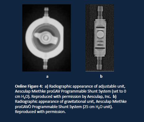
95 95
96 Pt has a shunt Programmable Non-Programmable Unknown Does it need to be checked after MRI? (ProGAV and Delta shunts don t need to be checked) Yes, it needs to be checked No, it does not need to be checked Scan patient Find out (A) type of shunt and (B) whether shunt is programmable or non-programmable. 1. Look in EPIC, implants, old operative notes, MR screening sheets, media 2. Interview patient again, Does anyone manage the shunt? Is it adjusted with a magnetic wand over the area? 3. Call where shunt was placed and try to get records. Does the patient have an appointment set up with someone (device rep or Dr Office) post-mri? NO YES For outpatients, set up a post-mri appointment, with neurosurgery or pts device manager, For inpatients, call for Neurosurgery consult ( ). All Information available Follow programmable or non-programmable process Information not available or incomplete Ask Neuroradiology to confirm whether shunt is programmable or non-programmable on shunt series X-rays. Dial next to the valve= programmable. Neurosurgery consult as back up for Neuroradiology. 96 Reschedule patient if shunt type is still unknown.
97 Appendix A Dotarem (Guebert) Fill out form located here Eovist (Bayer) bayer. Fill out form appendix B Appendix B Fax completed form to Spontaneous Adverse Event Report Form short version Sales Consultant: Voic #: Local case ID: Date informed of Adverse Event: Date of receipt of information: Initial report Follow-up report Follow-up information requested: Yes No GIVE INFORMATION ON THE PATIENT WHO HAS EXPERIENCED THE ADVERSE EVENT. Initials Gender male Age [years] FEMALE WHICH ADVERSE EVENT(S) HAS THE PATIENT EXPERIENCED? Date: Date: Date: Date: 97
98 Was the patient hospitalized? Yes No Did the patient die? Yes No DESCRIBE DETAILS OF THE ADVERSE EVENT(S). For contrast agents, please describe the procedure. (e.g. MRI, CT) TRADE NAME/ Generic Name WHICH BAYER DRUG(S) WERE INVOLVED? Example: Testobay/Testofloxacin Form u- lation Table t Tota l daily dose 200 mg Dose regime n 2x100 mg Route of application Lot number Date from-to or duratio n oral May 04 Indication for use urinary tract infection Name WHO HAS REPORTED THE ADVERSE EVENT(S)? Address Phone/Fax/ Physician Consumer Other specify: A Product Technical Complaint has been forwarded to Technical Complaints department Yes No IF MORE DETAILED INFORMATION ARE AVAILABLE FILL IN THE FULL VERSION OF THE SPONTANEOUS REPORT FORM, PLEASE. Appendix C MRI Safety Websites 98
99 Includes the List updated yearly list of MR tested devices MR Safety Papers, guidelines and information MR Safety Courses ACR website section on MRI Safety ASTM standards for testing Appendix D yid=1623&command=core.download patient screening form yid=1625&command=core.download companion screening form 99

100 Appendix E Lowering SAR and B1rms Values Increase Decrease Use Use Sparingly or Avoid TR Flip Angle Parallel Imaging FS Sequence Concatenations Slices Low Sar Mode if STIR Sequence available Slice Thickness Phase Resolution Gradient Echo Extra SAT bands sequences Averages Flip Angle Spin Echo/FSE sequences How to check SAR and B1RMS on Siemens Set up your sequence with a Pause During the Pause click on the SAR button and go to the Prediction Tab Click on the Line you re interested in B1rms or whole body SAR (ex highlighted above in yellow) Look at the read out (the green line highlighted above in RED) B1RMS number is displayed, *SAR value may be displayed in w/lb multiply by 2.2 to convert to w/kg 100
MRI Safety Policies. September
 MRI Safety Policies September- 1 YALE-NEW HAVEN HOSPITAL DEPT. OF DIAGNOSTIC RADIOLOGY POLICY AND PROCEDURE MANUAL SUBJECT: CONTACT INFORMATION MRI Safety Manual YNHH Fitkin MRI 789 Howard Ave, New Haven
MRI Safety Symposium. ACR Safe Practice Guidelines. An Overview of the ACR Guidance Document on MR Safe Practices. Zachary W. Friis, Ph.D.
 MRI Safety Symposium An Overview of the ACR Guidance Document on MR Safe Practices Zachary W. Friis, Ph.D.,DABR IT HAS BEEN RECOGNIZED THAT THERE ARE MANY POTENTIAL RISK IN THE MR ENVIRONMENT. NOT TO JUST
UBC MRI Research Centre
 THE UNIVERSITY OF BRITISH COLUMBIA UBC MRI Research Centre 3T Facility SAFETY POLICY July 2, 2008 The following document contains important safety information with respect to the 3T Facility at the UBC
CHINA BASIN 3T MRI Safety
 CHINA BASIN 3T MRI Safety Part I General Information 1. Before anyone (staff, subject, and visitor) may enter the magnet room, a screening form must be completed and reviewed by the research technologist,
TTNI Safety Policy. d. Controlled Drugs: Controlled substances are NOT allowed at this time.
 TTNI Safety Policy 1. Regulatory Requirements for the Conduct of Human Studies a. IRB and TTNI Approval: The TTNI Protocol Review Committee and the Texas Tech University Institutional Review Board (IRB)
3T/7T MRI FACILITY. SOP Number: Emergency Code Blue. Revision Chronology. Associate Director Signature: Date: Version Number Date Changes
 3T/7T MRI FACILITY SOP Number: 140.03 Title Emergency Code Blue Revision Chronology Version Number Date Changes 140.01 28 July 2008 New 140.02 21 January 2013 Updated emergency procedures 140.03 27 October
PATIENT REGISTRATION. Street City State Zip WORK INJURY/ ACCIDENT
 PATIENT REGISTRATION, Last First M.I. SEX: Male Female DOB: / _/ AGE: MARITAL STATUS: SS#: - - PHYSICIAN: ADDRESS: Street City State Zip (HOME) (WORK) TEL: - - TEL: - _- CELL: - _- EMAIL: PRIMARY INSURANCE:
UBC MRI Research Centre 7T Facility SAFETY POLICY
 THE UNIVERSITY OF BRITISH COLUMBIA UBC MRI Research Centre 7T Facility SAFETY POLICY June 13, 2007 The following document contains important safety information with respect to the 7T Facility at the UBC
PATIENT INFORMATION: CONTACT INFORMATION: EMERGENCY CONTACT: EMERGENCY PHONE: RESPONSIBLE PARTY (IF OTHER THAN PATIENT)
") PATIENT REGISTRATION PATIENT INFORMATION: NAME:,, (M.I.) ADDRESS:,, (Street) (City) (State) (Zip) SEX: MALE FEMALE DOB: / / AGE: MARITAL STATUS: SS #: / / REFERRING PHYSICIAN: CONTACT INFORMATION: (CELL):
PATIENT REGISTRATION PATIENT INFORMATION: NAME:,, (M.I.) ADDRESS:,, (Street) (City) (State) (Zip) SEX: MALE FEMALE DOB: / / AGE: MARITAL STATUS: SS #: / / REFERRING PHYSICIAN: CONTACT INFORMATION: (CELL):
Facility for Education & Research in Neuroscience (FERN) Magnetic Resonance Imaging Center Standard Operating Procedures Version 1.6, December 6, 2017
 Magnetic Resonance Imaging Center Standard Operating Procedures Version 1.6, December 6, 2017") Facility for Education & Research in Neuroscience (FERN) Magnetic Resonance Imaging Center Standard Operating Procedures Version 1.6, December 6, 2017 Facility for Education & Research in Neuroscience
Facility for Education & Research in Neuroscience (FERN) Magnetic Resonance Imaging Center Standard Operating Procedures Version 1.6, December 6, 2017 Facility for Education & Research in Neuroscience
STANDARD OPERATING PROCEDURE FOR GENERAL SAFETY
 STANDARD OPERATING PROCEDURE FOR GENERAL SAFETY SOP Number: 3T MRI 200.03 Version Number & Date: 3rd version; 01 Feb 2009 Effective Date: 01 Feb 2009 Superseded Version Number & Date (if applicable): 200.02
MRI Patient Screening and History
 Griffin Imaging, LLC 220 Rock Street Griffin, GA 30224 (770) 229-4660 Fax:: (770) 229-4632 Specializing In Open MRI, CT & Ultrasound MRI Patient Screening and History Patient Information Sheet PATIENT
Objective: Emergency Access Number Always use the code words, not the actual emergency!
 Emergency Codes Objective: At the end of this self-study module, participants will be able to demonstrate knowledge of all emergency codes and their responsibilities during each code. All codes are initiated
Using Body Mechanics
 Promotion of Safety Using Body Mechanics Muscles work best when used correctly Correct use of muscles makes lifting, pulling, and pushing easier Prevents unnecessary fatigue and strain and saves energy
UM Center for Brain Imaging Research (CBIR) MR Safety Manual.
 MR Safety Manual.") UM Center for Brain Imaging Research (CBIR) MR Safety Manual www.mdbrain.org SAFETY TRAINING Access to the imaging center: MR Operator: This individual has passed MRI safety and equipment training to ensure
UM Center for Brain Imaging Research (CBIR) MR Safety Manual www.mdbrain.org SAFETY TRAINING Access to the imaging center: MR Operator: This individual has passed MRI safety and equipment training to ensure
IMAGING RESEARCH FACILITY (IRF) STANDARD OPERATING PROCEDURES. Version 4.4 August 26, 2013
 STANDARD OPERATING PROCEDURES. Version 4.4 August 26, 2013") IMAGING RESEARCH FACILITY (IRF) STANDARD OPERATING PROCEDURES Version 4.4 August 26, 2013 Psychological and Brain Sciences, 1101 East 10th Street Indiana University Bloomington, IN, 47405-7007 IUB Imaging
IMAGING RESEARCH FACILITY (IRF) STANDARD OPERATING PROCEDURES Version 4.4 August 26, 2013 Psychological and Brain Sciences, 1101 East 10th Street Indiana University Bloomington, IN, 47405-7007 IUB Imaging
Emergency Codes - Ouellette Campus
 The Emergency Codes # is: 555 CODE RED - FIRE 555 CODE PURPLE - HOSTAGE SITUATION OR PERSON WITH WEAPON CODE BLUE - CARDIAC ARREST CODE GREEN - EVACUATION CODE YELLOW - MISSING PATIENT CODE BLACK - CODE
2017 Annual Mandatory Education. Sarasota Memorial Health Care System
 2017 Annual Mandatory Education Sarasota Memorial Health Care System Self-Study Module Questionnaire The goals of Annual Mandatory Education are to provide employees with information pertinent to their
REGISTRATION INFORMATION
 REGISTRATION INFORMATION PATIENT INFORMATION (PLEASE USE FULL LEGAL NAME) Last: First: MI: Sex: DOB: SSN# Marital Status: Home Phone: Address: Cell Phone: City: State: Zip: Employer: Work Phone: Emergency
Facility Name: Patient Registration. Name: Address: Home: Work: Mobile: Race: Gender: Marital Status: Emergency Contact Information
 Facility Date: MRN/Jacket: Patient Registration Address: Home: Work: Mobile: Email: Date of Birth: Race: Gender: Marital Status: SSN: Employer: Registered Location: Physician: Emergency Contact Information
University of California, San Diego Consent to Act as a Research Subject
 5 pages University of California, San Diego Consent to Act as a Research Subject National Institutes of Health (NIH) Nonalcoholic Steatohepatitis Clinical Research Network (NASH CRN) Farnesoid X Receptor
July 1, 2017 EMERGENCY PROCEDURES FOR DEPARTMENT OF ALLIED HEALTH SCIENCES IN BONDURANT HALL
 EMERGENCY PROCEDURES FOR DEPARTMENT OF ALLIED HEALTH SCIENCES IN BONDURANT HALL These procedures set forth the steps to be followed in the event of an emergency. It is important that all employees thoroughly
The 2005 Australian MRI Safety Survey
 MRI Safety MR Imaging Original Research The 2005 Australian MRI Safety Survey Nicholas J. Ferris 1,2 Helen Kavnoudias 3 Christy Thiel 3 Stephen Stuckey 4 Ferris NJ, Kavnoudias H, Thiel C, Stuckey S OBJECTIVE.
Contents. 1. Rationale. 2. Defining areas of responsibility. 3. Professional responsibilities. 4. Staffing of MR units. 5. Static magnetic field B 0
 ++ Contents 1. Rationale 2. Defining areas of responsibility 3. Professional responsibilities 4. Staffing of MR units 5. Static magnetic field B 0 6. Time-varying (gradient) magnetic fields 7. Radio-frequency
Required Contingency Plans for CMHCM Providers
 Required Contingency Plans for CMHCM Providers 1. Medical Emergency 2. Missing Consumer 3. Power Outage 4. Water Shortage 5. Fire 6. Bad Weather 7. Chemical/Shelter in Place 8. Choking 9. Death of Consumer
Nursing Student Orientation. Emergency Codes
 Nursing Student Orientation Emergency Codes EMERGENCY CODES CODE RED - FIRE CODE PURPLE HOSTAGE OR PERSON WITH A WEAPON CODE BLUE CARDIAC ARREST CODE GREEN - EVACUATION CODE YELLOW MISSING PATIENT CODE
Policies and Procedures Manual
 Policies and Procedures Manual BROWN UNIVERSITY MRI RESEARCH FACILITY Institute for Brain Science Sidney Frank Hall for Life Sciences Prepared by MRF Staff and Associates June 2011 Approved! 1! Rev. 6/1/2011
Macfeat Early Childhood Lab School Emergency Plan Withers Building Room 41 Rock Hill, SC (803)
") Emergency Plan Macfeat Early Childhood Lab School Emergency Plan Withers Building Room 41 Rock Hill, SC 29733 (803) 323-2219 The director may be contacted for further information or explanation of this
Emergency Plan Macfeat Early Childhood Lab School Emergency Plan Withers Building Room 41 Rock Hill, SC 29733 (803) 323-2219 The director may be contacted for further information or explanation of this
By then end of this course the participant will be able to: 0 Identify the emergency codes and what extension to use to activate the system.
 Agenda: By then end of this course the participant will be able to: 0 Identify the emergency codes and what extension to use to activate the system. 0 Explain the staff role in keeping RCHSD a safe place
Emergency Preparedness
 In the interest of maintaining a safe environment for all visitors at Stanford University, it is important for your program s staff and participants to know the following procedures in the unlikely event
KENTUCKY HOSPITAL ASSOCIATION OVERHEAD EMERGENCY CODES FREQUENTLY ASKED QUESTIONS
 KENTUCKY HOSPITAL ASSOCIATION OVERHEAD EMERGENCY CODES FREQUENTLY ASKED QUESTIONS Question - Why have standard overhead emergency codes? Answer Lessons learned from recent disasters shows that the resources
IVROP JOB SHADOW PROGRAM ORIENTATION
 IVROP JOB SHADOW PROGRAM ORIENTATION Hospital Incident Command System (HICS) Emergency Codes Hospital Emergency Incident Command System Emergency Codes HEICS Emergency Codes These codes are part of the
SAMPLE: Environmental Rounds and Safety Assessment Tool
 SAMPLE: Environmental Rounds and Safety Assessment Tool Area/Department Evaluated: Date: Security and Incident Management Y N N/A Comments 1. Are emergency telephone numbers posted by all stationary phones?
Mediastinal Venogram and Stent Insertion
 Mediastinal Venogram and Stent Insertion Radiology Department Patient information leaflet This leaflet tells you about the procedure known as a mediastinal venogram. It explains what is involved and the
ACR Guidance Document for Safe MR Practices: 2007
 Kanal et al. MRI Original Research 06_06_1616.fm 3/1/07 ACR Guidance Document for : 2007 Emanuel Kanal 1 A. James Barkovich 2 Charlotte Bell 3 James P. Borgstede 4 William G. Bradley, Jr. 5 Jerry W. Froelich
Emergency Operations Plan
 Emergency Operations Plan Twin Valley Community Local School District 100 Education Drive District Office West Alexandria, Ohio 45381 (937) 839-4688 Twin Valley Community Local School Emergency Operations
Polices and Procedures Manual
 Polices and Procedures Manual UNIVERSITY of CALIFORNIA SANTA BARBARA BRAIN IMAGING CENTER Psychology East Building Room B08 Prepared by BIC Staff Director: Scott Grafton MD 805-975-5272 grafton@psych.ucsb.edu
PATIENT SAFETY. Prevention of Error Physician and ADP Roles and Responsibilities
 PATIENT SAFETY Prevention of Error Physician and ADP Roles and Responsibilities Patient Safety At Navicent Health is our goal to provide the safest possible environment for our patients. Providing a safe
a. is used to administer an electric shock through the chest wall to the heart;
 Policy and Procedures for Use of Automatic External Defibrillators (AED) by CTHSS School Personnel and the Development of School AED Emergency Action Plans Overview: It has been well demonstrated that
IMPLEMENTATION PACKET
 EMERGENCY MEDICAL SERVICES AGENCY 300 North San Antonio Road Santa Barbara, CA 93110-1316 805/681-5274 FAX 805/681-5142 PUBLIC ACCESS DEFIBRILLATION IMPLEMENTATION PACKET Developed by: Marc Burdick, EMT-P,
Objective: Introduction: Abstract:
 Cost justification for using FDA Cleared MR Conditional Disposable EEG Electrodes for ceeg when 24/7 Neurodiagnostic Technologist Staff Coverage is not available Abstract: Leah Hanson, R.EEG/EPT Director
GREY NUNS COMMUNITY HOSPITAL ACTIVE ASSAILANT EMERGENCY RESPONSE PLAN
 GREY NUNS COMMUNITY HOSPITAL ACTIVE ASSAILANT EMERGENCY RESPONSE PLAN ACTIVE ASSAILANT EMERGENCY RESPONSE PLAN ALGORITHM Staff Member Discovering the incident Staff in close proximity to the incident Recognizes
LIMESTONE COUNTY SCHOOLS EMERGENCY MANAGEMENT GUIDE
 LIMESTONE COUNTY SCHOOLS EMERGENCY MANAGEMENT GUIDE ORGANIZATION PHONE ORGANIZATION PHONE Company 1 (XXX) XXX-XXXX Company 2 (XXX) XXX-XXXX Bomb Threat procedures: 1. Administration will evaluate the threat
JOB DESCRIPTION Position: Registered Radiologic Technologist
 JOB DESCRIPTION Position: Registered Radiologic Technologist POSITION TITLE: Registered Radiologic Technologist APPROVED BY: Medical Imaging Services Manager LATEST REVIEW OF JOB DESCRIPTION: 9/16 FORMER
Mediastinal Venogram and Stent Insertion
 Patient information leaflet Royal Surrey County Hospital NHS Foundation Trust Mediastinal Venogram and Stent Insertion Radiology This leaflet tells you about the procedure known as a mediastinal venogram.
Sample. A guide to development of a hospital blood transfusion Policy at the hospital level. Effective from April Hospital Transfusion Committee
 Sample A guide to development of a hospital blood transfusion Policy at the hospital level Name of Policy Blood Transfusion Policy Effective from April 2009 Approved by Hospital Transfusion Committee A
Life Safety for Students
 Life Safety for Students DISASTER RESPONSE Tift Regional Health System s Disaster Response includes an emergency code system and general actions each employee should take in the specified emergency situation.
Niagara County Emergency Services
 Niagara County Emergency Services STANDARD OPERATING PROCEDURES Subject: J. Francis Gaughn and Wheatfield Training Facilities Use by Fire Service or Law Enforcement Agencies Table of Contents Chapter 1...
Emergency Codes. ~( Code Triage
 Emergency Codes ~( Code Red ~( Code Green ~( Code Blue/Code Broselow ~( Code Gray ~( Code Pink ~( Code Brown ~( Code White ~( Code Yellow ~( Code Black ~( Code Triage Code Red (Code Green- Drill} Code
AUTOMATED EXTERNAL DEFIBRILLATOR IN THE SCHOOL SETTING
 Hitchcock Independent School District Carla Vickroy Superintendent AUTOMATED EXTERNAL DEFIBRILLATOR IN THE SCHOOL SETTING 2014-2015 1 TABLE OF CONTENTS AUTOMATIC EXTERNAL DEFIBRILLATOR (AED) PROGRAM...
MRI Safety: Inherent Dangers and Preventative Measures. Thursday, February 13 th, 2014
 MRI Safety: Inherent Dangers and Preventative Measures Thursday, February 13 th, 2014 Speaker Sue Dill Calloway RN, Esq. CPHRM, CCMSCP AD, BA, BSN, MSN, JD President Patient Safety and Healthcare Education
MRI Safety: Inherent Dangers and Preventative Measures
 MRI Safety: Inherent Dangers and Preventative Measures Thursday, February 13 th, 2014 Speaker Sue Dill Calloway RN, Esq. CPHRM, CCMSCP AD, BA, BSN, MSN, JD President Patient Safety and Healthcare Education
Indications for Calling A Code Blue or Pediatric Medical Emergency
 Code Blue/Pediatric Medical Emergency Code Blue is a term used to alert the Code Team and hospital staff of the significant deterioration in an individual s status (e.g. unresponsiveness, absence of blood
Radiologic Technologist Best Practices for MR Safety
 Radiologic Technologist Best Practices for MR Safety Lorenza Clausen, R.T.(R)(CT)(MR), MRSO(MRSC), CRT; Joy Cook, MS, R.T.(R)(CT)(MR); Amanda Garlock, MS, R.T.(R)(MR); Ashley Perkins, MHA, R.T.(R)(MR);
UNIVERSITY OF TOLEDO
 UNIVERSITY OF TOLEDO SUBJECT: CODE VIOLET VIOLENT SITUATION Procedure No: EP-08-015 PROCEDURE STATEMENT Code Violet will be initiated for serious situations involving any individual(s) exhibiting or threatening
LESSON ASSIGNMENT. Environmental Health and the Practical Nurse. After completing this lesson, you should be able to:
 LESSON ASSIGNMENT LESSON 6 Environmental Health and the Practical Nurse. LESSON ASSIGNMENT Paragraphs 6-1 through 6-5. LESSON OBJECTIVES After completing this lesson, you should be able to: 6-1. Select
FAMILY DISASTER PLAN. Name: Date: 4 STEPS OF SAFETY LOCAL OFFICE:
 FAMILY DISASTER PLAN Name: Date: It is recommended that every employee of the judicial branch have a personalized Family Disaster Plan. Families can - and do - cope with disaster by preparing in advance
The Practice Standards for Medical Imaging and Radiation Therapy. Radiography Practice Standards
 The Practice Standards for Medical Imaging and Radiation Therapy Radiography Practice Standards 2017 American Society of Radiologic Technologists. All rights reserved. Reprinting all or part of this document
PyroLance LLC. STANDARD OPERATING PROCEDURE
 PyroLance LLC. STANDARD OPERATING PROCEDURE Section: Subject: Approved: Demo/Training Live Fire Training Exercises SOP: 1.00 October 24, 2012 Execute d: Revised: Casparus Seyffert VP Operations Table of
WHAT IS AN EMERGENCY? WHY IT IS IMPORTANT TO PREPARE COMMUNICATIONS
 OVERVIEW The purpose of this plan is to provide for the carrying out of emergency functions to save lives; establish responsibilities necessary to performing these functions; prevent, minimize, and repair
Health and Safety Policy (Incorporating first aid and fire information)
") Health and Safety Policy (Incorporating first aid and fire information) It is the policy of Pollyteach to comply with the terms of the Health and Safety at Work Act 1974 and subsequent legislation and
Health and Safety Policy (Incorporating first aid and fire information) It is the policy of Pollyteach to comply with the terms of the Health and Safety at Work Act 1974 and subsequent legislation and
Emergency Response For Schools
 Emergency Response For Schools A resource to help School Administrator s manage the first 30 minutes during an emergency. to Chignecto Central Regional Centre for Education 1 Emergency Response for Schools
Environment of Care & Radiation Safety (EOC)
") Carolinas HealthCare System Blue Ridge Environment of Care & Radiation Safety (EOC) The Role of Accreditation and Regulatory Agencies The content of this training module was developed to meet criteria
Carolinas HealthCare System Blue Ridge Environment of Care & Radiation Safety (EOC) The Role of Accreditation and Regulatory Agencies The content of this training module was developed to meet criteria
University of Virginia Health System TABLE OF CONTENTS
 TABLE OF CONTENTS ACTIVE SHOOTER (RED ALERT)... Tab 1 BIOLOGICAL SPILL... Tab 2 BOMB THREAT... Tab 3 CHEMICAL SPILL... Tab 4 COMPUTER SYSTEMS... Tab 5 EARTHQUAKE... Tab 6 EVACUATION... Tab 7 FIRE... Tab
A AIRWAY Open the Airway B BREATHING Deliver two (2) Breaths. Code Blue Policy. Indications for Calling A Code Blue
 Breaths. Code Blue Policy. Indications for Calling A Code Blue") Code Blue Policy Code Blue is a term used to alert the Code Team and hospital staff of the significant deterioration in a patient s status (e.g. unresponsiveness, absence of blood pressure, status epilepticus)
Code Blue Policy Code Blue is a term used to alert the Code Team and hospital staff of the significant deterioration in a patient s status (e.g. unresponsiveness, absence of blood pressure, status epilepticus)
New Fire Safety Rules Summary Evvie Munley, LeadingAge
 New Fire Safety Rules Summary Evvie Munley, LeadingAge Following is the link to the Centers for Medicare and Medicaid Services (CMS) Final Rule, Medicare and Medicaid Programs; Fire Safety Requirements
JHU Civil Engineering Laboratory Manual Contents
 JHU Civil Engineering Laboratory Manual Contents 1. About the Manual... 2 2. THE JHU CE Laboratories... 2 3. Emergency Contacts... 3 4. Safety Regulations... 4 5. Laboratory Policy... 5 6. Annual Cleaning...
WHEELING-OHIO COUNTY BOARD OF HEALTH WHEELING-OHIO COUNTY HEALTH DEPARTMENT
 WHEELING-OHIO COUNTY BOARD OF HEALTH WHEELING-OHIO COUNTY HEALTH DEPARTMENT TITLE This Regulation shall be known as the Wheeling-Ohio County Health Department Tanning Bed Regulation and shall cover Ohio
Patient Safety: A Multidisciplinary Approach
 Patient Safety: A Multidisciplinary Approach Talk Objectives What is patient safety? Why is it important? Who is responsible for patient safety? What is a Culture of Safety? What can you do? What is Patient
SECTION 6 PERSONAL PROTECTIVE EQUIPMENT (PPE)
") SECTION 6 PERSONAL PROTECTIVE EQUIPMENT (PPE) 6.1 Personal Protective Equipment 6.2 Respiratory Protection Program 6.3 Information for Voluntary Use of Respirators 6.4 Hearing Conservation Program Reviewed:
SECTION 6 PERSONAL PROTECTIVE EQUIPMENT (PPE) 6.1 Personal Protective Equipment 6.2 Respiratory Protection Program 6.3 Information for Voluntary Use of Respirators 6.4 Hearing Conservation Program Reviewed:
Fire Captain (2142) Task List
 Task List") Fire Captain (2142) Task List OVERALL FUNCTION STATEMENT: Commands a Fire Department company, or unit involved in fire suppression, fire prevention, arson investigation, communications, training, the maintenance
Fire Captain (2142) Task List OVERALL FUNCTION STATEMENT: Commands a Fire Department company, or unit involved in fire suppression, fire prevention, arson investigation, communications, training, the maintenance
Chapter 10. Safety Needs. Copyright 2019 by Elsevier, Inc. All rights reserved.
 Chapter 10 Safety Needs Copyright 2019 by Elsevier, Inc. All rights reserved. Lesson 10.1 Define the key terms and key abbreviations in this chapter. Describe accident risk factors. Explain why you identify
HEALTH & SAFETY EDUCATION FOR THE WORKPLACE
 HEALTH & SAFETY EDUCATION FOR THE WORKPLACE Pamela L. Smith, Consultant and Facilitator Safety Consultant and Facilitator 1 INTRODUCTION TO HEALTH & SAFETY FOR SMALL BUSINESS TOPICS Workplace Safety Basic
EMERGENCY CONDITIONS & BASIC STAFF RESPONSE
 EMERGENCY CONDITIONS & BASIC STAFF PROBLEM CODE NAME DESCRIPTION INITIAL Admitted Adult Patient Missing Code Brown or has left where patient s leaving may be life-threatening to patient. Code Brown Procedure
UW MEDICINE PATIENT EDUCATION. Angiography: Kidney Exam. How to prepare and what to expect. What is angiography? DRAFT. Why do I need this exam?
 UW MEDICINE PATIENT EDUCATION Angiography: Kidney Exam How to prepare and what to expect This handout explains how to prepare and what to expect when having a kidney exam using angiography. What is angiography?
UW MEDICINE PATIENT EDUCATION. Angiography: Radiofrequency Ablation to Treat Solid Tumor. What to expect. What is radiofrequency ablation?
 UW MEDICINE PATIENT EDUCATION Angiography: Radiofrequency Ablation to Treat Solid Tumor What to expect This handout explains radiofrequency ablation and what to expect when you have this treatment for
CASPER COLLEGE MAGNETIC RESONANCE IMAGING PROGRAM ADMISSION GUIDE 2015/2016
 CASPER COLLEGE MAGNETIC RESONANCE IMAGING PROGRAM ADMISSION GUIDE 2015/2016 Revised 9/15 1 Table of Contents MRI Program Information.... 3 Program Admission Criteria/checklist...4 MRI Program Academic
Emergency Management
 Slide 1 Emergency Management Annual Training Module Welcome to the Emergency Management Learning Module. Emergency Management, Safety and Security are critical areas of operation within Lehigh Valley Health
Health and Safety Policy
 Health and Safety Policy Statement of intent: It is the policy of Step by Step School to comply with the terms of the Health and Safety at Work Act 1974 and subsequent legislation to provide and maintain
Adult Family Care Home Top Ten Health Deficiency Citations Statewide October 8, 2009 Year Date Range: January 1, 2008 through December 31, 2008
 Rank Tag Count Description Adult Family Care Home 1 F0401 182 Personnel records must include verification of freedom from communicable disease for the AFCH provider, each relief person, each adult household
Inferior Vena Cava (IVC) Filter Insertion
 Filter Insertion") Patient information leaflet Royal Surrey County Hospital NHS Foundation Trust Inferior Vena Cava (IVC) Filter Insertion Radiology This leaflet informs you about the procedure known as an Inferior Vena
Patient information leaflet Royal Surrey County Hospital NHS Foundation Trust Inferior Vena Cava (IVC) Filter Insertion Radiology This leaflet informs you about the procedure known as an Inferior Vena
Burn Intensive Care Unit
 Purpose The burn wound is especially susceptible to microbial invasion because of loss of the protective integument and the presence of devitalized tissue. Reduction of the risk of infection is of utmost
MR Safety in Clinical Practice
 1 2 MR Safety in Clinical Practice Which environment is safer? Wm. Faulkner, BS,RT(R)(MR)(CT),MRSO(MRSC ) Kristan Harrington, MBA,RT(R)(MR),MRSO(MRSC ) www.t2star.com/faulkner/mrso_coursematerial 3 4 1999:
Self-Assessment and Cross-Referencing for internationally trained magnetic resonance technologists
 Self-Assessment and Cross-Referencing for internationally trained magnetic resonance technologists Name: Date: This self-assessment tool is meant to assist you in identifying how your previous program
Big Sandy Fire Protection District/Simla Volunteer Fire Department Junior Firefighter Program Guidelines
 Big Sandy Fire Protection District/Simla Volunteer Fire Department Junior Firefighter Program Guidelines The Junior Firefighter Program was instituted by the Big Sandy Fire Protection District (Big Sandy
4. In most schools the plan should be that a witness calls the front office ASAP, and staff there will:
 1 Cardiac Emergency Response Plans 10 About: Cardiac Emergency Response Plans This plan should be in place for all schools, since sudden cardiac arrest can happen to anyone in the school, mostly to adults,
Radiation Dose Management Requirements from MACRA and Joint Commission, Potential Effects on Reimbursement
 Radiation Dose Management Requirements from MACRA and Joint Commission, Potential Effects on Reimbursement Radiation dose requirements are being slowly integrated into key performance indicators and metrics
SENIOR/ASSISTED LIVING FACILITY SPECIFIC QUESTIONNAIRE
 Corporate/Parent Name: SENIOR/ASSISTED LIVING FACILITY SPECIFIC QUESTIONNAIRE (please provide the following for each facility) Facility Specific Questionnaire Facility Description 1. Facility name: Location
Percutaneous Transhepatic Cholangiogram (PTC) and Biliary Stent
 and Biliary Stent") Percutaneous Transhepatic Cholangiogram (PTC) and Biliary Stent Radiology Department Patient information leaflet This leaflet informs you about the procedure known as a Percutaneous Transhepatic Cholangiogram
Percutaneous Transhepatic Cholangiogram (PTC) and Biliary Stent Radiology Department Patient information leaflet This leaflet informs you about the procedure known as a Percutaneous Transhepatic Cholangiogram
Patient Safety: Fall Prevention. Unlicensed Assistive Personnel
 Patient Safety: Fall Prevention Unlicensed Assistive Personnel Purpose and Objectives Purpose: Review the UCH Fall Prevention Program Objectives: 1. Present evidence about patient safety and falls. 2.
Patient Information Fracture Clinic
 Patient Information Fracture Clinic Orthopaedic Department Introduction This department deals with patients who have been diagnosed with a fracture or an injury within the A&E department or have been referred
The Practice Standards for Medical Imaging and Radiation Therapy. Cardiac Interventional and Vascular Interventional Technology. Practice Standards
 The Practice Standards for Medical Imaging and Radiation Therapy Cardiac Interventional and Vascular Interventional Technology Practice Standards 2017 American Society of Radiologic Technologists. All
Integrated Safe School Plan. Emergency Team Duties and Supply Lists. Incident Command System
 Emergency Team Duties and Supply Lists Incident Command System The Incident Command System (ICS) is used by first responder and government agencies to manage emergencies, crises, and disasters nationwide.
FY 18-Annual Education Module Test: Clinical 1. General Information
 FY 18-Annual Education Module Test: Clinical 1. General Information 1. What items should be included in a Fixit Ticket? a. Time b. Place c. Facts only 2. LEAN theory believes that staff who do the job
for the Wilderness CHECK: Check the Scene, the Resources and the Person person, other members of the group and any bystanders.
 Check Call Care for If you find yourself in an emergency, you should follow three basic emergency action principles: CHECK CALL CARE. These principles will help guide you in caring for the patient and
Standards. Successfully Preparing for Your Next AAAHC Accreditation Survey Annual Conference
 Successfully Preparing for Your Next AAAHC Accreditation Survey 2012 Annual Conference Guest Speaker Ray Grundman, MSN, MPA, CASC AAAHC Senior Director External Relations AAAHC Surveyor AAAHC - Past President
Using CAST for Adverse Event Investigation in Hospitals
 Using CAST for Adverse Event Investigation in Hospitals Meaghan O Neil March 27, 2014 Motivation As many as 98,000 people, die in hospitals each year as a result of medical errors that could have been
RMU Safety Update. Hazard Surveillance Inspections. RMU Safety on the Web. March 2006
 RMU Safety Update Published by the Robert Morris University Safety Department, (412) 262-8328 March 2006 Inside This Issue: Written Safety Policies...2 and Procedures...3 Members Partnerships Helped...4
POLCIE, AMBULANCE, FIRE DEPARTMENT DIAL FIRE, DISASTER, EVACUATE 3 BELLS
 FANNINDEL ISD EMERGENCY PROCEDURES MANUAL POLCIE, AMBULANCE, FIRE DEPARTMENT 911 DIAL FIRE, DISASTER, EVACUATE 3 BELLS FANNIN COUNTY SHERIFF 903-583-2143 DELTA COUNTY SHERIFF 903-395-2146 FANNINDEL ISD
STANDARD OPERATING GUIDELINE Civil Disturbances
 Purpose Fire Ground Safety Initiative STANDARD OPERATING GUIDELINE Civil Disturbances This standard operating guideline has been developed to provide basic operating guidelines for the personnel responding