Clinical Skills in Hospitals Project Chest tube management
|
|
|
- Brittany Norton
- 6 years ago
- Views:
Transcription
 Module 2: Pleurocaths Module 3: Underwater seal drains Module 4: Management")
1 Clinical Skills in Hospitals Project Chest tube management Module 1: Intercostal catherters (ICCs) Module 2: Pleurocaths Module 3: Underwater seal drains Module 4: Management scenarios
2
3 Clinical Skills in Hospitals Project Chest tube management Module 1: Intercostal catherters (ICCs) Module 2: Pleurocaths Module 3: Underwater seal drains Module 4: Management scenarios
4 ii Chest tube management Acknowledgments The authors wish to acknowledge the following people for their important contributions to this project: Dr Stuart Dilley, Mr Naveed Alam, Dr Robert O Brien, Julian Van Dijk, Santina Cotela and Dr Neil Cunningham from St Vincent s Hospital, Melbourne, for their tireless efforts as the primary authors of Chest tube management. Mr Matthew Williams at St Vincent s Education Centre for his contributions to this package. For further information please contact the project manager, Julian Van Dijk: (03) julian.vandijk@svhm.org.au State of Victoria 2009 This publication is copyright. No part may be reproduced by any process except in accordance with the provisions of the Copyright Act The Clinical Skills Centres in Hospitals Training Materials ( Materials ) were developed for the Department of Human Services ( DHS ) for use by clinical educators ( Educators ). Any Educator wishing to use the Materials must review the Materials prior to use and ensure that they are suitable for the Educator s purpose, including the needs of the Educator s intended training recipients ( Training Recipients ). Some aspects of the Materials may not be relevant to some Educators or Training Recipients. Educators and Training Recipients must not represent themselves as employees or agents of DHS in their use of the Materials. Except at the time of publication, neither DHS nor the State of Victoria warrants the accuracy of the information contained in the Materials. Neither DHS nor the State of Victoria makes any warranty as to the appropriateness of the information to the circumstances of Educators or Training Recipients. The Materials may not include all of the information required by Educators or Training Recipients in providing or receiving clinical training. If in doubt, Educators and Training Recipients should seek their own advice about the interpretation of the Materials. Any Educator or Training Recipient using the Materials does so at their own risk and shall accept responsibility for any actions arising from their use. DHS may update the Materials at its own discretion, but is not responsible for the provision of updates to the Materials. No responsibility is taken for failure to update the Materials and it is the responsibility of the Educator to ensure that the information contained in the Materials is up to date and reflects current medical practice, law and guidelines.
5 Chest tube management iii Preface In 2007 the Department of Human Services commissioned St Vincent s Hospital Melbourne, to design and develop simulation-based training packages for clinical skills trainers in Victorian hospitals. The project provides Victorian health professionals specifically, hospital clinical educators with a resource to deliver simulation-based clinical skills training. The information in this manual complements current training programs and should be considered as a resource in the workplace, rather than the definitive resource on the topic. Every effort has been made to provide the most current literature references. Authors have consulted other health professionals and current programs when possible in development to ensure that the modules produced in this package are consistent with current health practices.
6
7 Chest tube management v Course delivery in condensed form Sample timetable for one-day workshop This is an example of how the modules in Chest tube management could be combined into a one-day workshop. A sample timetable is provided for a course consisting of Modules 1, 2, 3 and 4. Course 1 (Modules 1, 2, 3 and 4) Timing Activity Objective 8.30 to 8.45 Introduction to faculty and participants 8.45 to 9.15 Facilitated discussion Module 1: 1, 2, to ICC insertion skills session Module 1: 3 and to Summary of main points from Module 1 Module 1: all to Morning tea to Facilitated discussion Module 2: to Skill station using part task trainers Module 2: 2, to Scenario around pleural catheter insertion to Debrief of scenario Module 2: all to Summary of main points from Module 2 Module 2: all to 1.15 Lunch 1.15 to 1.35 Facilitated discussion Module 3: all 1.35 to 2.15 Skill station/case study Module 3: all 2.15 to 2.25 Summary of main points from Module 3 Module 3: all 2.25 to 2.40 Afternoon tea 2.40 to 3.00 Introduction and manikin familiarisation Module 4: to 3.10 Simulation 1 Module 4: all 3.10 to 3.40 Debrief Module 4: all 3.40 to 3.50 Simulation 2 Module 4: all 3.50 to 4.20 Debrief Module 4: all 4.20 to 4.30 Summary of main points from Module 4 Course evaluation
8
9 Chest tube management Introduction vii Chest tube management Introduction Chest tube management was developed as a teaching and learning tool for Victorian clinical educators. The information contained in each module was developed using evidence-based resources and examples of best practice. Where expert opinion varies, a discussion section is included. However, it is not within the scope of Chest tube management to address the full spectrum of local variations. Variations can occur in several areas, including practices relating to types of equipment used, infection control processes, practice guidelines and so on. Therefore, educators should, where appropriate, adapt content to reflect their local policies, procedures and protocols. This will ensure the relevancy of the package content to your learners. The modules are designed to be discrete courses in their own right. They are timetabled so they can be completed in a 1 2 hour timeframe. This timeframe was chosen after we received feedback from clinical educators requesting shorter courses, because health professionals often have limited time to educate away from patients. However, the packages may also be combined into a one- or two-day course. Chest tube management should be used as an educational tool to assist in the teaching of clinical skills. It is structured as a guide to assist clinical educators, and uses many concepts taught in the Clinical Skills in Hospitals Project (Train-the-Trainer courses). Educators are encouraged to build on this resource by adding their own scenarios which incorporate health service protocols, policies and other resources. Each module is designed as a lesson plan to incorporate the simulations into the teaching of clinical skills. Aims Chest tube management aims to make participants confident in their application of skills associated with identifying indications for inserting intercostal catheters and finebore cannulae, and appropriately inserting and managing these catheters and cannulae in different environments and settings. This package is intended for use with medical and nursing participants. Package structure Chest tube management contains four modules that provide learning opportunities for health professionals at all levels of experience and from medical and nursing disciplines. Modules 1 and 2 are regarded as fundamental. Modules 3 and 4 are more difficult and are regarded as intermediate.
10 viii Chest tube management Introduction Level of complexity Complex For participants with more than 4 years experience or who have completed Modules 1 4 Intermediate For participants in postgraduate years 3 4 or who have completed Modules 1 and 2 Fundamental For participants in postgraduate years 1 2 Package structure Underwater seal drains Intercostal catheters Chest tube management scenarios Pleurocaths Skills included in Chest tube management include chest tube insertion, needle aspiration, fine-bore catheter insertion (for example, Pleurocath) underwater seal drains and haemo-pneumothorax management. This package was designed to develop participants knowledge, skills and behaviours in the safe management of pneumothoraces and haemothoraces, including chest tube insertion and management. The modules expose participants to increasingly complex skills and knowledge and test their ability to combine these individual skills, work as a team and solve problems in more difficult situations. Educators delivering these modules should be aware of participants level of experience and choose appropriate modules. Modules presume an increasing level of knowledge as they progress, ranging from a fundamental knowledge of anatomy and physiology for the fundamental modules, up to detailed knowledge of haemopneuomothorax management for the complex modules. Novice participants (such as first-year graduates) are expected to start with the fundamental modules, and only move onto intermediate and more complex modules as they demonstrate proficiency. More experienced participants may start at the intermediate level if the educator is satisfied that they have the prior knowledge and skills. Individual educators are responsible for assessing each participant s baseline knowledge and determining which modules they need to complete. More specific descriptions of presumed knowledge are outlined in each module.
11 Chest tube management Introduction ix The design of these packages presumes that the clinical educators using them have knowledge and expertise in current best practice regarding the teaching of clinical skills and conducting facilitated discussions. Knowledge and expertise are presumed commensurate with the Department of Human Services basic and advanced Train-the-Trainer programs. Clinical educators are encouraged to refer to the Department of Human Services Clinical Skills Facilitators Manual for theory on: 1. Peyton s model for teaching clinical skills 2. leading small group discussions 3. giving feedback 4. crisis resource management skills.
12
13 Chest tube management Module 1: Intercostal catheters (ICCs) 1 Module 1: Intercostal catheters (ICCs) Introduction Chest tube management was developed as a teaching and learning tool for Victorian clinical educators. The information contained in each module was developed using evidence-based resources and examples of best practice. Where expert opinion varies, a discussion section is included. However, it is not within the scope of Chest tube management to address the full spectrum of local variations. Variations can occur in several areas, including practices relating to types of equipment used, infection control processes, practice guidelines and so on. Therefore, educators should, where appropriate, adapt content to reflect their local policies, procedures and protocols. This will ensure the relevancy of the package content to your learners. The modules are designed to be discrete courses in their own right. They are timetabled so they can be completed in a 1 2 hour timeframe. This timeframe was chosen after we received feedback from clinical educators requesting shorter courses, because health professionals often have limited time to educate away from patients. However, the packages may also be combined into a one- or two-day course. Chest tube management should be used as an educational tool to assist in the teaching of clinical skills. It is structured as a guide to assist clinical educators, and uses many concepts taught in the Clinical Skills in Hospitals Project (Train-the-Trainer courses). Educators are encouraged to build on this resource by adding their own scenarios which incorporate health service protocols, policies and other resources. Each module is designed as a lesson plan to incorporate the simulations into the teaching of clinical skills. Aims Chest tube management aims to make participants confident in their application of skills associated with identifying indications for inserting intercostal catheters and finebore cannulae, and appropriately inserting and managing these catheters and cannulae in different environments and settings. This package is intended for use with medical and nursing participants. Package structure Chest tube management contains four modules that provide learning opportunities for health professionals at all levels of experience and from medical and nursing disciplines. Modules 1 and 2 are regarded as fundamental. Modules 3 and 4 are more difficult and are regarded as intermediate.
14 2 Chest tube management Module 1: Intercostal catheters (ICCs) Level of complexity Complex For participants with more than 4 years experience or who have completed Modules 1 4 Intermediate For participants in postgraduate years 3 4 or who have completed Modules 1 and 2 Fundamental For participants in postgraduate years 1 2 Package structure Underwater seal drains Intercostal catheters Chest tube management scenarios Pleurocaths Skills included in Chest tube management include chest tube insertion, needle aspiration, fine-bore catheter insertion (for example, Pleurocath) underwater seal drains and haemo-pneumothorax management. This package was designed to develop participants knowledge, skills and behaviours in the safe management of pneumothoraces and haemothoraces, including chest tube insertion and management. The modules expose participants to increasingly complex skills and knowledge and test their ability to combine these individual skills, work as a team and solve problems in more difficult situations. Educators delivering these modules should be aware of participants level of experience and choose appropriate modules. Modules presume an increasing level of knowledge as they progress, ranging from a fundamental knowledge of anatomy and physiology for the fundamental modules, up to detailed knowledge of haemopneuomothorax management for the complex modules. Novice participants (such as first-year graduates) are expected to start with the fundamental modules, and only move onto intermediate and more complex modules as they demonstrate proficiency. More experienced participants may start at the intermediate level if the educator is satisfied that they have the prior knowledge and skills. Individual educators are responsible for assessing each participant s baseline knowledge and determining which modules they need to complete. More specific descriptions of presumed knowledge are outlined in each module.
15 Chest tube management Module 1: Intercostal catheters (ICCs) 3 The design of these packages presumes that the clinical educators using them have knowledge and expertise in current best practice regarding the teaching of clinical skills and conducting facilitated discussions. Knowledge and expertise are presumed commensurate with the Department of Human Services basic and advanced Train-the-Trainer programs. Clinical educators are encouraged to refer to the Department of Human Services Clinical Skills Facilitators Manual for theory on: 1. Peyton s model for teaching clinical skills 2. leading small group discussions 3. giving feedback 4. crisis resource management skills.
16
17 Chest tube management Module 1: Intercostal catheters (ICCs) 5 Module 1: Intercostal catheters (ICCs) Authors: Mr Naveed Alam, Dr Robert O Brien Aims This module teaches participants to identify the indications for the insertion of a chest tube, and prompts discussion and practice of the technique for insertion of intercostal catheters (ICCs). Presumed knowledge This module is targeted to medical professionals with minimal experience in inserting chest tubes. However, they are expected to have a basic knowledge of: 1. The anatomy of the chest wall and chest cavity 2. Infection control and working in sterile environments 3. Basic surgical skills (i.e. making incisions and suturing. This module gives nursing professionals the opportunity to increase their awareness of the procedure as performed by a medical practitioner. Objectives By the end of this module, participants should have: 1. discussed the anatomy of the chest wall and chest cavity 2. identified the indications for insertion of an ICC 3. discussed the recommended site and technique for placement and insertion of an ICC 4. practised the recommended site and technique for placement and insertion of an ICC. Background information for educators This module encourages participants to become familiar with ICCs. Educators should take note of the mix of health professionals in the group. While medical staff are responsible for insertion of pleural catheters, all health professionals should be aware of the indications, techniques and problems associated with ICC insertion. If the group comprises non-medical staff, the focus of discussion and learning sessions should be on familiarity with equipment and assisting medical staff with pleural catheter insertion. Overview Pleural space anatomy diseases. Chest tubes indications insertion.
18 6 Chest tube management Module 1: Intercostal catheters (ICCs) Drainage systems management. Anatomy of the pleural space The pleura has two layers: visceral on the lung surface parietal on the chest wall, mediastinum and diaphragm. Pleural space is a potential space: in normal circumstances, there is no space, and the visceral and parietal layers are in apposition space develops when there is disruption of the layers. Pleural space normally has minimal fluid in it (10 ml), which is used as a lubricant to minimise friction during respiration. Diseases of the pleural space Air pneumothorax (PTX) can be classified as open or closed. Open pneumothoraces: communicate with atmospheric pressure through a hole in the chest wall are usually secondary to trauma. Closed pneumothoraces: can be spontaneous, related to trauma or iatrogenic traumatic closed pneumothoraces are usually related to blunt trauma. Air pneumothorax (PTX) can also be classified as tension or non-tension. Tension pneumothoraces are defined by: hypotension secondary to lack of venous return and subsequent decreased cardiac output: normally, venous return to the heart is facilitated by the somewhat negative intrathoracic pressure when there is a tension PTX, the intrathoracic pressure becomes elevated signs include: ipsilateral absence of breath sounds hyper-resonance contralateral tracheal deviation i Tension pneumothoraces require immediate decompression do not wait for CXR to confirm tension pneumothoraces.
19 Chest tube management Module 1: Intercostal catheters (ICCs) 7 use large IV cannula (14 or 16 gauge) into the second intercostal space (ICS) in the mid-clavicular line after decompression, place normal chest tube (24 or 28 French). If a chest X-ray is taken, one can see: mediastinal shift depression of ipsilateral diaphragm complete collapse of the lung. Chest tubes for PTX generally should be guided apically, and can be placed in the fourth ICS in the anterior to mid-axillary line. Blood haemothorax usually secondary to trauma may also be related to malignancy. Often a well-placed chest tube is the most important step in the management of a haemothorax. Proper drainage of the pleural space enables expansion of the underlying lung, which can often tamponnade the source of bleeding. Immediate evacuation of greater than 1.5 litres of blood, or ongoing drainage of > 200 ml per hour for more than two hours are indications for operative exploration. Chest tubes can be placed by experienced practitioners in lower positions and guided posteriorly. If placed by inexperienced staff they should be placed in the fourth ICS in the mid-axillary line. Pus pyothorax or empyema often related to pneumonia other causes are post-traumatic and post-operative usually requires drainage, either with chest tubes or surgical procedures. Excess pleural fluid pleural effusion Pleural effusion can be transudative or exudative: transudates: can be related to congestive heart failure or liver failure often respond to medical management exudates: can be related to pneumonia or malignancy often require drainage.

20 8 Chest tube management Module 1: Intercostal catheters (ICCs) Chyle chylothorax usually related to surgery in the chest or malignancy management is complex, and often starts with drainage and dietary restrictions. Chest tube indications as noted above. Chest tube insertion set up Equipment list chest tube suture large gauge (0 silk or Prolene) long curved forcep (Roberts) scalpel local anaesthetic preparation drapes clamps. Figure 1: Table set-up Patient positioning Make the patient as comfortable as possible. It is generally easier to place a chest tube when the patient is in a lateral position with their arms folded together and a pillow between them in the praying position. This helps the patient to be comfortable and allows placement by creating a flat surface. If not possible, tubes may need to be placed with the patient in a supine position, or even semi-upright.
21 Chest tube management Module 1: Intercostal catheters (ICCs) 9 Chest tube insertion steps 1. local anaesthesia 2. incision 3. dissection 4. finger dilation 5. chest tube insertion 6. suture tube placement and connection to drainage system. Local anaesthetic Be generous with the local anaesthetic. Dilute the agent so that you can use approximately 40 ml. Lignocaine with adrenaline can be given up to 7 mg per kg body weight, for example, 49 ml of 1% solution for a 70 kg man. The maximum dose of lignocaine without adrenaline is 3 5 mg/kg. Use 1 2 ml to anaesthetise the skin and raise a weal. Make your incision. Anaesthetise the deeper layers do not waste local anaesthetic on the superficial layers such as fat because they are insensate. Use a four-quadrant technique to anaesthetise the intercostal space where the tube will be inserted: anterior inferior, anterior superior, posterior inferior and posterior superior. First advance the needle to the inferior rib inject a few ml into the periosteum. Advance the needle above the rib, All the while gently withdrawing on the syringe. Proceed until you enter the pleural space. You will know you have entered the pleura when air (if it is a PTX) or pleural fluid (if it is an effusion) enters the syringe. Immediately upon entering the pleural space, pull back the needle while continuing to withdraw on the syringe until air or fluid no longer enters the syringe. Now you are in the thin layer of the pleura. Inject several ml. Repeat the procedure for the other quadrants. For the superior rib, ensure that you do not inject into the intercostal vessels. Chest tube insertion should be pain free. If the patient complains of pain, then administer more local anaesthetic.


22 10 Chest tube management Module 1: Intercostal catheters (ICCs) Figure 2: Inserting local anaesthetic into the incision area Incision Use a scalpel to make a 1.5 cm incision above the rib, below the interspace that will be used for insertion. A larger incision should be used for obese individuals, because the chest cavity will be deeper from the skin. Inexperienced practitioners should use a larger incision; e.g. 5cm Figure 3: Make a 1.5 cm incision Figure 4: Dissection Use a blunt, long, curved forcep to dissect directly over the rib, below the intercostal space. Try to keep the instrument closed, and push gently into the pleural space. Spread the instrument open on the way out of the pleural space to make a tract large enough for a finger. Dilate the tract at 90 degrees as well, to ensure it is large enough. This is the preferred method (spreading on the way into the pleural space might create numerous tracts with blind endings, which makes the insertion of the actual tube more difficult).


23 Chest tube management Module 1: Intercostal catheters (ICCs) 11 Take care not to plunge deep into the chest, but just enter the pleural space to avoid injury to deep structures. Finger dilation Figure 5: Blunt dissection using curved forceps So that multiple tracts are not required, and that the tract created is not lost, ensure that either the forcep, your finger or the tube is in the pleural space through the hole at all times. Gently insert your finger into the pleural cavity. The lung can often be palpated. If you can easily insert a finger, then the tube will also be easy to insert. Figure 6: Finger dilation


24 12 Chest tube management Module 1: Intercostal catheters (ICCs) Chest tube insertion Remove your finger and insert the tube into the pleural space. If the tract is well established, then the tube should feather in nicely. If there is difficulty, enlarge the tract. The forcep can be used to hold the tract open while the tube is inserted. The forcep can also be used to clamp the end of the tube and direct it into the pleural space. Figure 7: Tube insertion Suturing the tube in place Connect the tube to the underwater drainage system. Use a large suture to close the wound and secure the tube. Do not spiral the suture up the tube ( roman sandal ), because if it slides down it will become loose. Figure 8: Suturing process
25 Chest tube management Module 1: Intercostal catheters (ICCs) 13 Drainage systems Classic three-bottle system Three bottles are connected in sequence to form the drainage system: 1. The first bottle is for collection. 2. The second bottle is the water seal the one-way valve that allows air to leave the pleural space but not enter it. 3. The third bottle is for suction control. Newer systems New drainage bottles use dry apparatus to replace either one or both of the second and third bottles. Dry one-way valves and dry suction are available. Management and troubleshooting The quantity and quality of drainage are important (for example, 200 ml every 24 hours of serosanguinous fluid). Air leaks should be documented and graded.the following scale can be used: continuous air leak air leak with passive expiration air leak with forced expiration (cough) no air leak. Swinging of the tube denotes patency. Tubes that do not swing are blocked, or the underlying lung is completely expanded and is occluding the apertures. Drains should not be clamped, except in special circumstances. Drains are usually clamped to determine if there is a very small air leak. Chest tubes should only be clamped in controlled circumstances by staff who fully understand the physiology.
26 14 Chest tube management Module 1: Intercostal catheters (ICCs) Learning activities Suggested learning activities and timetables are outlined below. Timing Activity Objective 30 minutes Facilitated discussion 1, 2 and 3 45 minutes Chest tube insertion demonstration and practice 3 and 4 10 minutes Summary All 10 minutes Evaluation Total time = 1 hour 35 minutes Facilitated discussion The facilitator should lead a discussion amongst participants about the issues covered in the background information. The facilitator should not give a didactic lecture, but instead promote open discussion and knowledge sharing amongst participants. Participants should be encouraged to describe any real-life experiences they have encountered. Major issues which the facilitator should ensure are covered include: the anatomy of the chest wall and chest cavity the indications for insertion of an ICC the recommended site and technique for placement and insertion of an ICC. PowerPoint slides are available for the facilitator to use to summarise these main points at the end of the discussion, or as triggers if participants have not identified the major issues. Skills stations The skills stations allow participants to practise completing inserting a chest tube. Participants should be guided through the skill using Peyton s four-step model 5. Feedback should be provided at the completion of the skill and as the facilitators walk around the room. The program and resources required assume two facilitators for every 12 participants, a ratio of 1:6. Depending on the professional mix of the group (that is, medical and nursing), facilitators should place appropriate emphasis on those assisting and doing, as deemed appropriate for individual institutions. For more information on how to insert a chest tube, watch the instructional video included or go to the New England Journal of Medicine s Videos in Clinical Medicine Chest-Tube Insertion presentation at
27 Chest tube management Module 1: Intercostal catheters (ICCs) 15 Summary The summary session reinforces content covered in the learning activities, and is an opportunity for participants to reflect on what they have covered. No new material should be introduced. Major points to recap in the summary include: the anatomy of the chest wall and chest cavity the indications for insertion of a chest tube recommended site and technique for placement and insertion of a chest tube. Participants should be offered access to equipment and educators in the future to allow them to practise these skills if they need to improve their skill level or confidence. Participants may be encouraged to observe or assist experienced colleagues performing these skills in controlled settings to put these skills into a clinical context. Resource list The following resource list assumes three facilitators for every 12 participants, a ratio of 1:4. As a minimum, the following resources are needed to conduct this module. Resource Quantity Additional comments Chest tube 13 May need various sizes Suture large gauge (0 silk or Prolene) 13 Used in skill station Long curved forcep (Roberts) 13 Used in skill station Scalpel and blades 13 Used in skill station Water to simulate local anaesthetic 13 Used in skill station Syringe 13 Used in skill station Mats 13 Used in skill station Clamps 13 Used in skill station Pork spare ribs 13 Have the layer of skin removed so that subcutaneous fat may be removed before the session Evaluations 12 One for each participant
28 16 Chest tube management Module 1: Intercostal catheters (ICCs) Evaluation A formal evaluation has been specifically developed for this module. It incorporates the objectives of the module and the perceptions of the participants about whether they have increased their understanding by working through the module. It is highly recommended that this formal evaluation be copied and completed by all participants at the completion of the module. A range of informal evaluation tools may also be used in conjunction with this evaluation throughout the module, including those available in the Department of Human Services Clinical Skills Facilitators Manual from the basic course conducted in References 1. Alam, N Inter Costal Catheter Insertion Course, St Vincent s Hospital Medical Education Unit, St Vincent s Hospital, Melbourne, Australia 2. 'Chest-Tube Instertion' (online video) 2007 N Engl J Med Videos in Clinical Medicine 357: e15 (accessed 4 June 2008) 3. Royal Australasian College of Surgeons Early Management of Severe Trauma Course (EMST) Melbourne, Australia
29 Chest tube management Module 1: Intercostal catheters (ICCs) 17 Resources Facilitator feedback form The following form should be used to assist you in giving feedback after each participant has practised their ICC skills at the skill station. Feedback using the Pendleton model Pendleton s model of feedback assists learners to maximize their potential at different stages of training, raise their awareness of strengths and areas for improvement, and identify actions to be taken to improve performance. Pendleton s rules are structured in such a way that the learner identifies the positives first, in order to create a safe environment. This is followed by the facilitator or group reinforcing these positives and discussing skills to achieve them. Different techniques are then suggested. The advantage of this method is that the learner s strengths are discussed first. Avoiding a discussion of weaknesses right at the beginning prevents defensiveness and allows reflective behaviour in the learner. Below is a series of questions to assist you in this technique: 1. Ask the learner how they feel. 2. Ask the learner what went well and why (this can be combined with question 1 and 3). 3. Tell the learner what went well and why. 4. Ask the learner what could have been done better and why. 5. Tell the learner what could have been done better and why. 6. Summarise the learner s strengths and identify up to three things to concentrate on. Note: This form does not need to be given to the participant it is a guide for you, the group facilitator.
30 18 Chest tube management Module 1: Intercostal catheters (ICCs) Module 1: Intercostal catheters (ICCs) evaluation Thank you for participating in this module. As part of our commitment to quality improvement the following questionnaire will be used to plan future implementation of this module. We appreciate your time completing this evaluation. 1. Overall How would you rate this module? poor fair good very good outstanding 2. Learning objectives Please consider whether this module was successful in meeting the following learning objectives: Chest tube management Learning objectives of Module 1: Intercostal catheters (ICCs) Strongly disagree Disagree Slightly agree Agree Strongly agree Discussed the anatomy of the chest wall and chest cavity Identified the indications for insertion of an ICC Discussed the recommended site and technique for placement and insertion of an ICC Practised the recommended site and technique for placement and insertion of an ICC 3. Important learning outcomes What are the three most important things you have learned from this module?
31 Chest tube management Module 1: Intercostal catheters (ICCs) Module implementation Please indicate to what extent you agree or disagree with each of the following statements in relation to the implementation of the module. Strongly disagree Disagree Slightly agree Agree Strongly agree The facilitator respected my experience The facilitator encouraged my participation I was able to ask the facilitator questions The facilitator was able to answer my questions The feedback I received was clear The feedback I received will assist me in my future performance There was adequate time for the introduction I have increased my confidence in my knowledge and skill for ICC insertion I have identified future learning needs in this topic area 5. Future module implementation Do you think the module should be altered in any way? yes no If yes, what recommendations do you have? Thank you


32 20 Chest tube management Module 1: Intercostal catheters (ICCs) PowerPoint Presentation
33 Chest tube management Module 2: Pleurocaths 21 Module 2: Pleurocaths Introduction Chest tube management was developed as a teaching and learning tool for Victorian clinical educators. The information contained in each module was developed using evidence-based resources and examples of best practice. Where expert opinion varies, a discussion section is included. However, it is not within the scope of Chest tube management to address the full spectrum of local variations. Variations can occur in several areas, including practices relating to types of equipment used, infection control processes, practice guidelines and so on. Therefore, educators should, where appropriate, adapt content to reflect their local policies, procedures and protocols. This will ensure the relevancy of the package content to your learners. The modules are designed to be discrete courses in their own right. They are timetabled so they can be completed in a 1 2 hour timeframe. This timeframe was chosen after we received feedback from clinical educators requesting shorter courses, because health professionals often have limited time to educate away from patients. However, the packages may also be combined into a one- or two-day course. Chest tube management should be used as an educational tool to assist in the teaching of clinical skills. It is structured as a guide to assist clinical educators, and uses many concepts taught in the Clinical Skills in Hospitals Project (Train-the-Trainer courses). Educators are encouraged to build on this resource by adding their own scenarios which incorporate health service protocols, policies and other resources. Each module is designed as a lesson plan to incorporate the simulations into the teaching of clinical skills. Aims Chest tube management aims to make participants confident in their application of skills associated with identifying indications for inserting intercostal catheters and finebore cannulae, and appropriately inserting and managing these catheters and cannulae in different environments and settings. This package is intended for use with medical and nursing participants. Package structure Chest tube management contains four modules that provide learning opportunities for health professionals at all levels of experience and from medical and nursing disciplines. Modules 1 and 2 are regarded as fundamental. Modules 3 and 4 are more difficult and are regarded as intermediate.
34 22 Chest tube management Module 2: Pleurocaths Level of complexity Complex For participants with more than 4 years experience or who have completed Modules 1 4 Intermediate For participants in postgraduate years 3 4 or who have completed Modules 1 and 2 Fundamental For participants in postgraduate years 1 2 Package structure Underwater seal drains Intercostal catheters Chest tube management scenarios Pleurocaths Skills included in Chest tube management include chest tube insertion, needle aspiration, fine-bore catheter insertion (for example, Pleurocath) underwater seal drains and haemo-pneumothorax management. This package was designed to develop participants knowledge, skills and behaviours in the safe management of pneumothoraces and haemothoraces, including chest tube insertion and management. The modules expose participants to increasingly complex skills and knowledge and test their ability to combine these individual skills, work as a team and solve problems in more difficult situations. Educators delivering these modules should be aware of participants level of experience and choose appropriate modules. Modules presume an increasing level of knowledge as they progress, ranging from a fundamental knowledge of anatomy and physiology for the fundamental modules, up to detailed knowledge of haemopneuomothorax management for the complex modules. Novice participants (such as first-year graduates) are expected to start with the fundamental modules, and only move onto intermediate and more complex modules as they demonstrate proficiency. More experienced participants may start at the intermediate level if the educator is satisfied that they have the prior knowledge and skills. Individual educators are responsible for assessing each participant s baseline knowledge and determining which modules they need to complete. More specific descriptions of presumed knowledge are outlined in each module.
35 Chest tube management Module 2: Pleurocaths 23 The design of these packages presumes that the clinical educators using them have knowledge and expertise in current best practice regarding the teaching of clinical skills and conducting facilitated discussions. Knowledge and expertise are presumed commensurate with the Department of Human Services basic and advanced Train-the-Trainer programs. Clinical educators are encouraged to refer to the Department of Human Services Clinical Skills Facilitators Manual for theory on: 1. Peyton s model for teaching clinical skills 2. leading small group discussions 3. giving feedback 4. crisis resource management skills.
36
37 Chest tube management Module 2: Pleurocaths 25 Module 2: Pleurocaths Author: Dr Neil Cunningham Aims This module teaches health professionals to insert, or assist in the insertion of, a pleural catheter confidently and competently; and to recognise and act on important complications. Presumed knowledge This module is aimed at medical and nursing health professionals involved in inserting, using and maintaining ICCs. They are expected to have a basic knowledge of: 1. chest wall anatomy surface, intercostal muscles, pleura 2. thoracic and abdominal anatomy pleura, lung, heart, diaphragm, spleen, liver and kidneys. Objectives By the end of this module, participants should have: 1. reviewed the indications for pleural aspiration and insertion of pleural catheters 2. reviewed the potential complications of pleural aspiration/catheter insertion 3. practised pleural aspiration and insertion of pleural catheters using part task trainers 4. practised dealing with pleural catheter insertion and management on a simulated patient. Background information for educators This module allows participants to become familiar with pleural catheters. Educators should take note of the mix of health professionals in the group. While medical staff are responsible for insertion of pleural catheters, all health professionals should be aware of the indications, techniques and problems associated with pleural catheter insertion. If the group comprises non-medical staff, the discussion and learning sessions should focus on familiarity with equipment and assisting medical staff with pleural catheter insertion.
38 26 Chest tube management Module 2: Pleurocaths Types of pneumothorax Trauma related: open a pneumothorax with an open wound communicating to external atmosphere tension a build-up of pleural air sufficient to place pressure on mediastinal structures haemopneumothorax a combination of blood and air in the pleural space. Non-trauma related: primary pneumothoraces that occur in otherwise healthy people without lung disease secondary pneumothoraces that occur in people with underlying lung disease. Fluid accumulation Various fluids can accumulate in the pleura: pus (empyema) effusion: transudate protein < 3 g per ml exudate protein > 3 g per ml blood chyle. Options for draining the air or fluid (for therapeutic or diagnostic purposes) intercostal catheter (chest drain) can be used for drainage of traumatic or nontraumatic air or fluid needle aspiration suitable as an emergency procedure (in the setting of tension pneumothorax) or as an aspiration technique in diagnostic or therapeutic thoracocentesis pleurocath a fine-bore tube inserted into the pleura through a bevelled hollow metal cylinder pneumocath a fine-bore tube inserted into the pleura using the Seldinger technique.
39 Chest tube management Module 2: Pleurocaths 27 Indications for aspiration This is also covered in detail in Chest tube management Module 3: Underwater seal drains. Indications for chest drain insertion 1 : pneumothorax: in any ventilated patient tension pneumothorax after initial needle relief persistent or recurrent pneumothorax after simple aspiration large secondary spontaneous pneumothorax in patients over 50 years malignant pleural effusion empyema and complicated parapneumonic pleural effusion traumatic haemopneumothorax postoperative for example, thoracotomy, oesophagectomy, cardiac surgery. When to use a fine-bore pleural catheter rather than a chest tube Primary pneumothorax 2, 3 Conservative management should be employed for small closed, mildly symptomatic spontaneous pneumothoraces. Aspiration Initial aspiration can be performed with fine-bore pleural catheters. The catheter can be left in place while full expansion of lung occurs. Connection to suction via an underwater seal or one-way valve system can be performed if full expansion has not occurred. Aspiration compared to ICC2, 4 7 A variation in approaches to dealing with non-traumatic pneumothoraces 6 exists, and significant lack of adherence to pneumothorax treatment guidelines is evident in practice. 7 The current evidence-based recommendations are below. Aspiration (using either needle or pleural catheter) is as effective as intercostal catheter insertion in the treatment of primary pneumothorax when considering recurrence rates and success of lung re-expansion. Patients are less likely to be hospitalised, have shorter hospital stays and lower pain scores after aspiration. Secondary pneumothorax 5, 8 Secondary pneumothoraces occur in patients with pre-existing lung disease (such as emphysema). Successful lung re-expansion following aspiration is less than that seen in primary pneumothoraces. Most secondary pneumothoraces can be successfully aspirated. Aspiration is no less effective than ICC placement in recurrence rates. Large secondary pneumothoraces should be considered high risk for failure following aspiration, and high risk for recurrence.
40 28 Chest tube management Module 2: Pleurocaths Pleural effusion Symptomatic pleural effusion is suitable for drainage by needle (cannula), pleurocath or intercostal catheter. Pus or heavily blood-stained fluid may require an intercostal catheter, due to higher risk of catheter blockage. Malignant pleural effusion 9 Repeated pleural aspiration for symptomatic relief is recommended for patients with short life expectancies. There is a high recurrence rate, so pleurodesis is recommended otherwise. Fine-bore pleural catheters result in less discomfort and scarring than large chest tubes, and are ideally suited to this type of patient. Primary pneumothorax Start Breathless and/or rim of air > 2cm on chest radiograph? NO YES Aspiration YES Successful NO Consider repeat aspiration YES Successful NO Intercostal drain Successful YES Remove 24 hours after full re-expansion/cessation of air leak without clamping NO Referral to chest physician within 48 hours Suction Referral to thoracic surgeon after 5 days Consider discharge Figure 1:Treatment of primary pneumothoraces 5
41 Chest tube management Module 2: Pleurocaths 29 Secondary pneumothorax Start Breathless + age > 50 years + rim of air > 2cm on chest radiographic? NO Aspiration Successful YES NO YES Intercostal drain Successful YES Admit to hospital for stays NO Referral to chest physician after 48 hours Suction Successful YES Remove 24 hours after full re-expansion/cessation of air leak Consider discharge NO Early discussion with surgeon after 3 days Figure 2: Treatment of secondary pneumothoraces 5
42 30 Chest tube management Module 2: Pleurocaths Steps involved in inserting a needle or pleural catheter 1. Pleurocath insertion for pneumothorax 2. Pleurocath insertion for pleural effusion 3. Pneumocath insertion for pneumothorax 4. Emergency needle aspiration in tension pneumothorax 5. Needle aspiration in primary pneumothorax 6. Needle aspiration for investigation of pleural effusion. 1. Pleurocath insertion for pneumothorax Indication Consent Explain the procedure to the patient before starting. Equipment part task trainer 1% lignocaine 10 ml 23 g needle 10 ml syringe pleurocath scalpel three-way tap 50 ml syringe two large tegaderm option of Heimlich flutter valves connected to a drainage bag or underwater seal suction kit. Premedication optional benzodiazepine or opiate in distressed patients diazepam 10 mg oral or 1 5 mg IV morphine 1 5 mg IV. Confirm insertion site confirm patient s identity confirm correlation between clinical signs and available imaging (CXR/CT). The clinical signs in a pneumothorax are reduced or absent breath sounds on auscultation, with hyper-resonance to percussion on the affected side. In large pneumothoraces, there will be tracheal deviation away from the affected side.
43 Chest tube management Module 2: Pleurocaths 31 Insert catheter Insert the catheter through the safe triangle, which is bordered by the ribline of the fourth or fifth intercostal space and the midaxillary line. This minimises risk of puncture of internal mammary artery, and scarring of breast and muscle tissue. Position patient semi-recumbent with arm (of affected side) behind head. Preparation of equipment Aseptic technique: sterile gloves gown equipment and skin cleansing (iodine/chlorhexidine) there is no indication for IV antibiotics in non-trauma, non-empyema cases. Local anaesthesia: 1% lignocaine infiltrated to skin/intercostal/pleural levels. Catheter insertion procedure Local anaesthesia: 1% lignocaine to skin/intercostal/pleural levels pleural level will be noted by aspiration of bubbles. Remove the syringe, leaving the needle in place within the anaesthetised track. Make a nick in the skin to assist entry of catheter. Remove needle and insert tip of metal cannula into the safe triangle, which is bordered by the ribline of the fourth or fifth intercostal space and the midaxillary line, using a corkscrew motion to move through the layers. Once into the pleural space do not advance the metal cannula any further; instead, advance the flexible catheter into the pleural space to the desired length. Remove the metal cannula and hold onto the flexible catheter at the skin. Apply tegaderm dressings sandwiched around the catheter. Remove stopper from end of catheter. The catheter is first covered in a sterile fashion, sandwiched between two large tegaderm dressings. A flexible catheter will kink if folded directly to the skin, and can be securely fastened using a medicine cup, split up on one side. This allows the catheter to emerge perpendicular to the skin.
44 32 Chest tube management Module 2: Pleurocaths Confirm position: clinically (air expelled from catheter/needle) radiologically (repeat CXR if leaving the catheter in to confirm appropriate length of tube in pleural cavity). Drainage Either: attach three-way tap and 50 ml syringe and aspirate until resistance is felt or attach dry (Heimlich flutter valves connected to a drainage bag) or wet (underwater seal) suction. Suction/no suction No evidence exists to support routine use of suction in spontaneous pneumothorax. If needed, use the underwater seal at cm H 2 O. Remove catheter No evidence exists to support clamping during catheter removal. Advise patient about the Valsalva manoeuvre or expiration during removal. 2. Pleurocath insertion for pleural effusion Indication Consent Explain the procedure to the patient before starting. Equipment preparation 1% lignocaine 10 ml 23 g needle 10 ml syringe pleurocath scalpel three-way tap 50 ml syringe two large tegaderm option of Heimlich flutter valve connected to drainage bag. Premedication optional benzodiazepine or opiate in distressed patients diazepam 10 mg oral or 1 5 mg IV morphine 1 5 mg IV.
45 Chest tube management Module 2: Pleurocaths 33 Confirm insertion site confirm patient s identity confirm correlation between clinical signs and available imaging (CXR/CT). The clinical signs in a pleural effusion are reduced or absent breath sounds on auscultation, with dullness to percussion on the affected side. In large effusions there will be tracheal deviation away from the affected side. Insert catheter Insert the catheter through the safe triangle, which is bordered by the ribline of the fourth or fifth intercostal space and the midaxillary line. This minimises risk of puncture of internal mammary artery, and scarring of breast and muscle tissue. Alternatively, the posterior position can be used with the insertion site seventh intercostal space, aiming to place the catheter in a basal position. Position patient: semi-recumbent with arm (of affected side) behind head or seated upright for posterior insertion. Aseptic technique: sterile gloves gown equipment skin cleansing (iodine/chlorhexidine). There is no indication for IV antibiotics in non-trauma, non-empyema cases. Local anaesthesia: 1% lignocaine infiltrated to skin/intercostal/pleural levels. Catheter insertion procedure Local anaesthesia: 1% lignocaine to skin/intercostal/pleural levels pleural level will be noted by aspiration of fluid. Remove syringe, leaving the needle in place of anaesthetised track. Make a nick in the skin to assist entry of catheter. Remove needle and insert tip of metal cannula into the safe triangle, which is bordered by the ribline of the fourth or fifth intercostal space and the midaxillary line, or posteriorly, into the seventh intercostal space using a corkscrew motion to move through the layers.
46 34 Chest tube management Module 2: Pleurocaths Once in the pleural space, do not advance the metal cannula any further; instead, advance the flexible catheter into the pleural space to the desired length. Remove the metal cannula and hold onto the flexible catheter at the skin. Apply tegaderm dressings sandwiched around the catheter. Remove stopper from end of catheter. Cover aspiration site/secure drain tube Confirm position: clinically (fluid expelled from catheter) radiologically (repeat CXR if leaving the catheter in to confirm appropriate length of catheter in pleural cavity). Drainage Closed system using drainage bag or aspiration using three-way tap. Remove catheter No evidence exists to support clamping during catheter removal. Advise the patient about the Valsalva manoeuvre or expiration during removal. 3. Pneumocath insertion for pneumothorax Consent Explain the procedure to the patient before starting. Equipment 1% lignocaine 10 ml 23 g needle 10 ml syringe pneumocath kit scalpel three-way tap 50 ml syringe two large tegaderm. Premedication optional benzodiazepine or opiate in distressed patients diazepam 10 mg oral or 1 5 mg IV morphine 1 5 mg IV.
47 Chest tube management Module 2: Pleurocaths 35 Confirm insertion site confirm patient s identity confirm correlation between clinical signs and available imaging (CXR/CT). The clinical signs in a pneumothorax are reduced or absent breath sounds on auscultation, with hyper-resonance to percussion on the affected side. In large pneumothoraces there will be tracheal deviation away from the affected side. Insert catheter Insert the catheter through the safe triangle, which is bordered by the ribline of the fourth or fifth intercostal space and the midaxillary line. This minimises risk of puncture of internal mammary artery, and scarring of breast and muscle tissue. Position patient semi-recumbent with arm (of affected side) behind head. Preparation of equipment Aseptic technique: sterile gloves gown equipment skin cleansing (iodine/chlorhexidine). There is no indication for IV antibiotics in non-trauma, non-empyema cases. Local anaesthesia: 1% lignocaine infiltrated to skin/intercostal/pleural levels. Catheter insertion procedure Local anaesthesia: 1% lignocaine to skin/intercostal/pleural levels pleural level will be noted by aspiration of bubbles. Remove syringe, leaving the needle in place of the anaesthetised track. Make a nick in the skin to assist entry of catheter. Remove needle and insert trochar (sharp hollow needle) into the safe triangle, which is bordered by the ribline of the fourth or fifth intercostal space and the midaxillary line, using a corkscrew motion to move through the layers. Once in the pleural space, advance round-tipped guidewire throught the lumen of the trocar. Remove trocar and advance the flexible pneumocath over the guidewire. Remove guidewire.
48 36 Chest tube management Module 2: Pleurocaths Apply tegaderm dressings sandwiched around the catheter. For drainage of pneumothorax either: attach three-way tap and 50 ml syringe and aspirate until resistance is felt. Remove catheter No evidence exists to support clamping during catheter removal. Advise patient about the Valsalva manoeuvre or expiration during removal. 4. Emergency needle aspiration in tension pneumothorax Equipment 14 or 16g cannula Procedure This is an emergency you do not have time for the normal preparatory steps. Insert cannula into second/third intercostal space, midclavicular line. After the needle enters (when a hiss is observed coming from the cannula), withdraw the needle, leaving the cannula in place. The cannula inserted into the chest for aspiration should be manually held for the duration of the procedure. Prepare for insertion of intercostal catheter. 5. Needle aspiration in primary pneumothorax Equipment 16 g cannula attached to fluid filled syringe (water/saline) three-way tap 50 ml syringe. Procedure Local anaesthesia: 1% lignocaine to skin/intercostal/pleural levels. Insert cannula into second/third intercostal space, midclavicular line. After the needle enters (a bubbling will be observed in the fluid-filled syringe), withdraw the needle, leaving the cannula in place. Attach three-way tap to cannula and 50 ml syringe. Manually aspirate air until resistance is felt. A hollow needle inserted into the chest for aspiration/drainage should be manually held for the duration of the procedure.
49 Chest tube management Module 2: Pleurocaths Needle aspiration for investigation of pleural effusion 10 Equipment 21 g (green) needle 20 ml syringe. Procedure Remove 20 ml. Note appearance and odour. Order tests for: cytology protein LDH ph gram stain culture sensitivity AAFB stains. Ultrasound-guided aspiration is recommended if the effusion is small or loculated. Potential complications of insertion of a pleural catheter During procedure: difficulty due to previous operative scars, loculated collections, blocked catheter long-term disease (emphysema/asthma) rupture of bullae excessive bleeding (warfarin, haemophilia). After procedure: pneumothorax re-accumulation of air/fluid bleeding infection (local skin, empyema) pulmonary oedema liver, spleen, great vessel or heart puncture.
50 38 Chest tube management Module 2: Pleurocaths Learning activities Suggested learning activities and timetable are outlined below. Timing Activity Objective 30 minutes Facilitated discussion 1, 2 15 minutes Scenario skills station 1 3, 4 15 minutes Scenario skills station 2 3, 4 15 minutes Scenario skills station 3 3, 4 15 minutes Scenario skills station 4 3, 4 30 minutes Debrief 4 15 minutes Summary Total time = 2 hours 15 minutes Facilitated discussion The facilitator should lead a discussion amongst participants about the issues covered in the background information for example, indications for pleural catheter insertion, techniques of insertion and potential complications. The facilitator should not give a didactic lecture, but instead promote open discussion and knowledge sharing amongst participants. Participants should be encouraged to describe any real-life experiences they have encountered. PowerPoint slides are available for the facilitator to use to summarise these main points at the end of the discussion, or as triggers if participants have not identified the major issues. Skills stations 1. Pleurocath insertion for pneumothorax Equipment part task trainer 1% lignocaine 10 ml 23 g needle 10 ml syringe pleurocath scalpel three-way tap 50 ml syringe two large tegaderm option of Heimlich flutter valves connected to a drainage bag or underwater seal suction kit.
51 Chest tube management Module 2: Pleurocaths 39 Scenario 1: Spontaneous pneumothorax A 23-year-old female presents with breathlessness and left-sided pleuritic chest pain. You order a CXR, which shows a left-sided pneumothorax > 2 cm. You decide on manual aspiration. Scenario 2 (advanced scenario): Blocked catheter You are called to see the 23-year-old woman, in whom you placed a pleurocath and attached this to an underwater seal. After initial improvement in her symptoms, she complains of worsening breathlessness and shoulder tip pain. Her repeat CXR shows the catheter curled around several times adjacent to the diaphragm. The pneumothorax is still present. The underwater seal is not bubbling. What do you think is happening? What will you do next? 2. Pleurocath insertion for pleural effusion Equipment preparation: part task trainer 1% lignocaine 10 ml 23 g needle 10 ml syringe pleurocath scalpel three-way tap 50 ml syringe two large tegaderm option of Heimlich flutter valve connected to drainage bag. Scenario 3: Malignant effusion A 75-year-old man with disseminated pancreatic cancer is admitted to your ward. He is jaundiced and breathless with O 2 saturation 88% on 10 L O 2. He has absent breath sounds and is dull to percussion on the right side. His CXR shows a right-sided pleural effusion. You diagnose a pleural effusion and decide to place a pleuracath to drain this. Scenario 4 (advanced): Malignant effusion/hypotension You are called to see the 75-year-old man. He has drained 4.5 L of bloodstained fluid (which is still draining) and complains of feeling unwell. He is diaphoretic, HR 120, sbp 75, O 2 saturation 92% on 10 L O 2. He has improving breath sounds on the left side. He has a syncopal episode. What do you think is happening? What will you do next?
52 40 Chest tube management Module 2: Pleurocaths 3. Pneumocath insertion for pneumothorax Equipment: part task trainer 1% lignocaine 10 ml 23 g needle 10 ml syringe pneumocath kit scalpel three-way tap 50 ml syringe two large tegaderm. Scenario 1: Spontaneous pneumothorax A 23-year-old female presents with breathlessness and left-sided pleuritic chest pain. You order a CXR which shows a left-sided pneumothorax > 2 cm. You decide on manual aspiration. 4. Emergency needle aspiration in tension pneumothorax Equipment: mannequin with tension pneumothorax facility 16 g cannula. Case study: Tension pneumothorax A 24-year-old male patient in the emergency department presents with head and chest injuries, including left-sided flail chest. His GCS is 3 on arrival, and he has just been intubated. Suddenly his blood pressure drops (sbp 60), he becomes tachycardic (HR 130) and breath sounds on the left side are absent. His collar had been removed for intubation and you notice distended neck veins. His trachea is deviated to the right, and he has have muffled heart sounds. You diagnose a tension pneumothorax. Treat the tension pneumothorax.
53 Chest tube management Module 2: Pleurocaths Needle aspiration in primary pneumothorax Equipment: part task trainer 16 g cannula attached to fluid filled syringe (water/saline) three-way tap 50 ml syringe. 6. Needle aspiration for investigation of pleural effusion Equipment: part task trainer 21 g (green) needle 20 ml syringe. Case study: Malignant effusion A 75-year-old man with disseminated pancreatic cancer is admitted to your ward. He is jaundiced and breathless initially, his O2 saturation improves to 98% on 10 L O2, and his breathlessness settles. He has absent breath sounds, and is dull to percussion on the right side. His CXR shows a right-sided pleural effusion. You diagnose a pleural effusion and decide to perform a diagnostic tap. Summary The summary session reinforces content covered in the learning activities, and is an opportunity for participants to reflect on what they have covered. No new material should be introduced. Important points to include in the summary are: small, asymptomatic spontaneous pneumothorax is suitable for conservative management large, symptomatic spontaneous pneumothorax is suitable for aspiration by needle, pleurocath, pneumocath or intercostal catheter pleural effusions are suitable for aspiration by needle, pleurocath or intercostal catheter follow procedural steps before and after procedure to prevent complications tension pneumothorax needs immediate treatment.
54 42 Chest tube management Module 2: Pleurocaths Evaluation A formal evaluation has been specifically developed for this module. It incorporates the objectives of the module and the perceptions of the participants about whether they have increased their understanding by working through the module. It is highly recommended that this formal evaluation be copied and completed by all participants at the completion of the module. A range of informal evaluation tools may also be used in conjunction with this evaluation throughout the module, including those available in the Department of Human Services Clinical Skills Facilitators Manual from the basic course conducted in References 1 Laws, D., Neville, E., and Duffy, J., BTS guidelines for the insertion of a chest drain. Thorax 58 (90002), ii53 (2003). 2 Physicians, American College of Chest, Consensus Panel Recommendations Primary Spontaneous Pneumothorax, Available at: (2008). 3 Kramer, A. W. et al., Acquisition of communication skills in postgraduate training for general practice. Med Educ 38 (2), 158 (2004). 4 Noppen, Marc et al., Manual Aspiration versus Chest Tube Drainage in First Episodes of Primary Spontaneous Pneumothorax: A Multicenter, Prospective, Randomized Pilot Study. Am. J. Respir. Crit. Care Med. 165 (9), 1240 (2002); Wakai A, O'Sullivan RG, McCabe G., Simple aspiration versus intercostal tube drainage for primary spontaneous pneumothorax in adults. Cochrane Database of Systematic Reviews (1) (2007); Vedam, H. and Barnes, D. J., Comparison of large- and small-bore intercostal catheters in the management of spontaneous pneumothorax. Internal Medicine Journal 33 (11), 495 (2003). 5 Henry, M., Arnold, T., and Harvey, J., BTS guidelines for the management of spontaneous pneumothorax. Thorax 58 (90002), ii39 (2003). 6 Kelly, Anne-Maree, Review of management of primary spontaneous pneumothorax: Is the best evidence clearer 15 years on? Emergency Medicine Australasia 19 (4), 303 (2007). 7 Kelly, A. M. and Clooney, M., Deviation from published guidelines in the management of primary spontaneous pneumothorax in Australia. Internal Medicine Journal 38 (1), 64 (2008). 8 Physicians, American College of Chest, Consensus Panel Recommendations Secondary Spontaneous Pneumothorax, Available at: (2008). 9 Davies, R. J. O. and Gleeson, F. V., Introduction to the methods used in the generation of the British Thoracic Society guidelines for the management of pleural diseases. Thorax 58 (90002), ii1 (2003). 10 Maskell, N. A. and Butland, R. J. A., BTS guidelines for the investigation of a unilateral pleural effusion in adults. Thorax 58 (90002), ii8 (2003).
55 Chest tube management Module 2: Pleurocaths 43 Resources Facilitator feedback form The following form should be used to assist you in giving feedback after each participant has practised their Pleurocaths skills at the skill station. Feedback using the Pendleton model Pendleton s model of feedback assists learners to maximize their potential at different stages of training, raise their awareness of strengths and areas for improvement, and identify actions to be taken to improve performance. Pendleton s rules are structured in such a way that the learner identifies the positives first, in order to create a safe environment. This is followed by the facilitator or group reinforcing these positives and discussing skills to achieve them. Different techniques are then suggested. The advantage of this method is that the learner s strengths are discussed first. Avoiding a discussion of weaknesses right at the beginning prevents defensiveness and allows reflective behaviour in the learner. Below is a series of questions to assist you in this technique: 1. Ask the learner how they feel. 2. Ask the learner what went well and why (this can be combined with question 1 and 3). 3. Tell the learner what went well and why. 4. Ask the learner what could have been done better and why. 5. Tell the learner what could have been done better and why. 6. Summarise the learner s strengths and identify up to three things to concentrate on. Note: This form does not need to be given to the participant it is a guide for you, the group facilitator.
56 44 Chest tube management Module 2: Pleurocaths Module 2: Pleurocaths evaluation Thank you for participating in this module. As part of our commitment to quality improvement the following questionnaire will be used to plan future implementation of this module. We appreciate your time completing this evaluation. 1. Overall How would you rate this module? poor fair good very good outstanding 2. Learning objectives Please consider whether this module was successful in meeting the following learning objectives: Chest tube management Learning objectives of Module 2: Pleurocaths Strongly disagree Disagree Slightly agree Agree Strongly agree Reviewed the indications for pleural aspiration and insertion of pleural catheters Reviewed the potential complications of pleural aspiration/catheter insertion Practised pleural aspiration and insertion of pleural catheters using part task trainers Practiced dealing with pleural catheter insertion and management on a simulated patient 3. Important learning outcomes What are the three most important things you have learned from this module?
57 Chest tube management Module 2: Pleurocaths Module implementation Please indicate to what extent you agree or disagree with each of the following statements in relation to the implementation of the module. Strongly disagree Disagree Slightly agree Agree Strongly agree The facilitator respected my experience The facilitator encouraged my participation I was able to ask the facilitator questions The facilitator was able to answer my questions The feedback I received was clear The feedback I received will assist me in my future performance There was adequate time for the skills stations There was adequate time for the facilitated discussions There was adequate time for the simulations I have increased my confidence in performing a Plural Aspiration I have identified future learning needs in this topic area 5. Future module implementation Do you think the module should be altered in any way? yes no If yes, what recommendations do you have? Thank you


58 46 Chest tube management Module 2: Pleurocaths PowerPoint presentation
59 Chest tube management Module 3: Underwater seal drains 47 Module 3: Underwater seal drains Introduction Chest tube management was developed as a teaching and learning tool for Victorian clinical educators. The information contained in each module was developed using evidence-based resources and examples of best practice. Where expert opinion varies, a discussion section is included. However, it is not within the scope of Chest tube management to address the full spectrum of local variations. Variations can occur in several areas, including practices relating to types of equipment used, infection control processes, practice guidelines and so on. Therefore, educators should, where appropriate, adapt content to reflect their local policies, procedures and protocols. This will ensure the relevancy of the package content to your learners. The modules are designed to be discrete courses in their own right. They are timetabled so they can be completed in a 1 2 hour timeframe. This timeframe was chosen after we received feedback from clinical educators requesting shorter courses, because health professionals often have limited time to educate away from patients. However, the packages may also be combined into a one- or two-day course. Chest tube management should be used as an educational tool to assist in the teaching of clinical skills. It is structured as a guide to assist clinical educators, and uses many concepts taught in the Clinical Skills in Hospitals Project (Train-the-Trainer courses). Educators are encouraged to build on this resource by adding their own scenarios which incorporate hospital/health service protocols, policies and other resources. Each module is designed as a lesson plan to incorporate the simulations into the teaching of clinical skills. Aims Chest tube management aims to make participants confident in their application of skills associated with identifying indications for inserting intercostal catheters and finebore cannulae, and appropriately inserting and managing these catheters and cannulae in different environments and settings. This package is intended for use with medical and nursing participants. Package structure Chest tube management contains four modules that provide learning opportunities for health professionals at all levels of experience and from medical and nursing disciplines. Modules 1 and 2 are regarded as fundamental. Modules 3 and 4 are more difficult and are regarded as intermediate.
60 48 Chest tube management Module 3: Underwater seal drains Level of complexity Complex For participants with more than 4 years experience or who have completed Modules 1 4 Intermediate For participants in postgraduate years 3 4 or who have completed Modules 1 and 2 Fundamental For participants in postgraduate years 1 2 Package structure Underwater seal drains Intercostal catheters Chest tube management scenarios Pleurocaths Skills included in Chest tube management include chest tube insertion, needle aspiration, fine-bore catheter insertion (for example, Pleurocath) underwater seal drains and haemo-pneumothorax management. This package was designed to develop participants knowledge, skills and behaviours in the safe management of pneumothoraces and haemothoraces, including chest tube insertion and management. The modules expose participants to increasingly complex skills and knowledge and test their ability to combine these individual skills, work as a team and solve problems in more difficult situations. Educators delivering these modules should be aware of participants level of experience and choose appropriate modules. Modules presume an increasing level of knowledge as they progress, ranging from a fundamental knowledge of anatomy and physiology for the fundamental modules, up to detailed knowledge of haemopneuomothorax management for the complex modules. Novice participants (such as first-year graduates) are expected to start with the fundamental modules, and only move onto intermediate and more complex modules as they demonstrate proficiency. More experienced participants may start at the intermediate level if the educator is satisfied that they have the prior knowledge and skills. Individual educators are responsible for assessing each participant s baseline knowledge and determining which modules they need to complete. More specific descriptions of presumed knowledge are outlined in each module.
61 Chest tube management Module 3: Underwater seal drains 49 The design of these packages presumes that the clinical educators using them have knowledge and expertise in current best practice regarding the teaching of clinical skills and conducting facilitated discussions. Knowledge and expertise are presumed commensurate with the Department of Human Services basic and advanced Train-the-Trainer programs. Clinical educators are encouraged to refer to the Department of Human Services Clinical Skills Facilitators Manual for theory on: 1. Peyton s model for teaching clinical skills 2. leading small group discussions 3. giving feedback 4. crisis resource management skills.
62
63 Chest tube management Module 3: Underwater seal drains 51 Module 3: Underwater seal drains Authors: Santina Cotela, Julian Van Dijk Aims This module increases medical, nursing and physiotherapy participants knowledge and skills in the management of an underwater seal drainage system (UWSD). Presumed knowledge Participants are required to have completed Chest tube management Module 1: Intercostal catheters, and are expected to have a basic knowledge of: 1. chest wall anatomy and pleural cavity 2. the principles of pneumothorax causation and decompression. Objectives By the end of this module, participants should have: 1. discussed the indications for the use of a UWSD system 2. identified and discussed the mechanical principles of a three-chamber UWSD system 3. discussed patient management requiring a UWSD system 4. identified how to assess a functioning UWSD system 5. identified how to determine ineffective functioning of a UWSD 6. discussed troubleshooting strategies 7. participated in the clinical skills stations and case studies. Background information for educators Intercostal catheters (ICCs, or chest tubes) are inserted into the patient to relieve the pleural cavity of air and/or fluid. ICC tubes come in several sizes. The size of the catheter depends on the size of the patient, and whether there is a need to drain air and/or fluid. The ICC is inserted into the patient s pleural cavity by doctors trained in the insertion of ICC through the intercostal muscle at the intercostal space level that is appropriate to drain the air and/fluid for the individual patient. The appropriate placement of the ICC is important to facilitate drainage and permit re-expansion of the lung, restoring haemodynamic stability by minimising mediastinal shift, through the re-establishment of the negative intrapleural pressure. This is achieved by connecting the ICC to an underwater seal drain (UWSD).


64 52 Chest tube management Module 3: Underwater seal drains Figure 1: A standard wet UWSD system Figure 2: Collection chamber The UWSD is a specialised drain comprising three chambers: the collection chamber, the water seal chamber and the suction chamber. The collection chamber allows for the monitoring of the volume, rate and nature of the drainage. Drainage of fluid via an underwater seal drain occurs due to gravity; therefore, the drain should always be kept below chest level. The water seal chamber acts as a one-way valve. The air is evacuated via the water seal due to the pressure created by normal respiration and coughing. However, atmospheric air cannot penetrate the water seal and therefore cannot re-enter the pleural cavity during the next inspiration. Initially, when an ICC is inserted into a collapsed lung, the bubbling in the intercostal chamber may be continuous. However, as the lung re-expands, this bubbling (air leak) may become intermittent or occur only on cough. At this stage the patient may be taken off low-wall suction if the consultant decides, and the air leak monitored again under the new circumstances. The suction chamber regulates suction in order to re-expand the lung. Suction pressure is determined by the level of water in the suction control chamber, and affected by attaching low pressure wall suction, which is increased until gentle continuous bubbling is attained. Figure 3: Water seal Figure 4: Suction chamber
65 Chest tube management Module 3: Underwater seal drains 53 To achieve full expansion of the lung so that visceral and parietal pleurae are opposite can take varying amounts of time, depending on what condition is being treated and the patient s general lung health before ICC insertion. The decision to remove an ICC is made by the medical team, upon assessment of drainage, air-leak and CXR. Generally, an ICC may be in situ for approximately a week. Nursing The objectives of nursing care during UWSD system use are to: maintain a respiratory rate of between per minute prevent infection drain the site ensure the patient is pain free and comfortable maintain patient safety while the drain is in situ. Actions and rationales to achieve care objectives Observe the patient for signs of respiratory distress, that is, rapid respiration, pallor, cyanosis, sweating or pressure on the chest. These may be symptoms of tension pneumothorax or pulmonary embolism. Observe the patient for any signs of surgical emphysema, which is the presence of air or gas in the subcutaneous tissues of the body following the insertion of an ICC. This can indicate inappropriate ICC placement. Observe the patient for verbal and non-verbal signs of pain. Administer adequate analgesia as prescribed to minimise incisional pain and general discomfort, to try to prevent chest infection and promote re-expansion of the lung. A daily chest X-ray is essential in monitoring the progress of the lung expansion, and monitoring the positioning of the ICC. Monitor drainage into the UWSD. Monitor the colour and amount of drainage to determine whether it is residual blood in the pleural cavity or acute bleeding into the cavity, which may warrant a return to theatre. The longer the ICC is in situ the less fluid drainage should occur, and the colour should change from a haemoserous to a more serous colour. Documentation of drainage levels should occur as per the medical team s directions. Check the patency of the system. The absence of oscillations (swing or tidal) may indicate obstruction of the drainage system by clots or kinks, loss of negative pressure or complete re-expansion of the lung. The fluid in the tube, or in the tube of the underwater seal drainage chamber, should swing up and down this merely indicates normal breathing and system patency. Stripping or milking of ICC is a method sometimes used if the ICC is blocked. However, this method is controversial
66 54 Chest tube management Module 3: Underwater seal drains and should only be performed with medical permission, because it is known to create transient high levels of negative pressure within the pleural cavity. Air leaks should be documented and graded. The following scale can be used: continuous air leak air leak with passive expiration air leak with forced expiration (cough) no air leak. This is observed by the amount of bubbling occurring in the water seal chamber. Check that all ICC and UWSD connections are secured correctly. This should prevent air from entering the pleural space. If accidental disconnection occurs, reconnect as soon as possible. If necessary, submerge the drain in water or apply clamps for as short a time as possible. Keep the drainage system below chest level to prevent fluid from re-entering the chest cavity. Drains should not be clamped except in special circumstances. Drains are usually clamped to assess if there is a very small air leak. Chest tubes should only be clamped in controlled circumstances by staff who fully understand the physiology. Observe the ICC insertion site and dressings for signs of infection. Ensure that all dressings and system changes are performed using a sterile technique. Ensure that the underwater seal chamber and the suction chambers are only filled with sterile water. It is preferable that patient be nursed in a semi-fowler s position (sitting) to assist with breathing and the gravitational drainage of fluid from the pleural cavity. Learning activities Suggested learning activities and timetables are outlined below. Timing Activity Objective 20 minutes Facilitated discussion minutes Skills stations/case studies minutes Summary 5 minutes Evaluation Total time = 70 minutes
67 Chest tube management Module 3: Underwater seal drains 55 Facilitated discussion The facilitator should lead a discussion amongst participants about the issues covered in the background information, for example, the mechanics of the UWSD. The facilitator should not give a didactic lecture, but instead promote open discussion and knowledge sharing amongst participants. Participants should be encouraged to describe any real-life experiences they have encountered. Key concepts to cover include: functional principles of the UWSD system functions of each of the three chambers assessment of a normally functioning UWSD system assessment of an abnormally functioning UWSD system troubleshooting procedures to maintain UWSD patency patient care requirement during the use of an UWSD system. Where possible, demonstrations should use the UWSD equipment during the facilitated discussion. This will facilitate for the skills stations that are set around simple case scenarios. Skills stations The three skills stations have been set around cases studies. These allow participants opportunities to demonstrate and practise the skills required for the safe operation of the UWSD systems. The skills stations are designed to present a care requirement discussed in the background information. Set these up as either simple work stations, or if manikins that function with ICCs are available, set up the stations using this recourse. Simulation of the swing and bubble can be achieved in some manikins, which are recommended if available. The UWSD systems to be used for this activity are those commonly in use at the educators facility. Changes may be required to the skills stations to meet the skill requirements for this module. Clinical educators should practise the skills stations before the session to determine what can be realistically simulated. The operating protocol for the clinical educators health service should be integrated into the skills stations regarding scope of practice appropriate for the participants present.
68 56 Chest tube management Module 3: Underwater seal drains Making a pleural simulator Facilitators can make a simple pleural simulator to allow participants to view both normal and abnormal functioning of an UWSD system. This simulator will work with both wet and dry systems. Requirements: one old-style suction vessel with two openings or tubes (Figure 5: Pleural simulator setup, below, shows an old glass suction bottle with a rubber lid and two tubes through that lid) one normal wet or dry UWSD system. a small ICC Tube 9 (a size 20 F is shown in Figure 5) one balloon (a large party balloon works well) Blu-tack (in case of air leaks around the base of the suction bottle lid). Procedure: Set up the low-wall suction and UWSD as per normal setup. Using the suction bottle, remove the lid. Place the balloon over the end of one of the tubes and sleek the top to the tube to guarantee no air leaks. This becomes the simulated lung that will start to re-expand in the bottle when suction is applied. Ensure that the tube connected to the balloon is open to normal air pressure on the outside of the suction container. Using the other tube, or entrance into the suction bottle, thread or place an ICC in through the tube until it is seen clearly in the bottle. On the outside ICC connection point, attach and the UWSD tubing as per your health service guidelines. Apply either tape or Blu-tack to the entrance point of the ICC, entering the bottle to ensure a good seal. The suction bottle housing the ICC and balloon should be sealed so that a negative pressure is achieved when the low-wall suction is applied. This will be obvious, because the balloon will inflate. Once the seals have been checked, start the low-wall suction activating the system. With this module, you can apply leaks to the system by setting up poor connections, allowing participants to problem solve the issue and fix them. To simulate a haemothorax or pleural aspirate, fill the suction container and allow the bottom of the ICC to go below the fluid level.


 should have a leak in the connection between the ICC and the UWSD tubing. Sleek should be made available to fix this problem.")
69 Chest tube management Module 3: Underwater seal drains 57 Open to air normal air pressure Figure 5: Pleural simulator setup Skill station 1 This skill station allows participants to perform a normal assessment to determine if the UWSD is working correctly. The UWSD will be working normally. Skill station 1 You are looking after a 35-year-old man who required an ICC on the right-hand side of his chest. As part of you patient assessment, you assess the UWSD system. What do you assess to determine if the system is working? Skill station 2 This skill station allows participants to determine whether there is an air leak, and find a leak in the system that they can fix. The drain (simulated) should have a leak in the connection between the ICC and the UWSD tubing. Sleek should be made available to fix this problem. Case study 2 You are called to assess Mr Browns, who had an ICC inserted three days ago. He appears to be short of breath and restless. As part of your assessment, you review the UWSD. What do you find? What will you do? Skill station 3 This skill station allows participants to determine that the collection chamber needs to be replaced. The UWSD requires that the collection chamber is filled to capacity. It will also require a second chamber to be fitted. Case study 3 You are looking after a day 2 thoracic patient who required an ICC and drainage of a hemothorax. On assessment, you notice that the collection chamber is full. Replace the chamber as per your health service protocols.
70 58 Chest tube management Module 3: Underwater seal drains Evaluation A formal evaluation has been specifically developed for this module. It incorporates the objectives of the module and the perceptions of the participants about whether they have increased their understanding by working through the module. It is highly recommended that this formal evaluation be copied and completed by all participants at the completion of the module. A range of informal evaluation tools may also be used in conjunction with this evaluation throughout the module, including those available in the Department of Human Services Clinical Skills Facilitators Manual from the basic course conducted in References 1. Anaesthesia UK Underwater chest drain: aspx?articleid= Ashford & St Peter s Hospitals NHS Trust Procedure for Care of Patient with an Underwater Seal Drain: Policies-a/Procedure-for-care-of-patient-with-u.doc 3. Trauma.org Chest Trauma Intercostal Chest Drains: archive/thoracic/chestdrain.html Resources Resources Quantity Additional comments Facilitators 1:3 Group size up to 12 3 UWSD systems 3 Manikins 1 2 Capable of supporting ICC and UWSD UWSD chamber 1 2 Skill station 3 Sleek tape 2 rolls ICC drains 3 Low-wall suction system 1 3 To facilitate correct function
71 Chest tube management Module 3: Underwater seal drains 59 Facilitator feedback form The following form should be used to assist you in giving feedback after each participant has practised their UWSD skills at the skill station. Feedback using the Pendleton model Pendleton s model of feedback assists learners to maximize their potential at different stages of training, raise their awareness of strengths and areas for improvement, and identify actions to be taken to improve performance. Pendleton s rules are structured in such a way that the learner identifies the positives first, in order to create a safe environment. This is followed by the facilitator or group reinforcing these positives and discussing skills to achieve them. Different techniques are then suggested. The advantage of this method is that the learner s strengths are discussed first. Avoiding a discussion of weaknesses right at the beginning prevents defensiveness and allows reflective behaviour in the learner. Below is a series of questions to assist you in this technique: 1. Ask the learner how they feel. 2. Ask the learner what went well and why (this can be combined with question 1 and 3). 3. Tell the learner what went well and why. 4. Ask the learner what could have been done better and why. 5. Tell the learner what could have been done better and why. 6. Summarise the learner s strengths and identify up to three things to concentrate on. Note: This form does not need to be given to the participant it is a guide for you, the group facilitator.
72 60 Chest tube management Module 3: Underwater seal drains Module 3: Underwater seal drains evaluation Thank you for participating in this module. As part of our commitment to quality improvement the following questionnaire will be used to plan future implementation of this module. We appreciate your time completing this evaluation. 1. Overall How would you rate this module? poor fair good very good outstanding 2. Learning objectives Please consider whether this module was successful in meeting the following learning objectives: Chest tube management Learning objectives of Module 3: Underwater seal drains Strongly disagree Disagree Slightly agree Agree Strongly agree Discussed the indications for the use of a UWSD system Identified and discussed the mechanical principles of a three-chamber UWSD system Discussed patient management requiring an UWSD system Identified how to assess a functioning UWSD system Identified how to determine ineffective functioning of a UWSD Discussed troubleshooting strategies Participated in the clinical Skills stations and case studies 3. Important learning outcomes What are the three most important things you have learned from this module?
73 Chest tube management Module 3: Underwater seal drains Module implementation Please indicate to what extent you agree or disagree with each of the following statements in relation to the implementation of the module. Strongly disagree Disagree Slightly agree Agree Strongly agree The facilitator respected my experience The facilitator encouraged my participation I was able to ask the facilitator questions The facilitator was able to answer my questions The feedback I received was clear The feedback I received will assist me in my future performance There was adequate time for the introduction There was adequate time for the simulations I have increased my confidence in managing chest tubes and UWSD systems I have identified future learning needs in this topic area 5. Future module implementation Do you think the module should be altered in any way? yes no If yes, what recommendations do you have? Thank you


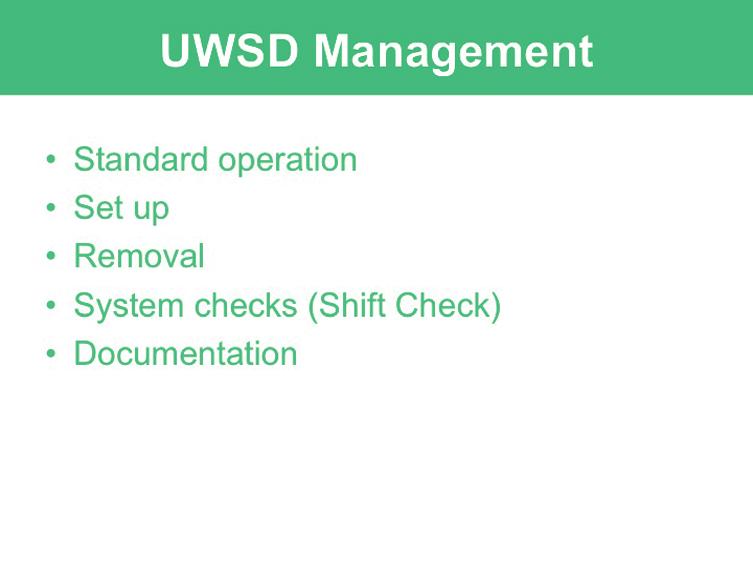

74 62 Chest tube management Module 3: Underwater seal drains PowerPoint presentation
75 Chest tube management Module 4: Management scenarios 63 Module 4: Management scenarios Introduction Chest tube management was developed as a teaching and learning tool for Victorian clinical educators. The information contained in each module was developed using evidence-based resourcees and examples of best practice. Where expert opinion varies, a discussion section is included. However, it is not within the scope of Chest tube management to address the full spectrum of local variations. Variations can occur in several areas, including practices relating to types of equipment used, infection control processes, practice guidelines and so on. Therefore, educators should, where appropriate, adapt content to reflect their local policies, procedures and protocols. This will ensure the relevancy of the package content to your learners. The modules are designed to be discrete courses in their own right. They are timetabled so they can be completed in a 1 2 hour timeframe. This timeframe was chosen after we received feedback from clinical educators requesting shorter courses, because health professionals often have limited time to educate away from patients. However, the packages may also be combined into a one- or two-day course. Chest tube management should be used as an educational tool to assist in the teaching of clinical skills. It is structured as a guide to assist clinical educators, and uses many concepts taught in the Clinical Skills in Hospitals Project (Train-the-Trainer courses). Educators are encouraged to build on this resource by adding their own scenarios which incorporate hospital/health service protocols, policies and other resources. Each module is designed as a lesson plan to incorporate the simulations into the teaching of clinical skills. Aims Chest tube management aims to make participants confident in their application of skills associated with identifying indications for inserting intercostal catheters and finebore cannulae, and appropriately inserting and managing these catheters and cannulae in different environments and settings. This package is intended for use with medical and nursing participants. Package structure Chest tube management contains four modules that provide learning opportunities for health professionals at all levels of experience and from medical and nursing disciplines. Modules 1 and 2 are regarded as fundamental. Modules 3 and 4 are more difficult and are regarded as intermediate.
76 64 Chest tube management Module 4: Management scenarios Level of complexity Complex For participants with more than 4 years experience or who have completed Modules 1 4 Intermediate For participants in postgraduate years 3 4 or who have completed Modules 1 and 2 Fundamental For participants in postgraduate years 1 2 Package structure Underwater seal drains Intercostal catheters Chest tube management scenarios Pleurocaths Skills included in Chest tube management include chest tube insertion, needle aspiration, fine-bore catheter insertion (for example, Pleurocath) underwater seal drains and haemo-pneumothorax management. This package was designed to develop participants knowledge, skills and behaviours in the safe management of pneumothoraces and haemothoraces, including chest tube insertion and management. The modules expose participants to increasingly complex skills and knowledge and test their ability to combine these individual skills, work as a team and solve problems in more difficult situations. Educators delivering these modules should be aware of participants level of experience and choose appropriate modules. Modules presume an increasing level of knowledge as they progress, ranging from a fundamental knowledge of anatomy and physiology for the fundamental modules, up to detailed knowledge of haemopneuomothorax management for the complex modules. Novice participants (such as first-year graduates) are expected to start with the fundamental modules, and only move onto intermediate and more complex modules as they demonstrate proficiency. More experienced participants may start at the intermediate level if the educator is satisfied that they have the prior knowledge and skills. Individual educators are responsible for assessing each participant s baseline knowledge and determining which modules they need to complete. More specific descriptions of presumed knowledge are outlined in each module.
77 Chest tube management Module 4: Management scenarios 65 The design of these packages presumes that the clinical educators using them have knowledge and expertise in current best practice regarding the teaching of clinical skills and conducting facilitated discussions. Knowledge and expertise are presumed commensurate with the Department of Human Services basic and advanced Train-the-Trainer programs. Clinical educators are encouraged to refer to the Department of Human Services Clinical Skills Facilitators Manual for theory on: 1. Peyton s model for teaching clinical skills 2. leading small group discussions 3. giving feedback 4. crisis resource management skills.
78
79 Chest tube management Module 4: Management scenarios 67 Module 4: Management scenarios Author: Dr Stuart Dilley Aims This module allows participants to practise and consolidate their familiarity with the individual skills (pleural aspiration, chest tube insertion and management of underwater seal drains) learned in the basic modules of the Chest tube management package in a logical and coordinated fashion in a controlled setting. Presumed knowledge This module is targeted to health professionals who are competent in the component skills of inserting and managing intercostal catheters, underwater seal drains and performing needle aspiration of the pleural space. Participants should have already practised these skills in the skills stations, but may not have had an opportunity to apply them to a clinical scenario. If participants do not yet feel confident with the individual skills, they should be redirected to Chest tube management Module 1: Intercostal catheters, Module 2: Pleurocaths and Module 3: Underwater seal drains. Objectives By the end of this module, participants should have: 1. reviewed the options for intervention in patients with pneumothoraces and haemothoraces 2. assessed and appropriately managed a simulated patient (manikin) requiring intervention for these conditions 3. determined the likely cause of a malfunctioning chest tube on a simulated patient (manikin) and initiated appropriate management to rectify this problem. Background information for educators Much of the background information pertaining to pleural aspiration, chest tube insertion and management of underwater seal drains was covered in Chest tube management Module 1: Intercostal catheters, Module 2: Pleurocaths and Module 3: Underwater seal drains. Participants undertaking Module 4: Management scenarios should have the knowledge and skills covered in these earlier modules. This module gives participants the opportunity to put these skills together as a coordinated response in a team environment.
80 68 Chest tube management Module 4: Management scenarios Learning activities Suggested learning activities and timetables are outlined below. Timetable 1 is designed for 12 participants working in two groups of six. Timetable 2 is designed for six participants working together. Timetable 1 Timing Activity Objective 20 minutes Introduction, manikin familiarisation 1 Group 1 Group 2 10 minutes Simulation 1 Simulation 2 All 30 minutes Debrief Debrief All 10 minutes Simulation 2 Simulation 1 All 30 minutes Debrief Debrief All 10 minutes Summary All 10 minutes Evaluation Total time = 2 hours Timetable 2 Timing Activity Objective 20 minutes Introduction, manikin familiarisation 1 10 minutes Simulation 1 All 30 minutes Debrief All 10 minutes Simulation 2 All 30 minutes Debrief All 10 minutes Summary All 10 minutes Evaluation Total time = 2 hours Introduction The facilitator should lead a brief discussion amongst participants to refresh or clarify any issues relating to the management of pneumothoraces or haemothoraces, and introduce the simulation training to follow. This should not be a comprehensive lecture on chest tube management. Time should be allocated to allow for a familiarisation session with the manikin.
81 Chest tube management Module 4: Management scenarios 69 Skills stations The skills stations allow participants to practise their skills in the recognition and management of pneumothoraces as a team, in a simulated environment. Participants are exposed to clinical scenarios and should manage these within the confines of the skills relevant to their health profession. The program assumes two facilitators for 12 participants. Participants should be divided into two groups of six (Timetable 1). Three participants each participate in one scenario and observe a second. Those not participating in the scenario observe and participate in the debriefing session. The debriefing period should include all six participants that is, the active participants and their observers. These scenarios can be run with smaller groups. If only six participants are present, the scenarios can be run consecutively (Timetable 2). Ideally, these scenarios should be run on human patient simulators that allow for the insertion of intercostal catheters (ICCs). However, they may be run on more basic simulators that do not allow actual insertion of ICCs. Regardless, the focus of the simulation and the debrief is not necessarily on the technical skill, but on the decision making process and the management plans. Simulation 1: Tension pneumothorax Scenario design In this scenario, a tall, thin 40-year-old male presents to the emergency department with pleuritic chest pain and shortness of breath. He shows signs of a tension pneumothorax that requires recognition, needle aspiration and preparation for formal chest tube insertion. Case history Patient details Sex Age Past history Social history History of present illness Male 40 years Previously well Lives with wife, two young kids Sudden onset of left-sided * chest pain at rest, with progressively increasing shortness of breath. Initially he went to his GP, who sent him for a CXR, but after the X-ray, his symptoms got worse and he was transported by ambulance directly from the X-ray facility to the ED. Presenting symptoms Sharp pain in left * side of chest, short of breath, difficulty breathing and talking. * Side of symptoms may vary, depending on resources/manikins available to educators.
82 70 Chest tube management Module 4: Management scenarios Resources General Setting/environment Patient attire Monitoring Supporting documentation required Hospital emergency department Street clothes: shirt, trousers ECG, pulse oximetry, non-invasive BP ED observation and treatment chart Equipment Equipment Number Sourced from Manikin ideally capable of simulating needle aspiration of pleural space and insertion of ICC 1 Hospital bed or ED trolley 1 Hospital gown 1 Pillow, blanket 1 Patient treatment chart 1 Hudson mask and tubing 1 Oxygen supply 1 Resuscitation trolley ALS drugs and airway equipment 1 IV cannulae CXR showing pneumothorax 1 Various sizes, including 14G for needle decompression Pleural aspiration set as used at specific institution Pleurocath and so on ICC insertion set and intercostal tubes as used at specific institution Underwater seal drain set as used at specific institution Heimlich valve if available at specific institution 1 set 1 set 1 1
83 Chest tube management Module 4: Management scenarios 71 Roles Participant 1 You are a health professional working in the emergency department of your hospital. An experienced ED nurse has begun to assess a 40-year-old man in the resuscitation cubicle with chest pain and shortness of breath, and calls for your assistance. You have two colleagues on which to call. The resuscitation cubicle is stocked with equipment and drugs as appropriate for a real ED. Participants 2 and 3 You are health professionals working in the hospital s ED. Your colleague (Participant 1) has gone to attend to a patient with chest pain in the resuscitation cubicle. Your colleague may call on you for assistance in managing the patient. A nurse is also present (faculty member) to assist with locating equipment and medications. Faculty role play: ED resus nurse You are a nurse working in the resuscitation area of the ED. You are assessing a 40-year-old man with chest pain and shortness of breath, who has just arrived having had a GP-arranged CXR performed at an outside X-ray facility. You are concerned about this man s shortness of breath and blood pressure and ask for assistance by calling in Participant 1 and directing them to the CXR. You assist all participants in finding equipment and appropriate drugs if needed, but do not assemble the underwater seal drain. Faculty role play: senior clinician You are a senior clinician working in the ED. If participants experience difficulties, it is appropriate to enter the scenario and offer assistance. Otherwise, at the conclusion of the scenario, you arrive to take a handover of the patient.
84 72 Chest tube management Module 4: Management scenarios Simulator programming considerations System Baseline state Change in State 1 Change in State 2 Resolution CVS HR 120 ST HR 100 ST HR 120 ST HR 90 SR BP 80/50 mmhg BP 110/60 mmhg BP 80/50 mmhg BP 120/60 mmhg Respiratory RR 30 RR 18 RR 30 RR 16 O 2 saturation 80% room air (RA) O 2 saturation 97% on oxygen O 2 saturation 90% RA O 2 saturation 99% on oxygen Left-sided* pneumothorax decreased air entry, absent breath sounds, hyper-expanded Left-sided* PTX persists Trachea midline Left-sided* PTX persists Trachea deviated away from side of PTX PTX signs gone Trachea midline Tracheal deviation away from side of PTX Neurologic GCS 15 GCS 15 GCS 15 GCS 15 Distressed More comfortable Distressed Comfortable Response to participant intervention Remain in state until needle decompression or chest tube insertion Needle decompression go to State 1 Chest tube insertion ** go to resolution No chest tube within 5 minutes go to State 2 Chest tube insertion ** go to resolution Chest tube insertion to resolution go * Side of symptoms may vary, depending on resources/manikins available to educators. ** Some manikins may not allow actual insertion of a chest tube. In this instance, the focus of the simulation should be recognising the need for intervention and going through the process of insertion and setting up the underwater seal drain.
85 Chest tube management Module 4: Management scenarios 73 Debriefing points: signs and symptoms of tension pneumothorax need for rapid decompression of tension pneumothorax need for progression from needle decompression to formal chest tube sites of insertion for needle decompression and chest tubes. Simulation 2: Malfunctioning intercostal catheter Scenario design In this scenario, a 60-year-old female on the surgical ward following chest tube (ICC) insertion for traumatic haemo-pneumothorax experiences increasing shortness of breath. Her chest tube appears to be blocked and her pneumothorax is re-accumulating. Participants should recognise that the ICC is not functioning, take steps to try and remedy this (which they cannot) and plan to insert a second ICC when they are unsuccessful. Case history Patient details Sex Age Past history Female 60 years Smoker of 40 years Current medications: salbutamol puffer 2 puffs PRN ipratropium puffer 2 puffs bd Social history History of present illness Lives at home with husband Presented to hospital two days ago after a motor vehicle accident CXR revealed a large left * pneumothorax and pleural effusion which required the insertion of an ICC A large quantity of blood-stained pleural fluid has drained Presenting symptoms Increasing pain in left * side of chest, associated increasing shortness of breath * Side of symptoms may vary, depending on resources/manikins available to educators.
86 74 Chest tube management Module 4: Management scenarios Resources General Setting/environment Patient attire Monitoring Supporting documentation required Hospital surgical ward Hospital gown/pyjamas ECG, pulse oximetry, non-invasive BP Ward observation and treatment chart Equipment Equipment Number Sourced from Manikin ideally capable of simulating needle aspiration of pleural space and insertion of ICC 1 Hospital bed or trolley 1 Hospital gown 1 Pillow, blanket 1 Patient treatment chart 1 Hudson mask and tubing 1 Oxygen supply 1 Resuscitation trolley ALS drugs and airway equipment 1 IV cannulae CXR showing ICC 1 Pleural aspiration set as used at specific institution Pleurocath and so on ICC insertion set and intercostal tubes as used at specific institution Underwater seal drain set as used at specific institution Heimlich valve if available at specific institution Various sizes, including 14G for needle decompression 1 set 1 set 1 1
87 Chest tube management Module 4: Management scenarios 75 Roles Participant 1 You are a health professional working in the surgical ward of your hospital. An experienced nurse has begun to review a 60-year-old woman with shortness of breath, and calls for your assistance. The patient had been admitted to the surgical ward two days earlier after a motor vehicle accident. An ICC was inserted for a haemo-pneumothorax and a large bloody pleural effusion was drained. You have two colleagues on which to call. The ward s resuscitation trolley is stocked with equipment and drugs as appropriate for an average surgical ward. Participants 2 and 3 You are health professionals working in the hospital s surgical ward. Your colleague (Participant 1) has gone to attend to a patient with shortness of breath. Your colleague may call on you for assistance in managing the patient. A nurse is also present (faculty member) to assist with locating equipment and medications. Faculty role play: ward nurse You are a nurse working in the surgical ward. You are assessing a 60-year-old woman with shortness of breath. She had been admitted to the surgical ward two days earlier after a motor vehicle accident. An ICC was inserted for a haemo-pneumothorax and a large bloody pleural effusion was drained. You are concerned about her shortness of breath and believe the ICC is not working properly. You ask for assistance by calling in Participant 1. You assist all participants in finding equipment and appropriate drugs if needed, but are unfamiliar with the workings of the underwater seal drain. Faculty role play: senior clinician You are a senior clinician working in the surgical ward. If participants experience difficulties, it is appropriate to enter the scenario and offer assistance. Otherwise, at the conclusion of the scenario, you arrive to take a handover of the patient.
88 76 Chest tube management Module 4: Management scenarios Simulator programming considerations System Baseline state Resolution CVS HR 100 SR BP 120/80 mmhg HR 100 SR BP 120/80 mmhg Respiratory RR 20 O 2 Saturation 90% RA Left-sided* pneumothorax decreased air entry, absent breath sounds, hyperexpanded ICC already inserted ** no bubbling, no swing with respiration, blood-stained fluid in collection chamber RR 20 O 2 Saturation 98% on oxygen Left-sided PTX persists if participants still preparing to insert second ICC Symptoms resolve if second ICC inserted Neurologic GCS 15 GCS 15 Response to participant intervention Remains in baseline state until all of the following have been done or discussed: provision of supplemental oxygen check ICC for patency check location of ICC consider need for CXR call for medical assistance if appropriate consider need for needle decompression consideration of insertion of second ICC * Side of symptoms may vary, depending on resources/manikins available to educators. ** For this scenario, it is not necessary to have a manikin that allows insertion of ICC. Simulating the presence of an ICC by taping the catheter to the manikin s chest wall, and then covering with appropriate dressings should suffice. In this instance, there is no swing or bubbling to observe in the UWS, therefore simulating a blocked ICC. Debriefing points: recognition of malfunctioning ICC awareness of likely causes of ICC malfunctions awareness of limits to intervention according to health professional status (medical, nursing, allied health) recognition of need for second ICC in some instances.
89 Chest tube management Module 4: Management scenarios 77 Summary The summary session reinforces content covered in the learning activities, and is an opportunity for participants to reflect on what they have covered. No new material should be introduced. Points to cover in the summary include: indications for pleural drainage and intercostal catheter insertion options for pleural drainage and intercostal catheter insertion recognition of tension pneumothorax and need for rapid response options for diagnosing and dealing with chest tube and UWS drain malfunctions. Participants should be encouraged to review the use of chest drains and their management in their own time to reinforce the skills acquired in this module. This should include exposure to real patients with chest drains in situ. Participants should be offered access to equipment and educators in the future if they need to practise or improve their skill level or confidence. Resource list The following resource list assumes two facilitators for every 12 participants, a ratio of 1:6. As a minimum, the following resources are needed to conduct this module. Resource Quantity Additional comments Equipment and resources as listed in scenario requirements above
90 78 Chest tube management Module 4: Management scenarios Evaluation A formal evaluation has been specifically developed for this module. It incorporates the objectives of the module and the perceptions of the participants about whether they have increased their understanding by working through the module. It is highly recommended that this formal evaluation be copied and completed by all participants at the completion of the module. A range of informal evaluation tools may also be used in conjunction with this evaluation throughout the module, including those available in the Department of Human Services Clinical Skills Facilitators Manual from the basic course conducted in 2007.
91 Chest tube management Module 4: Management scenarios 79 Resources Facilitator feedback form The following form should be used to assist you in giving feedback after each participant has practised their Pleurocaths skills at the skill station. Feedback using the Pendleton model Pendleton s model of feedback assists learners to maximize their potential at different stages of training, raise their awareness of strengths and areas for improvement, and identify actions to be taken to improve performance. Pendleton s rules are structured in such a way that the learner identifies the positives first, in order to create a safe environment. This is followed by the facilitator or group reinforcing these positives and discussing skills to achieve them. Different techniques are then suggested. The advantage of this method is that the learner s strengths are discussed first. Avoiding a discussion of weaknesses right at the beginning prevents defensiveness and allows reflective behaviour in the learner. Below is a series of questions to assist you in this technique: 1. Ask the learner how they feel. 2. Ask the learner what went well and why (this can be combined with question 1 and 3). 3. Tell the learner what went well and why. 4. Ask the learner what could have been done better and why. 5. Tell the learner what could have been done better and why. 6. Summarise the learner s strengths and identify up to three things to concentrate on. Note: This form does not need to be given to the participant it is a guide for you, the group facilitator.
92 80 Chest tube management Module 4: Management scenarios Module 4: Management scenarios evaluation Thank you for participating in this module. As part of our commitment to quality improvement the following questionnaire will be used to plan future implementation of this module. We appreciate your time completing this evaluation. 1. Overall How would you rate this module? poor fair good very good outstanding 2. Learning objectives Please consider whether this module was successful in meeting the following learning objectives: Chest tube management Learning objectives of Module 4: Management scenarios Strongly disagree Disagree Slightly agree Agree Strongly agree Reviewed the options for intervention in patients with pneumothoraces and haemothoraces Assessed and appropriately managed a simulated patient (manikin) requiring intervention for these conditions Assessed and appropriately managed a simulated patient (manikin) experiencing complications of this management 3. Important learning outcomes What are the three most important things you have learned from this module?
93 Chest tube management Module 4: Management scenarios Module implementation Please indicate to what extent you agree or disagree with each of the following statements in relation to the implementation of the module. Strongly disagree Disagree Slightly agree Agree Strongly agree The facilitator respected my experience The facilitator encouraged my participation I was able to ask the facilitator questions The facilitator was able to answer my questions The feedback I received was clear The feedback I received will assist me in my future performance There was adequate time for the introduction There was adequate time for the simulations I have increased my confidence in managing chest tubes I have identified future learning needs in this topic area 5. Future module implementation Do you think the module should be altered in any way? yes no If yes, what recommendations do you have? Thank you

94 82 Chest tube management Module 4: Management scenarios PowerPoint presentation 1. 2.
STANDARDIZED PROCEDURE NEONATAL / PEDIATRIC THORACENTESIS (NEEDLE ASPIRATION) (Neonatal, Pediatric)
 (Neonatal, Pediatric)") I. Definition To insert a needle into the chest in order to evacuate air or fluid II. Background Information A. Setting: Inpatient neonatal / pediatric patients or outpatient during Emergency Transport
I. Definition To insert a needle into the chest in order to evacuate air or fluid II. Background Information A. Setting: Inpatient neonatal / pediatric patients or outpatient during Emergency Transport
Pleural procedures and thoracic ultrasound British Thoracic Society Pleural Disease Guideline 2010
 Pleural procedures and thoracic ultrasound British Thoracic Society Pleural Disease Guideline 2010 Tom Havelock, Richard Teoh, Diane Laws, Fergus Gleeson On behalf of the BTS Pleural Disease Guideline
Chest Drain Insertion
 Chest Drain Insertion Information for Patients What is a chest drain? The insertion of a chest drain is a sterile procedure that involves placing a small drain through your skin into the space between
RNSP: Advanced RN Intervention
 Policies and Procedures RNSP: Advanced RN Intervention Title: CHEST TUBES: IRRIGATION AND/OR ADMINISTRATION OF A MEDICATION TO THE PLEURAL SPACE I.D. Number: 1016 Authorization: [X] SHR Nursing Practice
POLICY ON INTERCOSTAL (CHEST) DRAINS
 DRAINS") POLICY ON INTERCOSTAL (CHEST) DRAINS Charles Pantin/Sriram Iyer/Marta BaboresPage 1 of 18 Policy Title: INTERCOSTAL (CHEST) DRAINS POLICY Executive Summary: This policy governs the insertion of intercostals
POLICY ON INTERCOSTAL (CHEST) DRAINS Charles Pantin/Sriram Iyer/Marta BaboresPage 1 of 18 Policy Title: INTERCOSTAL (CHEST) DRAINS POLICY Executive Summary: This policy governs the insertion of intercostals
STANDARDIZED PROCEDURE CENTRAL LINE PLACEMENT and TEMPORARY NONTUNNELLED CENTRAL VENOUS DIALYSIS CATHETER INSERTION (Adult, Peds)
") I. Definition: This protocol covers the task of central (venous) catheter placement and temporary nontunnelled central venous dialysis catheters by the Advanced Health Practitioner. The purpose of this
I. Definition: This protocol covers the task of central (venous) catheter placement and temporary nontunnelled central venous dialysis catheters by the Advanced Health Practitioner. The purpose of this
393 PICC INSERTION USING ULTRASONOGRAPHY AND MICRO INTRODUCER TECHNIQUE 06/10/03 1
 393 ULTRASONOGRAPHY AND MICRO INTRODUCER TECHNIQUE 06/10/03 1 POLICY: General Information: 1. RN s validated to insert PICCs with the additional training in the use of ultrasonography (U/S) and microintroduction
STANDARDIZED PROCEDURE BONE MARROW ASPIRATION (Adult,Peds)
") I. Definition: This protocol covers the task of bone marrow aspiration by an Advanced Health Practitioner. The purpose of this standardized procedure is to allow the Advanced Health Practitioner to safely
I. Definition: This protocol covers the task of bone marrow aspiration by an Advanced Health Practitioner. The purpose of this standardized procedure is to allow the Advanced Health Practitioner to safely
Early discharge with Exudrain wound drains NURSING INFORMATION ON THE CARE OF BREAST SURGERY PATIENTS AT HOME
 Early discharge with Exudrain wound drains NURSING INFORMATION ON THE CARE OF BREAST SURGERY PATIENTS AT HOME Home sweet home For the first few days after surgery many patients have two wound drains and
Formative DOPS: Percutaneous endoscopic gastrostomy (PEG)
") Date of procedure Trainee name Trainer name Formative DOPS: Membership no. (eg. GMC/NMC) Membership no. (eg. GMC/NMC) Outline of case Difficulty of case Easy Moderate Complicated Please tick appropriate
Date of procedure Trainee name Trainer name Formative DOPS: Membership no. (eg. GMC/NMC) Membership no. (eg. GMC/NMC) Outline of case Difficulty of case Easy Moderate Complicated Please tick appropriate
STANDARDIZED PROCEDURE LUMBAR DRAIN INSERTION (Adults, Peds)
") I. Definition The purpose of this standardized procedure is for the Advanced Health Practitioner to safely place a lumbar drain. II. Background Information A. Setting: The setting (inpatient vs outpatient)
I. Definition The purpose of this standardized procedure is for the Advanced Health Practitioner to safely place a lumbar drain. II. Background Information A. Setting: The setting (inpatient vs outpatient)
A lifetime of specialist care. Managing your chest drain at home
 A lifetime of specialist care Managing your chest drain at home Contents Introduction 3 Patient details 3 How to contact us 4 Going home with a chest drain 5 How do I look after my chest drain? 5 General
Mediastinal Venogram and Stent Insertion
 Patient information leaflet Royal Surrey County Hospital NHS Foundation Trust Mediastinal Venogram and Stent Insertion Radiology This leaflet tells you about the procedure known as a mediastinal venogram.
Video Assisted Thoracoscopy (VATS) Information for patients Thoracic Surgery
 Information for patients Thoracic Surgery") Video Assisted Thoracoscopy (VATS) Information for patients Thoracic Surgery X X X This leaflet has been written to provide information about your procedure. We hope it answers some of your questions or
Video Assisted Thoracoscopy (VATS) Information for patients Thoracic Surgery X X X This leaflet has been written to provide information about your procedure. We hope it answers some of your questions or
Mediastinal Venogram and Stent Insertion
 Mediastinal Venogram and Stent Insertion Radiology Department Patient information leaflet This leaflet tells you about the procedure known as a mediastinal venogram. It explains what is involved and the
Family Medicine Residency Surgery Rotation
 Family Medicine Residency Surgery Rotation Rotation Goal The overall goal for the educational experience provided in the areas of general surgery, trauma surgery, office orthopedic surgery and sports medicine,
Subacute Care. 1. Define important words in the chapter. 2. Discuss the types of residents who are in a subacute setting
 175 26 Subacute Care 1. Define important words in this chapter 2. Discuss the types of residents who are in a subacute setting 3. List care guidelines for pulse oximetry 4. Describe telemetry and list
Medical technologies guidance Published: 21 March 2018 nice.org.uk/guidance/mtg37
 Thopaz+ portable digital system for managing chest drains Medical technologies guidance Published: 21 March 2018 nice.org.uk/guidance/mtg37 NICE 2018. All rights reserved. Subject to Notice of rights (https://www.nice.org.uk/terms-and-conditions#notice-ofrights).
Having a portacath insertion in the x-ray department
 Having a portacath insertion in the x-ray department This leaflet provides information about a portacath insertion, including the benefits, risks and any alternatives. It also explains what you can expect
STANDARDIZED PROCEDURE HEPATIC ARTERY INFUSION OF CHEMOTHERAPY (Adults, Peds)
") I. Definition Hepatic arterial infusion (HAI) of chemotherapy is accomplished by a small drug delivery system or pump that is implanted in a subcutaneous pocket in the lower abdomen. The pump reservoir
I. Definition Hepatic arterial infusion (HAI) of chemotherapy is accomplished by a small drug delivery system or pump that is implanted in a subcutaneous pocket in the lower abdomen. The pump reservoir
Laparoscopic Radical Nephrectomy
 Urology Department Laparoscopic Radical Nephrectomy Information Aims of this leaflet To give information on the intended benefits and potential risks of kidney surgery To guide you in the decisions you
All about Your Implanted Venous Access Device (IVAD, Port )
") All about Your Implanted Venous Access Device (IVAD, Port ) Your doctor has chosen an Implanted Venous Access Device (IVAD) for you based on your treatment needs. Because the IVAD can remain in place for
All about Your Implanted Venous Access Device (IVAD, Port ) Your doctor has chosen an Implanted Venous Access Device (IVAD) for you based on your treatment needs. Because the IVAD can remain in place for
Post-operative "Fast-Track" pathways for lung resection. Dennis A. Wigle Division of Thoracic Surgery Mayo Clinic
 Post-operative "Fast-Track" pathways for lung resection Dennis A. Wigle Division of Thoracic Surgery Mayo Clinic Post-operative "Fast-Track" pathways for lung resection Dennis A. Wigle Division of Thoracic
Vascular Access Department Insertion of a tunnelled Central Venous Catheter Information for patients
 Vascular Access Department Insertion of a tunnelled Central Venous Catheter Information for patients What is a tunnelled central venous catheter (CVC)? A CVC is a long, narrow tube (catheter) that is put
Percutaneous Transhepatic Biliary Drainage Interventional Radiology
 Percutaneous Transhepatic Biliary Drainage Interventional Radiology Your doctor has scheduled a percutaneous transhepatic biliary drainage to be done in the Interventional Radiology (IR) Department on
Introduction to physical examination & general survey.
 In The Name of God (A PROJECT OF NEW LIFE COLLEGE OF NURSING KARACHI) Introduction to physical examination & general survey. Shahzad Bashir RN, BScN, DCHN, MScN (Std. DUHS) Instructor New Life College
All About Your Peripherally Inserted Central Catheter (PICC)
") All About Your Peripherally Inserted Central Catheter (PICC) General Information Intravenous (IV) therapy is the delivery of fluid directly into a vein. An intravenous catheter is a hollow tube that is
All About Your Peripherally Inserted Central Catheter (PICC) General Information Intravenous (IV) therapy is the delivery of fluid directly into a vein. An intravenous catheter is a hollow tube that is
LAPAROSCOPIC SIMPLE REMOVAL OF THE KIDNEY
 Procedure Specific Information What is the evidence base for this information? This publication includes advice from consensus panels, the British Association of Urological Surgeons, the Department of
Curriculum For The LMA Supreme
 Curriculum For The LMA Supreme Course Description This course is designed to provide instruction in a procedure for the use of the LMA Supreme by the EMT-Intermediate `99 and Paramedic. Prerequisites 1.
Ambulatory Chest Drainage System Chest Bag
 Ambulatory Chest Drainage System Chest Bag PATIENT MANAGEMENT GUIDE Provided as a service to patients by Smiths Medical Ambulatory Chest Drainage System (Chest Bag) Introduction You have left hospital
Axillary Node Dissection
 Axillary Node Dissection Breast Care Centre Information for patients Name of Consultant: Date of surgery: Key worker: Direct line: 0116 250 2513 Monday - Friday 9 am - 4.30 pm (except Bank Holidays) What
Wyoming STATE BOARD OF NURSING
 David D. Freudenthal Governor Wyoming STATE BOARD OF NURSING Mary Kay Goetter, PhD, RNC, NEA-BC Executive Director 1810 Pioneer Avenue Cheyenne, Wyoming 82002 Phone: 307-777-7601 FAX: 307-777-3519 http://nursing.state.wy.us
CONSENT FORM UROLOGICAL SURGERY
 CONSENT FORM for UROLOGICAL SURGERY (Designed in compliance with consent form 1) PATIENT AGREEMENT TO INVESTIGATION OR TREATMENT Patient Details or pre-printed label Patient s NHS Number or Hospital number
Certified PICC Ultrasound Inserter CPUI Renewal Application
 APPLICATION FOR CPUI CERTIFICATION RENEWAL This form renews CPUI PICC Certification by documentation and does not establish competency. Competency must be established by the employer and facility. Name
STANDARDIZED PROCEDURE SKIN BIOPSY (Adult, Peds)
") I. Definition Skin biopsy is the removal of a small piece of tissue, under local anesthetic, from a lesion suspected of malignancy, other dermatitis, or for clinical research purposes. The technique to
I. Definition Skin biopsy is the removal of a small piece of tissue, under local anesthetic, from a lesion suspected of malignancy, other dermatitis, or for clinical research purposes. The technique to
Going home with a redivac drain after surgery
 Going home with a redivac drain after surgery This leaflet explains about going home with a redivac drain following your surgery. If you have any further questions, please speak to the nurse or doctor
Assisting with the Bedside (Percutaneous) Removal of Chronic Peritoneal Dialysis Catheters
 Removal of Chronic Peritoneal Dialysis Catheters") Assisting with the Bedside (Percutaneous) Removal of Chronic Peritoneal Dialysis Catheters ORIGIN DATE: APRIL 27, 2009 REVISED DATE: NOVEMBER 2013 This procedure is posted on the BC Provincial Renal Agency
Assisting with the Bedside (Percutaneous) Removal of Chronic Peritoneal Dialysis Catheters ORIGIN DATE: APRIL 27, 2009 REVISED DATE: NOVEMBER 2013 This procedure is posted on the BC Provincial Renal Agency
PICU tracheostomy protocol
 PICU tracheostomy protocol This protocol is based on the joint Royal Brompton & Harefield NHS Trust and Great Ormond Street Hospital for Children NHS Trust, Great Ormond Street Hospital Manual of Children
Surgery for Pneumothorax
 Oxford University Hospitals NHS Trust Thoracic Surgery Surgery for Pneumothorax Information for patients Welcome to the Oxford Heart and Lung Centre The information in this booklet will help to prepare
STANDARDIZED PROCEDURE VENTRICULAR SEPTAL DEFECT (VSD) CLOSURE ASSIST (Neonatal, Peds)
 CLOSURE ASSIST (Neonatal, Peds)") I. Definition A ventricular septal defect is an abnormal opening in the wall (septum) that divides the two lower chambers of the heart (ventricles). A ventricular septal defect closure is a procedure performed
I. Definition A ventricular septal defect is an abnormal opening in the wall (septum) that divides the two lower chambers of the heart (ventricles). A ventricular septal defect closure is a procedure performed
LAPAROSCOPIC RADICAL REMOVAL OF THE KIDNEY AND URETER
 Procedure Specific Information What is the evidence base for this information? This publication includes advice from consensus panels, the British Association of Urological Surgeons, the Department of
1. Communicate to the UAP any special information needed prior to the administration of the medication.
 Objectives At the completion of this module, unlicensed assistive personnel (UAP) should be able to: 1. administer medications by intradermal injection. 2. document medication administration in the client
SURGICAL RESIDENT CURRICULUM FOR THE DIVISION OF CARDIOTHORACIC SURGERY
 SURGICAL RESIDENT CURRICULUM FOR THE DIVISION OF CARDIOTHORACIC SURGERY Residency Years Included: PGY1_X_ PGY2_X_ PGY3 PGY4 PGY5 Fellow I. The Clinical Mission of the Division of Cardiothoracic Surgery
About your PICC line. Information for patients Weston Park Hospital
 About your PICC line Information for patients Weston Park Hospital This booklet explains what a PICC line is, how it is inserted and some general advice on its use and care. What is a PICC line? A Peripherally
Inferior Vena Cava (IVC) Filter Insertion
 Filter Insertion") Patient information leaflet Royal Surrey County Hospital NHS Foundation Trust Inferior Vena Cava (IVC) Filter Insertion Radiology This leaflet informs you about the procedure known as an Inferior Vena
Patient information leaflet Royal Surrey County Hospital NHS Foundation Trust Inferior Vena Cava (IVC) Filter Insertion Radiology This leaflet informs you about the procedure known as an Inferior Vena
This leaflet can be made available in other formats including large print, CD and Braille and in languages other than English, upon request.
 Information for patients This leaflet can be made available in other formats including large print, CD and Braille and in languages other than English, upon request. Your doctor has recommended you have
Peripherally Inserted Central Catheter (PICC)
") University Teaching Trust Peripherally Inserted Central Catheter (PICC) IV Team 0161 206 0459 All Rights Reserved 2017. Document for issue as handout. Contents l What is a PICC? l Why do I need a PICC?
University Teaching Trust Peripherally Inserted Central Catheter (PICC) IV Team 0161 206 0459 All Rights Reserved 2017. Document for issue as handout. Contents l What is a PICC? l Why do I need a PICC?
Policies & Procedures
 Policies & Procedures Title: ENTERAL FEEDING TUBE WITH A STYLET: ASSISTING WITH INSERTION OF: CARE OF, REMOVAL OF Authorization [X] SHR Nursing Practice Committee ID Number: 1109 Source: Nursing Date Reaffirmed:
Section G - Aseptic Technique. Version 5
 Section G - Aseptic Technique Version 5 Important: This document can only be considered valid when viewed on the Trust s Intranet. If this document has been printed or saved to another location, you must
Percutaneous Transhepatic Cholangiogram (PTC) and Biliary Stent
 and Biliary Stent") Patient information leaflet Royal Surrey County Hospital NHS Foundation Trust Percutaneous Transhepatic Cholangiogram (PTC) and Biliary Stent Radiology This leaflet informs you about the procedure known
Patient information leaflet Royal Surrey County Hospital NHS Foundation Trust Percutaneous Transhepatic Cholangiogram (PTC) and Biliary Stent Radiology This leaflet informs you about the procedure known
Having an Oesophageal Dilatation
 Having an Oesophageal Dilatation Information for Patients In this leaflet: Introduction 2 What is an Oesophageal Dilatation?...2 What are the benefits of an Oeosphageal Dilatation? 2 Are there any risks?.2
Australian and New Zealand College of Anaesthetists (ANZCA)
") PS08 2016 Australian and New Zealand College of Anaesthetists (ANZCA) Statement on the Assistant for the Anaesthetist 1. PURPOSE The purpose of this document is to recognise the importance of and to promote
PS08 2016 Australian and New Zealand College of Anaesthetists (ANZCA) Statement on the Assistant for the Anaesthetist 1. PURPOSE The purpose of this document is to recognise the importance of and to promote
Information for Patients
 Having a Radiologically Inserted Gastrostomy (RIG) Information for Patients In this leaflet: Introduction.. 2 What is a RIG feeding tube?. 2 Why do I need a RIG?. 2 Are there any risks?.2 Are there any
The Ohio State University Department of Orthopaedics. Residency Curriculum. PGY1 Rotations
 The Ohio State University Department of Orthopaedics Residency Curriculum PGY1 Rotations Goals and Objectives Anesthesiology Rotation PGY1 Level I. Core Competency Areas By the end of the PGY1 rotation
Percutaneous nephrolithotomy (PCNL)
") Percutaneous nephrolithotomy (PCNL) This leaflet explains more about the PCNL procedure to clear kidney stones, including the benefits, risks and any alternatives and what you can expect when you come
Percutaneous nephrolithotomy (PCNL) This leaflet explains more about the PCNL procedure to clear kidney stones, including the benefits, risks and any alternatives and what you can expect when you come
STANDARDIZED PROCEDURE REMOVAL OF EXTERNAL VENTRICULAR DRAINAGE CATHETER OR INTRACRANIAL PRESSURE DEVICE (Adult, Peds)
") I. Definition This procedure will take place when a neurosurgery physician deems appropriate. The purpose of this standardized procedure is to allow the Advanced Health Practitioner to safely remove an
I. Definition This procedure will take place when a neurosurgery physician deems appropriate. The purpose of this standardized procedure is to allow the Advanced Health Practitioner to safely remove an
Sentinel Node Biopsy for Breast Cancer
 Sentinel Node Biopsy for Breast Cancer Breast Care Centre Information for Patients Name of Consultant: i... Date of surgery:... Key worker:... Direct line: 0116 250 2513 Monday - Friday 9 am - 4 pm (except
Percutaneous Transhepatic Cholangiogram (PTC) and Biliary Stent
 and Biliary Stent") Percutaneous Transhepatic Cholangiogram (PTC) and Biliary Stent Radiology Department Patient information leaflet This leaflet informs you about the procedure known as a Percutaneous Transhepatic Cholangiogram
Percutaneous Transhepatic Cholangiogram (PTC) and Biliary Stent Radiology Department Patient information leaflet This leaflet informs you about the procedure known as a Percutaneous Transhepatic Cholangiogram
RADICAL REMOVAL OF THE KIDNEY AND URETER
 Procedure Specific Information What is the evidence base for this information? This publication includes advice from consensus panels, the British Association of Urological Surgeons, the Department of
INFORMATION FOR PATIENTS WHO ARE PREPARING FOR LUNG RESECTION SURGERY
 St James s Hospital Department of Cardiothoracic Surgery INFORMATION FOR PATIENTS WHO ARE PREPARING FOR LUNG RESECTION SURGERY R 255 JULY 2014 CONTENTS Your lungs and how they work...1 Why do I need surgery?...1
Patient Information. Having a Laparoscopy
 Patient Information Having a Laparoscopy This information has been written to explain your operation, and the benefits and risks. The medical and nursing staff will be happy to answer any questions you
STANDARDIZED PROCEDURE FEMORAL VENOUS BLOOD DRAW (Adult, Peds)
") I. Definition The Femoral venous blood draw (FVBD) is the procedure of performing a needle stick into the femoral vein for the purpose of drawing blood work that will assist in lab monitoring. II. Background
I. Definition The Femoral venous blood draw (FVBD) is the procedure of performing a needle stick into the femoral vein for the purpose of drawing blood work that will assist in lab monitoring. II. Background
SARASOTA MEMORIAL HOSPITAL NURSING PROCEDURE
 SARASOTA MEMORIAL HOSPITAL NURSING PROCEDURE TITLE: ISSUED FOR: HEMODIALYSIS TEMPORARY CATHETER (INSERTION, DRESSING CHANGE, REMOVAL, MEDICATION AND BLOOD DRAWS, DISCONTINUATION OF MEDS AND IV FLUIDS)
ASEPTIC TECHNIQUE LEARNING PACKAGE
 ASEPTIC TECHNIQUE LEARNING PACKAGE Staff Name:... Date:... Table of Contents What is Aseptic technique? 3 Core infection control components 3 Key parts 5 References 6 Aseptic technique questionnaire 7
ANTERIOR RESECTION WHAT ARE THE BENEFITS OF HAVING AN ANTERIOR RESECTION?
 WHAT IS AN ANTERIOR RESECTION? ANTERIOR RESECTION This is an operation that is designed to remove part of your lower large bowel and then join the bowel ends back together again. This is called an anastamosis.
@ncepod #tracheostomy
 @ncepod #tracheostomy 1 Introduction Tracheostomy: Remedy upper airway obstruction Avoid complications of prolonged intubation Protection & maintenance of airway The number of temporary tracheostomies
LAPAROSCOPIC RECONSTRUCTION OF THE PELVIS OF THE KIDNEY
 Procedure Specific Information What is the evidence base for this information? This publication includes advice from consensus panels, the British Association of Urological Surgeons, the Department of
Placement and Care of Your Gastrojejunostomy Tube (GJ Tube) Interventional Radiology
 Interventional Radiology") Placement and Care of Your Gastrojejunostomy Tube (GJ Tube) Interventional Radiology Your healthcare team recommended that you undergo gastrojejunostomy tube (GJ tube) placement. This procedure will be
Placement and Care of Your Gastrojejunostomy Tube (GJ Tube) Interventional Radiology Your healthcare team recommended that you undergo gastrojejunostomy tube (GJ tube) placement. This procedure will be
STANDARDIZED PROCEDURE NEONATAL / PEDIATRIC INTRAOSSEOUS LINE PLACEMENT (Neonatal, Pediatric)
") I. Definition To place a large bore needle into the bone marrow for the purpose of emergency access for fluids and medications. II. Background Information A. Setting: Inpatient neonatal / pediatric patients
I. Definition To place a large bore needle into the bone marrow for the purpose of emergency access for fluids and medications. II. Background Information A. Setting: Inpatient neonatal / pediatric patients
Guidelines on Postanaesthetic Recovery Care
 Page 1 of 10 Guidelines on Postanaesthetic Recovery Care Version Effective Date 1 OCT 1992 2 FEB 2002 3 APR 2012 4 JUN 2017 Document No. HKCA P3 v4 Prepared by College Guidelines Committee Endorsed by
Totally Implantable Venous Access Devices (port) Information for patients. Cross section of a port
 Information for patients. Cross section of a port") Cystic Fibrosis Unit, Ward 26 0121 424 2000 Information for Patients Totally Implantable Venous Access Devices (port) Information for patients This leaflet tells you about the procedures for Totally Implantable
Cystic Fibrosis Unit, Ward 26 0121 424 2000 Information for Patients Totally Implantable Venous Access Devices (port) Information for patients This leaflet tells you about the procedures for Totally Implantable
Enhanced Recovery Programme for Nephrectomy (Kidney Removal)
") Enhanced Recovery Programme for Nephrectomy (Kidney Removal) This information leaflet will explain what will happen when you come to the hospital for your operation. The enhanced Recovery Programme is
Enhanced Recovery Programme for Nephrectomy (Kidney Removal) This information leaflet will explain what will happen when you come to the hospital for your operation. The enhanced Recovery Programme is
Replacement Of Balloon Retained Gastrostomy (BRG) Procedure Introduction and Aim
 Procedure Introduction and Aim") Reference Number: UHB 189 Version Number: 2 Date of Next Review: 10 th Dec 2018 Previous Trust/LHB Reference Number: Replacement Of Balloon Retained Gastrostomy (BRG) Procedure Introduction and Aim The
Reference Number: UHB 189 Version Number: 2 Date of Next Review: 10 th Dec 2018 Previous Trust/LHB Reference Number: Replacement Of Balloon Retained Gastrostomy (BRG) Procedure Introduction and Aim The
MIAMI DADE COLLEGE MEDICAL CAMPUS BENJAMIN LEON SCHOOL OF NURSING RN-BSN PROGRAM MANUAL OF CLINICAL PERFORMANCE
 MIAMI DADE COLLEGE MEDICAL CAMPUS BENJAMIN LEON SCHOOL OF NURSING RN-BSN PROGRAM MANUAL OF CLINICAL PERFORMANCE 1 INTRODUCTION Welcome to Miami Dade College Bachelor of Science in Nursing Program. The
PLACEMENT. Disclaimer
 1 TITLE: GUIDELINE FOR PERCUTANEOUS ENDOSCOPIC GASTROSTOMY TUBE Disclaimer PLACEMENT The Canadian Society of Gastroenterology Nurses and Associates (CSGNA) presents this guideline to be used as a reference
Hickman line insertion in the interventional radiology department
 Hickman line insertion in the interventional radiology department This leaflet explains more about what a Hickman line is, how it is inserted into the body and why your doctor has recommended this for
Chapter 10. medical and Surgical Asepsis. safe, effective Care environment. Practices that Promote Medical Asepsis
 chapter 10 Unit 1 Section Chapter 10 safe, effective Care environment safety and Infection Control medical and Surgical Asepsis Overview Asepsis The absence of illness-producing micro-organisms. Asepsis
Tenckhoff Catheter Insertion
 Tenckhoff Catheter Insertion Information for patients with chronic kidney disease (CKD) who have chosen to have peritoneal dialysis Renal Directorate Produced: May 2010 Review date: May 2012 This leaflet
Beth Israel Deaconess Medical Center Department of Anesthesia, Critical Care, and Pain Medicine Rotation: Post Anesthesia Care Unit (CA-1, CA-2, CA-3)
") Beth Israel Deaconess Medical Center Department of Anesthesia, Critical Care, and Pain Medicine Rotation: Post Anesthesia Care Unit (CA-1, CA-2, CA-3) Goals GOALS AND OBJECTIVES To analyze and interpret
Beth Israel Deaconess Medical Center Department of Anesthesia, Critical Care, and Pain Medicine Rotation: Post Anesthesia Care Unit (CA-1, CA-2, CA-3) Goals GOALS AND OBJECTIVES To analyze and interpret
Title Protocol for the Management of Chest Wall Injuries (over 12 years of age) in MIU s and WIC s.
 in MIU s and WIC s.") Document Control Title Protocol for the Management of Chest Wall Injuries (over 12 years of age) in MIU s and WIC s. Author Author s job title Professional Lead, Minor Injuries Unit Directorate Department
Document Control Title Protocol for the Management of Chest Wall Injuries (over 12 years of age) in MIU s and WIC s. Author Author s job title Professional Lead, Minor Injuries Unit Directorate Department
Contents. Welcome to the Cath Lab P4/5
 Contents Welcome to the Cath Lab Preparation Instructions : information to ensure you are ready for your procedure in the Cath Lab, set out for you as questions (Q) and answers (A) How the day will go
Laparoscopic partial nephrectomy
 Laparoscopic partial nephrectomy This leaflet is written to give you information and answer questions you may have about your surgery. If you have any further questions, please speak to your doctor or
STANDARDIZED PROCEDURE ARTERIAL CATHETER INSERTION (Adult)
") I. Definition: This protocol covers the task of arterial line insertion by an Advanced Health Practitioner. The purpose of this standardized procedure is to allow the Advanced Health Practitioner to safely
I. Definition: This protocol covers the task of arterial line insertion by an Advanced Health Practitioner. The purpose of this standardized procedure is to allow the Advanced Health Practitioner to safely
Registered Nurse Intravenous Therapy and Peripheral Cannulation Competency Framework
 Registered Nurse Intravenous Therapy and Peripheral Cannulation Competency Framework Name: Location: Date commenced: Contents Competency: Page No: Page 1. Core: Introduction Demonstrate knowledge that
Occluding the Fallopian Tubes
 From Minilaparotomy for Female Sterilization: An Illustrated Guide for Service Providers 2003 EngenderHealth 9 Occluding the Fallopian Tubes Since the introduction of female sterilization, numerous methods
MARSHALLTOWN MEDICAL & SURGICAL CENTER Marshalltown, Iowa
 MARSHALLTOWN MEDICAL & SURGICAL CENTER Marshalltown, Iowa CARE OF PATIENT POLICY & PROCEDURES Policy Number: 4.37 Subject: Implanted Venous Access Device (Infus-A-Port), Nursing Management Of (Indwelling
VANDERBILT UNIVERSITY MEDICAL CENTER MULTIDISCIPLINARY SURGICAL CRITICAL CARE PERCUTANEOUS TRACHEOSTOMY MANAGEMENT GUIDELINE
 PERCUTANEOUS TRACHEOSTOMY MANAGEMENT GUIDELINE I. PURPOSE: - To standardize the steps and processes involved in the performance of bedside percutaneous tracheostomies in the SICU. - This document should
Skin Tunnelled Catheter (STC), also known as Central line
, also known as Central line") Skin Tunnelled Catheter (STC), also known as Central line Intravenous Therapy Department Patient information leaflet What is a skin tunnelled catheter? A skin tunnelled catheter (STC) is a long flexible
Skin Tunnelled Catheter (STC), also known as Central line Intravenous Therapy Department Patient information leaflet What is a skin tunnelled catheter? A skin tunnelled catheter (STC) is a long flexible
Effective: Revised: April 15, 2016 SUCTIONING, MODIFIED STERILE TRACHEAL
 SUCTIONING, MODIFIED STERILE TRACHEAL Purpose: Tracheal suctioning is performed to remove secretions and maintain a patent airway. Additional Authority: Nevada Revised Statute, Nevada Nurse Practice Act,
THE ROY CASTLE LUNG CANCER FOUNDATION
 Surgery for lung cancer How will it be decided if I am suitable for surgery? Successful surgery for lung cancer, with the chance of cure, may only be possible after the surgeon has considered the following
Medical Appraisal Portfolio
 Why should I use It? The GUMeP is similar to the Foundation eportfolio Experience of the GUMeP as a student will help when you have to use it in Foundation Years Through undergraduate use you will develop
Surgical Treatment. Preparing for Your Child s Surgery
 Surgical Treatment Preparing for Your Child s Surgery If your child needs an operation, it will be performed at a hospital that has special expertise in heart surgery for children. This may be a hospital
Peripherally Inserted Central Catheter (PICC Line)
") Feedback We appreciate and encourage feedback. If you need advice or are concerned about any aspect of care or treatment please speak to a member of staff or contact the Patient Advice and Liaison Service
Feedback We appreciate and encourage feedback. If you need advice or are concerned about any aspect of care or treatment please speak to a member of staff or contact the Patient Advice and Liaison Service
INTRODUCTION. LEARNING OBJECTIVES (CanMEDS)
") OVERVIEW The Thoracic Surgery selective is based at Health Sciences Centre. Students participate in the surgical management of patients with lung cancer and esophageal cancer, as well as other conditions
OVERVIEW The Thoracic Surgery selective is based at Health Sciences Centre. Students participate in the surgical management of patients with lung cancer and esophageal cancer, as well as other conditions
Teaching Methods. Responsibilities
 Avera McKennan Critical Care Medicine Rotation Goals and Objectives Pulmonary/Critical Care Medicine Fellowship Program University of Nebraska Medical Center Written: May 2011 I) Rotation Goals A) To manage
The Children s Hospital. Gastrostomy. Information for parents and carers
 The Children s Hospital Gastrostomy Information for parents and carers What is a gastrostomy? A gastrostomy is a tube which is inserted through the abdominal wall (tummy) into the stomach. There are two
Effective Date: August 31, 2006 SUBJECT: PRESSURE SORE (DECUBITUS ULCER), PREVENTION AND TREATMENT
, PREVENTION AND TREATMENT") COALINGA STATE HOSPITAL NURSING POLICY AND PROCEDURE MANUAL SECTION - Treatments POLICY NUMBER: 420 Effective Date: August 31, 2006 SUBJECT: PRESSURE SORE (DECUBITUS ULCER), PREVENTION AND TREATMENT 1.
COALINGA STATE HOSPITAL NURSING POLICY AND PROCEDURE MANUAL SECTION - Treatments POLICY NUMBER: 420 Effective Date: August 31, 2006 SUBJECT: PRESSURE SORE (DECUBITUS ULCER), PREVENTION AND TREATMENT 1.
A PATIENT S GUIDE TO UNDERSTANDING ADVANCE HEALTH CARE DIRECTIVES. By Maureen Kroning EdD, RN
 A PATIENT S GUIDE TO UNDERSTANDING ADVANCE HEALTH CARE DIRECTIVES By Maureen Kroning EdD, RN Dedication This handbook is dedicated to patients, families, communities and the nurses that touch their lives
1. Infection Control, Centers for Disease Control and Prevention (CDC). (2002). Guideline for hand hygiene in health-care settings
. (2002). Guideline for hand hygiene in health-care settings") HOSPITAL CORPSMAN SKILLS BASIC (HMSB) MAY 8 Checklist (PCL) Clinical Skill: Intravenous Therapy Circle One: Initial Evaluation Re-Evaluation Command: A. INTRODUCTION Upon successful completion of this
HOSPITAL CORPSMAN SKILLS BASIC (HMSB) MAY 8 Checklist (PCL) Clinical Skill: Intravenous Therapy Circle One: Initial Evaluation Re-Evaluation Command: A. INTRODUCTION Upon successful completion of this
STANDARDIZED PROCEDURE INTRAVENTRICULAR CHEMOTHERAPY VIA OMMAYA RESERVOIR (Adult, Peds)
") I. Definition The administration of chemotherapy via Ommaya Reservoir into cerebrospinal fluid (CSF) for treatment of previously diagnosed central nervous system (CNS) involvement by leukemia and lymphoma
I. Definition The administration of chemotherapy via Ommaya Reservoir into cerebrospinal fluid (CSF) for treatment of previously diagnosed central nervous system (CNS) involvement by leukemia and lymphoma