DEPARTMENT OF SURGERY. Section of Trauma and Critical Care PROTOCOL MANUAL. Copyright 2011 Trauma Program Office
|
|
|
- Homer Lucas
- 6 years ago
- Views:
Transcription
1 DEPARTMENT OF SURGERY Section of Trauma and Critical Care PROTOCOL MANUAL 2011 Copyright 2011 Trauma Program Office 1
2 UTABLE OF CONTENTS SECTION 1: INTRODUCTION Introduction 6 SECTION 2: ED POLICIES & PROCEDURES Trauma Admission Policy 16 Adult Trauma Alert Policy 17 Adult Trauma Alert Activation Criteria 23 Pediatric Trauma Alert Policy 24 Pediatric Trauma Alert Activation Criteria 30 Trauma Resuscitation Roles & Responsibilities 31 Trauma Bay diagram 35 ED Trauma Labs 36 Care of Patient in Need of Immediate Surgery When OR is at Maximum Capacity 37 Optimal Logistics of Initial Trauma Evaluation 40 Nursing Care Record: Trauma/Critical Care Policy 41 SECTION3: ED PROTOCOLS ED Triage Algorithm 45 Termination of Resuscitation 46 ED Resuscitative Thoracotomy 47 I: Airway ED Rapid Sequence Intubation 50 II: Head & Face Indications for Head CT 56 Initial Management of Head Injury 57 Severe Closed Head Injury Algorithm 60 Facial Bone Fracture 61 III: Spine Evaluation Spine Evaluation and Clearance 62 Cervical Spine Clearance Algorithm 63 Initial Management of Spinal Cord Injury 64 Critical Pathway of Removal of Backboard and Cervical Collar 65 IV: Blunt and Penetrating Neck Trauma Blunt Cerebrovascular Injury Algorithm 66 Penetrating Neck Injury Evaluation 67 Penetrating Neck Algorithm 69 V: Thoracic Trauma Blunt Cardiac Injury 70 Blunt Cardiac Injury Algorithm 72 Blunt Injury to the Thoracic Aorta 73 Blunt Thoracic Injury with Suspected Injury to Thoracic Aortic Arch Or Arch Vessels 77 Suspected Thoracic Aortic Injury Algorithm 78 Penetrating Mediastinal Wound Algorithm 79 VI: Blunt and Penetrating Abdominal Trauma Thoraco-Abdominal Stab Wound Algorithm 80 Diagnostic Evaluation of Blunt Abdominal Trauma 81 Blunt Abdominal Trauma Algorithm 83 GSW to Abdomen, Flank, or Low Back Algorithm 84 Evaluation of Genitourinary Trauma 85 Genitourinary Trauma Algorithm 86
3 - 3 - VII: Pelvis Pelvic Fracture Radiologic Evaluation Algorithm 87 Pelvic Fracture Treatment Algorithm 88 VIII: Blunt and Penetrating Extremity Injury Traumatic Peripheral Vascular Injury 89 Extremity Blunt Trauma Vascular Injury Algorithm 90 Penetrating Extremity Vascular Injury Algorithm 91 Open Long Bone Extremity Fracture Protocol 92 IX: Miscellaneous Management of Injury in Pregnancy 93 Initial ED Management of the Pregnant Trauma Patient Algorithm 95 ED Evaluation of Burn Patients 96 Burn Service Care Algorithm 99 Burn Adult Resuscitation Algorithm 100 Massive Transfusion Protocol 101 Trauma Early Glutamine Administration & Enteral Feeding Algorithm 102 The Anti-Coagulated Trauma Patient 103 SECTION 4: UICU SPECIFIC PROTOCOLS Mechanical Wean / Extubation Guidelines 104 Ventilator Separation Protocol 106 Early Tracheostomy Protocol 107 Nosocomial Pneumonia Guidelines 108 Ventilator Associated Pneumonia Pathway 110 Analgesia & Sedation Guidelines 112 Neuromuscular Blockade Protocol 118 Abdominal Compartment Syndrome Assessment and Monitoring Guidelines 127 Guidelines for Operative Procedures in the ICU 131 Burn ICU High Dose Vitamin C Orders 134 ICU Anemia Management Protocol 135 SECTION 5: UICU AND FLOOR PROTOCOLS Fifth Floor Quality Improvement Project 137 Fifth Floor Practice Standards 147 Logroll Guidelines 151 Fever Evaluation 154 Guideline for Rib Management 160 Chest Tube Management Guidelines 163 DVT Risk Factors 165 DVT Prophylaxis Algorithm 166 Therapy for Documented DVT/PE 167 Algorithm for Diagnosis & Treatment of Pulmonary Embolism 168 Stress Ulcer Prevention Guidelines 169 Calculation of Injury Severity Score 172 Guideline for the Maintenance of Endoscopic Feeding Tubes 173 Enteral Feeding: Verification & Maintenance of Small-Bore Feed Tubes Policy 178 Bowel Regimen Protocol 180 Diarrhea Guidelines Algorithm 181 Constipation Guidelines Algorithm 182 Bladder Management Protocol for the Non-complicated Patient 184 3
4 - 4 - Bladder Management Algorithm 185 Central Venous Catheter, A-Line, PA Line Insertion, Maintenance, Removal Guidelines 186 Hypnosedative/Alcohol Withdrawal Guideline 190 Use of Neuraxial Pain Control Methods for Trauma and Acute Care Surgery 193 Pain Management Protocol for Hospitalized Patients 195 Rehabilitation Management Protocol 196 SECTION 6: DISCHARGE INSTRUCTIONS Blue Surgery Discharge Follow-up Protocol 197 Discharge Home Instructions & Inter-Facility Transfer 198 Discharge Pain Medication Protocol 200 Discharge Pain Medication Algorithm 201 SECTION 7: WITHDRAWAL OF CARE/POTENTIAL ORGAN DONATIONU/ Diagnosis of Death Policy 202 Notification of Coroner & Release of Medical Information Policy 205 Donation after Cardiac Death Policy 207 Organ & Tissue Procurement Policy 213 Potential Donor Management Guidelines 216 Withholding/Withdrawal of Care Policy 225 SECTION 8: DISASTER MANAGEMENT Mass Casualty Response Plan Policy 233 Mutual Aid Plan Policy 239 Decontamination Plans for Chemical, Biological Agent, Radiation Policy 243 4
5 - 5 - Revised 10/08 Preface The Faculty and Staff of the Trauma Program at the University of Kentucky Hospital are pleased to present the new Trauma Protocol Manual This latest revision has been updated and expanded to include more resource material relevant to the variety of disciplines involved in the care of the multi-injured trauma patient. The UK Trauma Program strives to deliver timely and effective care to the injured patient utilizing evidence-based guidelines and protocols. This manual outlines expectations and standards of care appropriate for Level 1 Trauma Center designation. To further enhance trauma care internally and in our service region, the Trauma Protocol Manual will be published on-line via the Department of Surgery Website, the Trauma Program Website and the University of Kentucky CareWeb. These sites may be accessed at the web addresses listed below. New and/or updated protocols/guidelines will be posted on the website. The UK Trauma Program recommends periodic review of the site to monitor for latest revisions. The protocols/guidelines are formatted so that they may be downloaded and printed. Hardcopies are available in the Trauma Program Office (H213) or by calling Faculty and house staff may obtain CD version by contacting Trish Cooper (3-5037) in the Trauma Program Office. Please direct inquiries to Lisa Fryman, RN, Trauma Nurse Coordinator/Program Manager at or lisa.fryman@uky.edu. Specific protocol discussion may be directed to Faculty by calling Dept of Surg, Trauma & Critical Care: Trauma Program: University of Kentucky CareWeb: 5
6 - 6 - SECTION 1: INTRODUCTION BLUE SURGERY (TRAUMA/EMERGENCY) SERVICE INTRODUCTION: The Blue Surgery (Trauma/Emergency) rotation is sponsored by the Division of General Surgery, Section of Trauma and Critical Care. The length of the rotation depends on the year of postgraduate training. The rotation is designed specifically to provide all residents with experience and didactic knowledge in comprehensive care of the injured adult (> 15 years of age) and adults requiring emergent general surgical intervention. ROTATION OBJECTIVES: 1. Provide clinical experience, instruction, and knowledge in the initial assessment/evaluation, resuscitation, surgical intervention, and management of all injured adults. 2. Provide clinical experience, instruction, and knowledge in the initial assessment/evaluation, resuscitation, surgical intervention, and management of adults with emergent general surgical illness and/or requiring emergent surgical intervention. 3. Provide clinical experience, instruction, and knowledge in the management of critically ill patients. ROTATION REQUIREMENTS: I. Patient Care Responsibilities A. Trauma Patients* The majority of trauma patients are admitted via the Emergency Department (ED). There will be direct inter-facility transfer of injured patients from referring the referring hospital to the ICU or floor. Occasionally, direct admits to the OR will bypass ED evaluation. Trauma patients can present as a referral from another hospital and physician, direct from the accident scene via helicopter (scene call), or unannounced by ground ambulance from Fayette County or the surrounding county EMS. *Refer to attached "Trauma Admission Policy" Trauma Expect: Patients referred and accepted by the trauma service from another hospital (ground or air transport) are considered trauma expects. Trauma expects can be referred for EM evaluation. Unless referred for EM evaluation, the Trauma/emergency surgery residents are immediately responsible for supervision of trauma expect patient care upon that patient's arrival in the ED. Local EMS Transports: Patients transported by local EMS providers become the responsibility of the Trauma/emergency surgery residents by one of two mechanisms: 1. Trauma consult called by the ED 2. Trauma Alert* called by ED. *The Trauma Alert system is discussed below under a separate heading. 6
7 - 7 - Initial assessment and evaluation of the multiply injured patient should proceed according to ATLS protocol. A review of your ATLS provider manual is highly recommended. Resident roles and responsibilities during the initial evaluation are outlined in the attached documents. Role assignment is pre-designated depending upon experience, skill proficiency, and resident knowledge base. The chief surgical resident in house (PGY4 or PGY5) assumes responsibility for the timely evaluation, management, and disposition of the trauma patient. This responsibility also includes the timely notification of the attending physician. Patient disposition should be determined within 60 minutes of ED admission. The entire diagnostic evaluation/disposition should not exceed 120 minutes. Should it become obvious at any point during the initial evaluation that the patient will require surgical intervention, it is imperative that the OR be contacted immediately. A surgical resident will accompany hemodynamically unstable patients outside the ED for all diagnostic procedures (i.e., CT scan, angiography, etc.). Physicians are not required to accompany "stable patients". It is the responsibility of the ED nursing staff to insure that all trauma patients will be accompanied by an RN during procedures done outside the ED. There is a policy that governs the RN responsibilities for transport. 1. Trauma Alert System The trauma alert notification system was designed to provide rapid and efficient mobilization of personnel and resources essential for resuscitation, evaluation, diagnosis and treatment of the multiply injured patient. The trauma alert system is divided into three levels in order to maximize the efficiency resource allocation. Trauma Alert A trauma alert will be called based on the outlined mandatory and/or potential criteria (see attached document). Patients receiving a trauma alert may be arriving via ground ambulance, air medical transport, or could be present in the ED and experience an acute deterioration in condition. Trauma Alert Red A second level of trauma alert called 'Trauma Alert Red' is present in order to provide immediate OR access for patients that have a high likelihood of requiring emergent life-saving surgical intervention. 'Trauma Alert Red' is reserved for injured patients with a prehospital report of hypotension following blunt injury and for patients who have sustained penetrating injury to the neck, thorax and/or abdomen. An operating room will be held for 30 minutes after the trauma alert red has been called. The chief surgical resident is responsible for the decision to release the OR suite as soon as possible after patient arrival. Trauma Alert Rotation Responsibility for trauma alert resuscitations will alternate weekly between emergency medicine and the trauma/emergency surgery service. The rotation schedule will be printed each month. When emergency medicine is supervising trauma alerts, only a senior surgical resident needs to attend the alert. This will allow the surgery team to be available in cases where immediate surgical intervention is required. This rotation applies only to trauma alerts. The trauma team will respond to all trauma alert reds and to all pediatric trauma alerts. 2. Trauma Labs There is a document outlining laboratory values that will be ordered when ordering trauma labs. A trauma panel is available. The labs ordered are based on the severity of the injuries. Blood Alcohol and urine drug screens are mandatory. Any questions regarding the necessity for additional lab values should be clarified with the chief surgical resident and communicated to the nurse. Refer to policy on trauma labs in the ED Policies/Procedures Section. 7
8 Documentation Trauma Admit Form The trauma admission form is to be completed IN FULL on ALL injured patients admitted to the trauma service or receiving consultation from the trauma service. This includes ALL ED and OPERATING ROOM MORTALITIES. Critical errors and frequently missing data are as follows: 1. Injury time. 2. Loss of consciousness. 3. Laboratory results including ETOH and urine drug screen results. 4. Procedures. 5. Primary diagnosis in detail (MVC is not considered an adequate medical diagnosis and will not be accepted). 6. Trauma service admitting attending physician. The trauma attending on service not the on-call attending physician should be listed as the admitting physician. 7. Referring physician and referring hospital. 8. For aeromedical scene work, the county where the scene work occurred. The trauma admission form, upon completion, will be added to the medical record. This precludes writing an admission narrative H&P. The original goes to the patient's medical record. The yellow copy should be placed in the Trauma Coordinator's mailbox in the general surgery corridor. Any missing trauma admit forms are the responsibility of the chief surgical resident on-call that day. Any trauma admission form submitted incomplete will be returned to the chief resident for completion within 24 hours. Missing data elements will be noted for completion. THE TRAUMA ADMIT FORMS SHOULD BE COMPLETED IN LEGIBLE ENGLISH. Documentation should not stop with the completion of the trauma admission form. Any and all significant changes in patient condition while in the ED should be documented completely and legibly in the medical record. Daily Census A daily census will be the responsibility of the off-going chief resident and his/her team. Updated census information should be complete for morning rounds. All patients admitted to or consulted by the service should be represented on the census. ED and OR mortalities should be listed on the weekly M&M list. Procedure Documentation All procedures (deep lines, chest tubes, arterial lines, intubation, DPL, LP, cutdown, etc.) should have a procedure note completed in SCM in detail. a. The attending physician will be notified prior to performing a procedure. We realize there are emergent situations that necessitate immediate performance of procedures that would preclude prior attending physician notification. b. Procedure notes should be completed for all procedures regardless of whether the attending is present or absent. Procedures such as Intubation, bronchoscopy, Groshong catheter removal, suture of lacerations, etc. should be documented. These procedure notes are used to provide necessary and complete documentation in the medical record for procedures performed. 8
9 - 9 - c. All procedures performed in the ED by trauma service residents should have a note completed. There has been some confusion about procedures performed in the Emergency Department after hours and on weekends. The supervising attending physician for emergency department patients is the attending surgery physician listed on the call schedule not the blue surgery attending on the service. There are occasions when the blue service attending is present after hours and on weekends and should be listed as the supervising physician. The latter circumstances should be obvious. A brief written note should appear in the progress notes that documents the procedure and indicates that a more detailed note will follow. For all procedures the following information must be provided: Name: Diagnosis: Reg Number: Indication: Date of Procedure: Resident Surgeon: Location: Service: (performing the procedure) Attending Surgeon: Preparation: Anesthetic: Progress Notes and Medical Chart Documentation Please remember that the medical chart is a legal document. Think before you write. Do not ventilate disagreements in the medical record. The attending faculty assumes the liability for your actions and your words. Daily progress, as well as any and all acute changes in patient condition should be documented in the chart completely, accurately and legibly with the appropriate date and time. 4. Trauma Admission Orders Computer trauma order sets ARE TO BE USED FOR ALL trauma service patients. There are formatted order sets for 1) ICU admission, 2) Mechanical Ventilation, 3) Admission to the FLOOR, and 4) TRANSFER from the ICU to the FLOOR. REMEMBER, PATIENT ADMISSION AND/OR TRANSFER CANNOT PROCEED WITHOUT COMPLETED ORDERS. PLEASE INSURE THAT ORDERS ARE COMPLETED IN A TIMELY FASHION. B. General Surgical Emergency Patients and UKMC Inpatient Consults The vast majority of emergency general surgery patients are admitted via the UK Emergency Department (ED). Occasionally, there will be direct inter-facility transfer from referring hospitals to the ICU or floor that will bypass ED evaluation. Emergency general surgery patients present either as: 1. A referral from another hospital and physician. *Trauma/Emergency Surgery residents are immediately responsible for supervision of general surgery referrals accepted from another hospital upon that patient's arrival in the UK ED. * Patients accepted in transfer by other general surgery services (Green and Gold) or Green and Gold patients that present in the ED for evaluation are and remain the primary responsibility of the Green or Gold Surgery service chief resident. 2. A consult from the ED attending. The Trauma/Emergency Surgery service is responsible for the evaluation of ALL general surgery UK ED** consults. 9
10 **The Trauma/Emergency service should and will evaluate all ED general surgery consults. After 6:00 am and before 5:00 pm, it is permissible to triage appropriately to other general surgery services (Green and Gold) but only after appropriate evaluation and reasonable diagnostic possibilities have been established. The Blue surgery attending must approve the transfer. The triage or transfer of service should be arranged between the chief surgical residents and/or between service attendings not between junior house officers. During regular working hours (8:00 am to 5:00 pm, Monday through Friday), all ICU surgical consults and in house UK emergency consults (including emergent Kentucky Clinic consults) are the responsibility of the Trauma/Emergency Surgery service. After regular working hours and on weekends, in-house UK or in-house VA emergency general surgery consults and VA ED consults are not the responsibility of the Blue Surgery service residents. These patients are the responsibility of the General Surgery ESS resident. The only exception to these rules is elective general surgery consults directed specifically to one of the Blue (Trauma/Emergency) Surgery attendings. The chief surgical resident in house (PGY4 or PGY5) assumes ultimate responsibility for the timely evaluation, management, and disposition of all general surgery emergency patients. This responsibility also includes the timely notification of the attending physician. NOTIFICATION OF CONSULTANTS Consultant(s) evaluation is frequently required for the complete evaluation and treatment of the multiply injured patient. Timely consultant notification and patient evaluation are necessary to minimize emergency department length of stay and to insure high quality patient care. The Section of Trauma and Critical Care has established the following guidelines. We expect the Trauma/Emergency Surgery service residents to adhere to these guidelines. Consultants should be notified promptly following completion of the secondary survey (<20 minutes after patient arrival) or sooner if their services are required (acute neurosurgical, face team, cardiothoracic, or orthopedic intervention). Consultants should respond to a page within 10 minutes. Consultants should be present for patient evaluation within 20 minutes of notification. Consultation should be performed by an upper level resident (PGY2 or higher) or faculty. Interns should not be notified for ED patient evaluation unless all other members of the consultant team are involved in priority patient care that precludes their presence. TRAUMA SERVICE WARD A single geographic location for all trauma patients will improve patient care, facilitate rounds, reduce phone calls, and reduce housestaff workload. Fifth floor west has been designated as the trauma/emergency surgery service ward. Overflow will be to the designated Orthopedic trauma ward on 5 South. Patients with multi-system trauma and significant or predominant orthopedic injuries should be admitted to 5 South. The admitting office is aware of the trauma service ward but may need prompting or direction for admission to 5 West and 5 South. ADMISSION OFFICE NOTIFICATION POLICY FOR THE BLUE SURGERY SERVICE The decision regarding hospital admission, level of care (ICU, floor, telemetry), and admitting service can be made rapidly (<20 minutes) for the vast majority of patients. With the exception of patients taken directly to the OR, the trauma/emergency surgery service will insure that the admitting process is initiated at the completion of the secondary survey or within 20 minutes of patient arrival. Admitting office notification should occur as soon as possible for a patient taken directly to the OR. Prompt notification of the admitting office will allow bed hunting/assignment to proceed simultaneously with ED evaluation thereby avoiding needless bed assignment delays. A working diagnosis, sex, and hospital area (ICU, telemetry, floor, etc.) are all the information needed to initiate a bed search. Once a bed has been assigned and before the patient is transferred from the ED, admitting must have the patient's name and the name of the admitting service attending 10
11 physician. Admitting office notification can be accomplished in one of two ways: 1. You may give a verbal order to the ED nurse caring for the patient; or 2. You may enter the information directly in the computer. Do not call admitting because this is time-consuming and inefficient! II. Call Coverage Responsibility Call coverage teams will consist of Senior Surgical Resident (PGY 4 or 5), a midlevel surgery resident (PGY2 or 3), and an intern. Night call and work hours will conform to the ACGME work hours and night call standards. During some months additional senior, midlevel and first year residents (EM, Pulmonary, Anesthesia, OB/GYN, PM&R, Family Medicine) will rotate on the service. These additional resident resources will be integrated into the service to provide additional coverage in compliance with ACGME work hour standards. A. Referring Physician Calls Receiving referring physician calls is a necessary part of resident education. Calls from a referring physician (including UKMDs) are the responsibility of the chief resident (PGY 4 or 5). Any other resident or intern receiving such a call should immediately forward the call to his/her chief resident. The chief resident should gather the following information: 1. Physician/referring hospital name 2. Patient name/age 3. Chief complaint/mechanism and time of injury 4. Preliminary diagnosis 5. Vital Signs including EMV and fluid volume administered. 6. Mode of transport/eta. During the initial discussion with the referring physician, the chief can and should make appropriate recommendations and suggestions regarding patient care prior to transport in order to ensure optimal transfer (e.g. additional IV access, immobilization, splinting, mode of transport - ground vs air medical). After accepting patients for transfer*, the chief surgical resident will notify ED triage and relay the following information on all expects: 1. Patient name/age/sex 2. Referring Physician and Hospital/ETA 3. Residents to be paged on arrival and pager number 4. Patient history/chief complaint 5. Specific diagnostics/interventions to be prepared *The air medical dispatcher will contact ED triage for all patients being transported by the air medical service. Referring physicians should be treated in a polite and courteous manner. REMEMBER THAT THEY ARE ASKING FOR OUR ASSISTANCE. MANY REFERRING HOSPITALS DO NOT HAVE THE RESOURCES TO CARE FOR THESE PATIENTS. ALL patients referred by an outside physician are to be accepted in transfer by the chief surgical resident unless otherwise instructed by the Trauma/Emergency surgery service attending on call (NO EXCEPTIONS). B. ICU Call Primary ICU calls for Blue Surgery Service patients are the responsibility of the PG2 or PG3 on call. The Critical Care Nursing Staff have been instructed to direct all calls to the PG2 or PG3 on call. Interns receiving ICU calls will refer them to an upper level resident. C. Floor Call Primary floor calls are the responsibility of the Blue Surgery Intern on call. Questions or problems regarding floor patients should be directed to the chief surgical resident on call. 11
12 III. Patient Rounds Patients rounds should occur twice daily on all Trauma/Emergency Surgical Service patients and consults. A. Morning Rounds The chief surgical resident assumes primary responsibility for the timing and conduct of rounds. In general, daily morning rounds begin at 6:00 am (7:00 am on weekends) in the Trauma ICU unless otherwise notified. Residents from each call team are to be present for rounds. Given that this is not always possible; a resident from each of the call teams should be present so that information transfer occurs in an orderly fashion. The intensivist (Surgical Critical Care) residents will make AM ICU rounds with the Blue Surgery Service. B. Attending Rounds Daily attending rounds will arranged between the faculty on service and the senior surgical residents. Residents should present patient information in a clear, concise, and detailed format facilitating the completion of rounds in a thorough but timely fashion. C. Discharge Planning Rounds Discharge planning with the Trauma Case Manager will occur Monday through Friday at 9:30am in physician dining area. These rounds are mandatory for the surgical intern on-call. These rounds are multidisciplinary, facilitate patient care, and insure timely patient discharge. The rounds will be done in conjunction with the orthopedic trauma service case manager and social worker. IV. Patient Discharges and Service Transfers A. Hospital Discharge* Patient discharge from the hospital should be timely and efficient. This process is facilitated by discharge planning rounds. Timely and cordial interaction with the nursing staff that provide discharge teaching and with the social worker who arranges extended care (i.e. Nursing Home, Rehabilitation, Coma Center, Home Health, etc.) is critical. The Trauma Case Manager will be responsible for coordinating discharge of the multiply injured patients. When patients are identified for discharge, the nursing staff should be notified on the day prior to discharge. Discharge orders and prescriptions will be completed the evening before or by 8:00am on the day of discharge. The following critical errors are often made in patient discharges: 1. Patients are not scheduled for subspecialty appointments prior to discharge (i.e. Neurosurgery, Orthopedics, ENT, Plastics, etc.) 2. Patients are not given adequate supplies or medication**. This is poor patient care, results in unnecessary patient calls, and is unfair to the patient and their family. Please make sure that patients are given adequate medication and supplies to make it to their first clinic appointment. Pre-printed prescriptions should be used. 3. Appropriate labs and X-rays are not being ordered for the first clinic visit. 4. The first blue surgery clinic appointment should be scheduled 2-4 weeks following discharge unless otherwise specified. Not all patients require a Trauma/Emergency Surgery service clinic appointment. Please check with chief surgical resident, attending physician, or case manager before scheduling a follow-up appointment. 5. The attending physician of record for the discharge summary is the attending on service when the patient is discharged from the hospital. *Please refer to the attached discharge documents and policies in this manual. 12
13 **Trauma service residents will not prescribe narcotic pain medications after hours or on weekends. Patients should be instructed to contact the clinic during regular working hours for narcotic pain medications. B. Service to Service Transfers Multiple or single system injury patients can be transferred to an appropriate subspecialty service when they are stable. Coordination of the transfer process is the primary responsibility of the chief surgical resident. The Blue Surgery Service should function as a consultative service after transfer of the patient when consultation is appropriate for good patient care. (See attached Trauma Admission Policy) V. Clinic Responsibility A. Trauma/Emergency Surgery Clinics There is one clinic for the service. The main clinic for the service is Tuesday 8:30 am 12 noon. Residents are expected to attend in accordance with the resident duty hour restrictions. All patients seen by a resident should be staffed by an attending physician unless otherwise instructed. B. Clinic Phone Calls During weekday working hours, clinic phone calls will be referred to the Trauma Case Manager (TCM). It is the Trauma Case Manager's responsibility to triage these phone calls appropriately. Residents will be contacted by the TCM for prescriptions or medication** renewals. If the patient needs to be seen by a physician, the TCM will notify the chief resident and the ED triage nurse. During evening hours and on weekends all patient phone calls will be directed to the Chief surgical resident on call. **Trauma service residents will not prescribe narcotic pain medications after hours or on weekends. Patients should be instructed to contact the clinic during regular working hours for narcotic pain medications. C. Elective OR Scheduling for clinic patients The Surgical Residents are ultimately RESPONSIBLE for ALL ELECTIVE SCHEDULING OF OR CASES FOR CLINIC PATIENTS. 1. All cases should be posted by completing the OR Case Posting form. The patient name, procedure, estimated time, CPT code, and attending on service at the time of surgery are the necessary information. 2. An 'Operating Room Case Posting Form' must be completed for all cases. This should be given to the Surgery Clinic Staff who will complete the process. 3. Same Day Surgery Patients and Outpatients should have and H&P*, consent*, appropriate preoperative labs*, and a referral to anesthesia clinic (when appropriate) prior to the day of surgery. (*This should be given to clinic personnel). 4. Patients scheduled for elective admission prior to surgery can have H&P, consent, and preoperative labs completed on the day of admission. 5. The Clinic should be notified regarding the booking of all elective cases, inpatient or outpatient. This allows the clinic personnel to maximize utilization of our OR time. VI. OR Scheduling The Trauma/Emergency Surgery service has elective operating room time every Thursday and Friday. Cases must be booked no later than 24 hours prior to Surgery. Do not wait until the last minute to book elective cases. The chief surgical resident is responsible for checking, verifying, and establishing case order. This should be accomplished along with the surgical attending on service. 13
14 A. Emergent The booking of emergent surgical cases is the primary responsibility of the chief surgical resident. B. Elective* The booking of elective surgical cases on Trauma/Emergency Surgery inpatients is the primary responsibility of the chief surgical resident. *The clinic personnel must be notified about elective inpatient booking so they can keep our schedule correct. *Scheduling for clinic patients is covered under clinic responsibilities. PERSONNEL Trauma Nurse Coordinator (TNC) Name: Lisa J. Fryman, RN Phone: Pager: The TNC is responsible for program administration, quality assurance activities, and systems problem solving. The TNC maintains and facilitates the trauma registry for the purpose of research and quality assurance. Trauma admission forms should be forwarded to the TNC. The TNC also coordinates ATLS and other education related activities. Clinically, the TNC evaluates quality of care, especially during the initial assessment and resuscitative phase. Trauma Case Manager (TCM) Name: Amie Hawkins, RN and Darcy Tekulve, RN Office: The TCM is responsible for: 1. Discharge planning -determines patient disposition early and makes appropriate referrals to outside agencies accordingly. -directs consultation of Rehabilitation Medicine and Physical Therapy as necessary. -along with nursing, identifies patients needing Home Health Referrals, outpatient equipment needs, and teaching needs. 2. Liaison between the Blue surgery service and nursing -keeps nursing abreast of the current plan of care and keeps medicine abreast of the nursing plan of care. -along with nursing, makes adjustments in the nursing plan of care to meet patient outcomes. -during weekdays takes all patient clinic calls and triages appropriately. -arranges for appropriate consults to PT, OT, RMS, and Psychiatry. -develops and monitors protocols related to the care of trauma patients. -assists interns in discharging patients by completing the discharge face sheet and arranging clinic appointments. TEACHING CONFERENCES I. Trauma and Critical Care Conference A. Trauma/Critical Care Conference* There is a mandatory weekly trauma/critical care conference. All team members are expected to attend unless patient care responsibilities preclude attendance. The conference schedule is available from any of the trauma service faculty. 14
15 LEGAL RESPONSIBILITY Many trauma patients become involved in civil or criminal cases. Consequently, residents often receive a subpoena to testify in these cases. Under most circumstances, the responsibility to testify in court belongs to the attending faculty member that supervised the case. Please bring all subpoenas involving testimony in these cases to the immediate attention of the Chief, Section of Trauma and Critical Care or to the Chief, Division of General Surgery. All other medical practice/legal issues should be brought to the immediate attention of the Chief, Division of General Surgery. TRAUMA/EMERGENCY SERVICE PROTOCOLS There are a number of protocols that govern the treatment and care of Trauma/Emergency Surgery Service patients. The surgical residents are expected to be familiar with these protocols and to adhere to them. All of the protocols are contained in the attached package for your review. 15
16 SECTION 2: ED POLICIES AND PROCEDURES Trauma Admission Policy This policy is designed to clarify protocols for trauma patient admissions to the University of Kentucky Hospital. A major trauma patient is any patient with significant injury to two or more systems or who, on the basis of mechanism of injury, has a high potential for injury to two or more systems. Major trauma patients also include those with evidence of physiological compromise that cannot be attributed to only or organ system. All trauma patients with injury to more than one system, or who are otherwise included under the definition of major trauma, will be admitted to the Trauma Service for a period of no less than twenty-four (24) hours. Admission to Services other than the Trauma service does not preclude close consultation with the Trauma Service; alteration in the condition of the patient will require evaluation by the Trauma Service and any appropriate consultative service. If after twenty-four hours the patient has a single system injury without injury to another system, transfer to the appropriate service will be instituted. The Trauma Service may be requested to function as a consultative service on the patient after transfer of the patient to another service. 16
17 University of Kentucky / UK HealthCare Policy and Procedure Policy #ED08-50 Title/Description: Adult Trauma Alert ( patient >14 years of age) Purpose: To effectively manage major trauma patients, the Trauma Alert Team must assemble quickly and mobilize the resources essential to diagnosis and treatment. Policy Procedure Persons and Sites Affected Policies Replaced Effective Date Review/Revision Dates PROCEDURE: To effectively manage major trauma patients, the Trauma Alert Team must assemble quickly and mobilize the resources essential to diagnosis and treatment. I. Criteria for Adult Trauma Alert In order to ensure that critically injured patients receive appropriate medical care, the Trauma Service has developed criteria to guide medical professionals in rendering trauma care. A. Mandatory Criteria 1. Trauma Alert Red A Trauma Alert Red will be issued for any trauma patient: Any with documented hypotension (Systolic B/P < 90) GSW to neck, chest or abdomen. GCS < 8 with mechanism attributed to trauma. Patient with respiratory compromise or obstruction: Indicates intubation of trauma patient from the scene Includes intubated patients transferred from referring hospital with ongoing respiratory comprise Does not include intubated patients from referring hospital who are stable from a respiratory standpoint Transfer trauma patients receiving blood to maintain vital signs Emergency Medicine Attending discretion 2. Trauma Alert A Trauma Alert will be issued if a patient exhibits one or more of the following criteria: Any intubated trauma patient Respiratory rate < 10 or > 30 Glasgow Coma Scale (EMV) < 12 Penetrating trauma to head 17
18 Stab wounds to neck, chest, abdomen, back or pelvis Combination of 2 nd or 3 rd degree burn > 15% BSA and multiple trauma Spinal Cord Injury Suspected or known Pregnant trauma patient > 24 weeks gestation Age > 65 with significant chest, abdomen, pelvic or extremity injury 2 or more proximal extremity fractures, open fracture and/or pelvic fracture Amputation above ankle or wrist Emergency Medicine Attending discretion B. Potential Criteria (High index of suspicion for major injury) The characteristics of the accidents or injuries listed below indicate that patient condition may necessitate a Trauma Alert. Evidence of high energy dissipation: a. Falls > 20 ft. b. Rollover MVC c. Crash speed change > 40 mph d. Motorcycle crash speed > 20 mph & separation of rider e. Pedestrian struck by motor vehicle f. Ejection of patient g. Same vehicle occupant fatality h. Intrusion into vehicle > 12 inches i. Blast injury Multiple system trauma involving more than one surgical specialty Pre-existing cardiac, pulmonary, or systemic medical disease Patient age > 55 years Victim extrication time > 20 minutes II. Initiation of Trauma Alert A. The Trauma Service authorizes the following individuals to initiate a Trauma Activation, if any mandatory or potential criteria are met during transport or upon arrival: Pre-hospital ambulance and Air Medical personnel Emergency Department charge nurse Surgical resident Emergency Department physician, Trauma Service senior resident, or attending may initiate a Trauma Alert at his/her discretion regardless of mandatory criteria met. B. Authorized personnel will initiate the appropriate Trauma Activation when: A patient who exhibits one or more mandatory criteria is scheduled to arrive at hospital < 15 minutes. A patient who exhibits one or more mandatory criteria arrives without previous notification. A patient s condition deteriorates acutely while in the Emergency Department. 18
19 Only the Senior Trauma/Emergency Physician can deactivate a Trauma Alert and dismiss Trauma Alert personnel from the Emergency Department. The Emergency Department Charge Nurse will document Trauma Alert deactivation on the Nursing Care Record and will ensure appropriate documentation on the Trauma Alert Log completed by the clerical staff in order to prevent unwarranted patient billing. III. Trauma Alert Activation Process A. Notification 1. When authorized personnel request a Trauma Alert Red/Trauma Alert the ED charge nurse will: Notify the ED Patient Relations Assistant (PRA) of Trauma Alert Red/Trauma Alert. Document the patient s name, time, and Trauma Alert indicator on the Trauma/Critical Care Flow Sheet. 2. The ED PRA will call the paging operator via STAT number and instruct him/her to issue a Trauma Alert or Trauma Alert Red supplying estimated time of arrival (ETA) and brief patient descriptors for mechanism of injury. Notify the Blood Bank by phone (Trauma Alert Red/Trauma Alert only) Document all notifications in the Trauma Alert Log and denote Trauma Alert Red or Trauma Alert. 3. When the paging operator receives instructions from the ED PRA, he/she will activate the Trauma Alert pager system to notify Trauma Team members. The Trauma Alert resuscitation management will be by Emergency Medicine with Trauma Service chief resident available. All Trauma Alert Reds will be managed by the Trauma Service. B. The Trauma Alert Team consists of: 1. Emergency Medicine Rotation Emergency Medicine Attending Emergency Medicine Residents Trauma Chief Resident OB Chief Resident (OB cases only) Emergency Department Nurses Emergency Department Technicians Emergency Department Paramedics Radiology Technologist Respiratory Therapist CT Scan Technologist (notified) Operating Room Charge Nurse (notified only) Ultrasound Technologist 19
20 Trauma Service Rotation Trauma Attending (Trauma Alert Red only) Trauma Chief Resident Trauma Service Primary Call Resident Anesthesiologist (Trauma Alert Red only) Emergency Medicine Resident Emergency Department Nurses Emergency Department Technicians Emergency Department Paramedics Radiology Technologist Respiratory Therapist CT Scan Technologist (notified) Operating Room Charge Nurse (notified only) Ultrasound Technologist IV. Trauma Alert Responsibilities A. Trauma Team Physicians When physicians assigned to the Trauma Team are notified that a Trauma Activation is in effect they will: Report to the ED within five minutes of notification. Assume roles outlined in Trauma Resuscitation Roles and Responsibilities. As the Trauma Charge Physician the Trauma Chief Resident/Senior Emergency Medicine Physician will coordinate response activity. The Trauma Service Primary Call Resident will notify capacity command center staff of potential admission. B. Trauma Alert Emergency Department Nurses When a Trauma Activation is issued the ED charge nurse and primary patient care nurse will: Designate a Trauma Nurse #1 and a Trauma Nurse #2. Designate a Trauma Nursing Care Technician. Designate a Blood Bank runner. Assign other personnel roles and responsibilities as designated in the Trauma Resuscitation Roles and Responsibilities. Assist with preparation of trauma resuscitation room for patient. Document the names and arrival times of Trauma Team personnel. Obtain Trauma Alert package with documentation forms. 20
21 C. Trauma Alert Radiology Personnel When a Trauma Activation is issued the Radiology technologist assigned to the team will: Deliver a portable Radiology machine to the Emergency Department within five minutes. Perform and process radiographs of trauma patients as quickly and efficiently as possible. Notify appropriate radiologist that the trauma patient s radiographs are ready. CT scan technologist will supply contrast for potential scanning and ensure CT scan availability for the patient. D. Blood Bank Personnel When the Blood Bank is notified by the ED clerk that a Trauma Activation has been issued Blood Bank personnel will: Prepare in a cooler 4 units of 0 negative blood, 0 positive is acceptable for males, within 10 minutes of notification. Prepare requested number of units of type-specific blood within 15 minutes of receiving patient s Type & Cross sample. When type-specific blood is prepared Blood Bank personnel will notify ED that blood is ready. Prepare requested number of units of cross-matched blood within 45 minutes after receipt of patient blood sample. When cross-matched blood is prepared Blood Bank personnel will notify ED that blood is ready. E. Operating Room Personnel When the Operating Room is notified via the pager that a Trauma Activation has been issued Operating Room personnel will: Determine Operating Room availability. Evaluate potential need for Operating Room Scrub and Circulating Teams. Evaluate potential need for Anesthesiology Team (attending and senior resident/crna). In the event of a Trauma Alert Red, an operating room will be designated and held for 30 minutes in anticipation of emergency operative intervention. The Attending Anesthesiologist will respond to the Emergency Department within 5 minutes of Trauma Alert Red notification to assist with airway management if necessary and to evaluate the patient for pending operative intervention. F. Laboratory Personnel When the laboratory is notified by the ED clerk that a Trauma Activation has been issued laboratory personnel will: Expedite analysis of all trauma labs. Call Emergency Department to report all panic level laboratory results. All other laboratory results will be reported to the ED via the computer. 21
22 G. Trauma Alert Respiratory Therapy Personnel When a Trauma Activation is issued the Trauma Team Respiratory Therapist will: Report to Emergency Department within 5 minutes of notification. Assist physicians with airway management and ventilatory support. Obtain, set up and manage ventilator. V. Trauma Alert Quality Assurance A. The Trauma Coordinator will review all Trauma Alert Red/Trauma Alert records for completeness of information and for details of the response efforts (trauma indications, arrival times of Trauma Alert Team members). B. The Trauma Coordinator will submit a quarterly report and summary of all Trauma Alerts to the Trauma Patient Care Committee. C. The Trauma Coordinator will present specific issues or concerns related to a case(s) for discussion and action planning. If the committee determines that response was sub-optimal, the Chairman will notify the department director or service chief of the area that delivered sub-optimal service. D. All Trauma Alert Red activations will be reviewed utilizing the following criteria: Formerly ED Response of trauma team members Appropriateness Forward progress/conduct of resuscitation Time to OR OR preparedness Patient outcome Persons and Sites Affected Enterprise Chandler Good Samaritan Kentucky Children s Ambulatory Department ED Policies Replaced Chandler HP Good Samaritan Kentucky Children s CH Ambulatory KC Other Effective Date: 12/89 Review/Revision Dates: 02/10 Approval by and date: Signature Name Penne Allison, RN, BSN, MSOM, Director Signature Name Roger Humphries, MD Medical Director Date Date 22
23 University of Kentucky Hospital Level I Trauma Center Adult Trauma Activation Criteria Criteria Trauma Alert Red Criteria Trauma Alert One or more of the following: One or more of the following: Confirmed SBP<90 at any time Gunshot wounds to the neck, chest or abdomen GCS <8 with mechanism attributed to trauma Intubated patients transferred directly from scene Patients with respiratory compromise or obstruction - Includes intubated patients who are transferred from another facility with ongoing respiratory compromise Does not include intubated patients from referring facility who are stable from a respiratory standpoint Transfer trauma patients receiving blood to maintain vital signs Emergency Medicine Attending discretion Response/Resources activated: Trauma Surgery Attending Trauma Surgery Chief Resident Anesthesiology Attending Emergency Medicine Resident ED Nurses ED Technician ED Paramedics Ultrasound Technologist Radiology Technologist CT Scan Technologist Respiratory Therapist Blood bank cooler of uncross-matched blood Operating Room Charge Nurse notified Operating Room made available Chaplain Any intubated trauma patient Respiratory Rate <10 or > 30 GCS < 12 Penetrating head trauma Stab wounds to neck, chest, back, abdomen or pelvis > 15% BSA with 2 nd or 3 rd degree burns and multiple trauma Spinal Cord Injury Suspected or known Pregnant trauma patient > 24 weeks Age > 65 with significant chest, abdomen, pelvic or extremity injuries 2 or more proximal extremity fractures, open fractures and/or pelvic fractures Amputation above ankle or wrist Emergency Medicine Attending discretion Potential Criteria Age > 55 with significant mechanism of injury Falls > 20 feet Rollover MVC Ejection of patient Extrication > 20 minutes Motorcycle crash speed > 20 mph & separation of rider Motor vehicle crash speed > 40 mph Same vehicle occupant fatality Pedestrian struck by motor vehicle Intrusion into vehicle > 12 inches Blast injury Multiple system trauma involving more than 1 surgical specialty Response/Resources activated: Emergency Medicine Attending Trauma Surgery Chief Resident Emergency Medicine Resident OB Chief Resident *if applicable ED Nurses ED Technicians ED Paramedics Radiology Technologist Ultrasound Technologist Respiratory Therapist CT Scan Technologist Blood Bank cooler of uncross-matched blood Chaplain Revised February 17,
24 Revised August 2008 UNIVERSITY OF KENTUCKY HOSPITAL POLICY NUMBER: CHANDLER MEDICAL CENTER FIRST ISSUED: 02/99 EMERGENCY DEPT. POLICY CURRENT AS OF: 10/08 SUBJECT: Pediatric Trauma Alert (patients < 14 years of age) PURPOSE: To establish guidelines for rapid assembly and mobilization of resources prior to and upon arrival of critically injured pediatric patients. PROCEDURE: To effectively manage pediatric major trauma patients, the Pediatric Trauma Alert Team must assemble quickly and mobilize the resources essential to diagnosis and treatment. I. Criteria for Pediatric Trauma Alert In order to ensure that critically injured pediatric patients receive appropriate medical care, the Trauma Service has developed criteria to guide medical professionals in rendering trauma care. A. Mandatory Criteria Trauma Alert Red A Pediatric Trauma Alert Red will be issued if any pediatric trauma patient exhibiting any of the criteria: Gunshot wound to the neck, chest, or abdomen Hemodynamic instability (Systolic B/P<90 for child >1 year of age or Systolic B/P <80 for child <1 year of age) after 2 boluses of 20ml/kg of lactated ringers (LR) Any pediatric trauma patient receiving/having received blood transfusion to maintain vital signs GCS < 8 with mechanism attributed to trauma Patient with respiratory compromise or obstruction Indicates intubation of trauma patient from the scene Includes intubated patients transferred from referring hospital with ongoing respiratory comprise Does not include intubated patients from referring hospital who are stable from a respiratory standpoint Emergency Medicine Attending Discretion 2. Trauma Alert A Trauma Alert will be issued if a patient exhibits one or more of the following criteria: Pediatric trauma score <13 Respiratory rate <10 or >30 Glasgow Coma Scale (EMV) < 10 Any intubated trauma patient Penetrating trauma to head Combination of 2 nd or 3 rd degree burn > 15% BSA and multiple trauma 24
25 Stab wounds to the neck, back, chest, abdomen or pelvis Emergency Medicine Attending Discretion Pediatric Surgery Attending Discretion B. The characteristics of the accidents or injuries listed below indicate that patient condition necessitate a Trauma Alert. Age < 14 with significant mechanism of injury Evidence of high energy dissipation: a. Falls > 15 feet b. Roll over MVC c. Ejection of the patient d. Motorcycle crash speed change > 20 mph with separation of rider e. Motor Vehicle crash speed > 40 mph f. Same vehicle occupant fatality g. Pedestrian struck by motor vehicle h. Blast injury Multiple system trauma involving more than one surgical specialty Extrication time > 20 minutes Two or more proximal extremity fractures Amputation above ankle or wrist level II. Initiation of Trauma Alert A. The Trauma Service authorizes the following individuals to initiate a Trauma Activation if any criteria are met during transport or upon arrival: Pre-hospital ambulance and Air Medical personnel Emergency Department charge nurse Surgical resident Emergency Department physician, trauma service senior resident, or trauma attending may initiate a Trauma Alert at his/her discretion regardless of mandatory criteria met B. Authorized personnel will initiate a Trauma Alert when: A patient who exhibits one or more criteria is scheduled to arrive at hospital < 15 minutes. A patient who exhibits one or more criteria arrives without previous notification. A patient s condition deteriorates acutely while in the Emergency Department. Only the Senior Trauma/Emergency Physician can deactivate a Trauma Alert and dismiss Trauma Alert personnel from the Emergency Department. The Emergency Department Charge Nurse will document Trauma Alert deactivation on the nursing care record and will ensure appropriate documentation on the Trauma Alert Log completed by the clerical staff in order to prevent unwarranted patient billing. 25
26 III. Trauma Alert Activation Process A. Notification: 1. When authorized personnel request a Trauma Alert Red/Trauma Alert the ED charge nurse will: Notify the ED Patient Relations Assistant (PRA) of Trauma Alert/Trauma Alert Red. Document the patient s name, time, and trauma alert indicator on the Trauma/Critical Care Flow Sheet. Page the Pediatric Surgery attending, chief resident, and resident. Place 6661 for Trauma Alert Red & 6662 for Trauma Alert as return # to communicate a pediatric trauma alert. Page the Emergency Surgical Services (ESS) at # The ED PRA will call the paging operator via STAT number and instruct him/her to issue a Trauma Alert/Trauma Alert Red supplying estimated time of arrival (ETA), and brief patient descriptors for mechanism of injury, and: Notify the Blood Bank by phone (Trauma Alert Red/Trauma Alert only) Document all notifications in the Trauma Alert Log and denote Trauma Alert/Trauma Alert Red. 3. When the paging operator receives instructions from the ED clerk, he/she will activate the Trauma Alert pager system to notify Trauma Alert Team members. B. The Pediatric Trauma Alert Team consists of: Pediatric Surgery Attending (Required for Trauma Reds) Pediatric Surgery Chief Resident Trauma Surgery Chief Resident Anesthesiology Attending (Required for Trauma Reds) Emergency Department Nurse(s) Emergency Medicine Attending & Residents Emergency Department Paramedics Emergency Department Technician Respiratory Therapist Radiology Technologist Ultrasound Technologist CT scan Technologist Blood Bank Cooler of Uncrossed matched blood Operating room Charge Nurse notified Operating room made available Chaplain The Pediatric Chief Surgery Resident and intern will respond within five minutes (15 minutes if out of house). Because the Pediatric Surgery Chief Resident may take call from home, the Trauma Service Chief Resident also responds to the Pediatric Trauma Alerts to provide direction of the 26
27 resuscitation until the Pediatric Surgery Chief Resident arrives, or to provide assistance to the Pediatric Surgery Chief Resident. Care of the patient and direction of the resuscitation will be assumed by the Pediatric Surgery Chief Resident upon his/her arrival. IV. Trauma Alert Responsibilities A. Trauma Team Physicians When physicians assigned to the Trauma Team are notified that a Trauma Activation is in effect they will: Report to the ED within five minutes of notification. Assume roles outlined in Trauma Resuscitation Roles and Responsibilities. As the Trauma Charge Physician, the Trauma Chief Resident/Senior Emergency Medicine Physician will coordinate response activity. The Trauma Service Primary Call Resident will notify capacity command center staff of potential admission. B. Trauma Alert Emergency Department Nurses When a trauma Alert is issued, the ED charge nurse and primary patient care nurse will: Designate a Trauma Nurse #1 and a Trauma Nurse #2 Notify the Pediatric Charge Nurse of the patient s pending arrival Designate a Trauma Nursing Care Technician Designate a Emergency Department Paramedic Designate a Blood Bank Runner and send for blood when Trauma Alert Red or Trauma Alert is initiated. Assign other personnel roles and responsibilities as designated in the Trauma Resuscitation Roles and Responsibilities Prepare trauma resuscitation room for patient Document the names and arrival times of Trauma Alert personnel C. Trauma Alert Radiology Personnel When a Trauma Alert is issued, the Radiology technician assigned to the team will: Deliver a portable Radiology machine to the Emergency Department within five minutes. Perform and process radiographs of trauma patients as quickly as possible. Notify radiologist that trauma patient s radiographs are ready for evaluation. The radiologist will respond within five minutes and assist with rapid interpretation of radiographs. CT Scan technologists will respond within five minutes, supply contrast for potential scanning, ascertain scanning needs, and ensure CT Scan and abdominal ultrasound availability for the patient. 27
28 D. Blood Bank Personnel When the Blood Bank is notified by the ED clerk that a Trauma Alert has been issued, Blood Bank personnel will: Prepare four units of 0 negative blood (0 positive is acceptable for males) within ten minutes of notification. Prepare requested number of units of type-specific blood within 15 minutes after receipt of patient blood sample, and notify ED when blood is ready. Prepare requested number of units of cross-matched blood within 45 minutes after receipt of patient blood sample, and notify ED when blood is ready. E. Operating Room Personnel When the Operating Room is notified via the pager that a Trauma Alert has been issued, Operating Room personnel will: Determine Operating Room availability. Evaluate potential need for Operating Room Scrub and Circulating Teams. Evaluate potential need for Anesthesiology Team (attending and senior resident/crna). In the event of a Trauma Alert Red, an operating room will be designated and held for 30 minutes in anticipation of emergency operative intervention. The Attending Anesthesiologist will respond to the Emergency Department within five (5) minutes of Trauma Alert Red notification to assist with airway management if necessary and to evaluate the patient for pending operative intervention. F. Laboratory Personnel When the laboratory is notified by the ED clerk that a Trauma Alert has been issued, laboratory personnel will: Expedite analysis of all trauma labs. Call Emergency Department to report all panic level laboratory results. All other laboratory results will be reported to the ED via the computer. G. Trauma Alert Respiratory Therapy Personnel When a Trauma Alert is issued, the Trauma Alert Team Respiratory Therapist will: Report to Emergency Department within five minutes of notification. Assist physicians with airway management and ventilatory support. Obtain, set up, and manage ventilator. V. Pediatric Trauma Alert Quality Assurance A. The Pediatric Trauma Coordinator will review all Trauma Alert Red/Trauma Alert records for completeness of information and for details of the response efforts 28
29 (trauma indications, person who initiated alert, arrival times of Trauma Alert, and ancillary personnel). B. The Pediatric Trauma Coordinator will submit a quarterly report and summary of all Trauma Alerts to the Pediatric Trauma Patient Care Committee. C. The Pediatric Trauma Coordinator will present specific issues or concerns related to a case(s) for discussion and action planning. If the committee determines that response was sub-optimal, the Chairman will notify the department director or service chief of the area that delivered sub-optimal service. D. All Pediatric Trauma Alert Red activations will be reviewed utilizing the following criterion: Response of trauma alert members Appropriateness Forward progress/conduct of resuscitation Time to OR OR preparedness All Pediatric Trauma Alert activations quality monitoring results will be forwarded to the Chief of Pediatric Trauma. Formerly
30 University of Kentucky Hospital Level I Trauma Center Pediatric Trauma Activation Criteria Trauma Alert Red Trauma Alert Criteria One or more of the following: Criteria One or more of the following: Gunshot wounds to the neck, chest or abdomen. Confirmed age specific hypotension at any time. Any pediatric trauma who remains HDUS after 2 Boluses of 20 ml/kg Isotonic Crystalloid. Any pediatric patient receiving/received blood transfusion. GCS <8 with mechanism attributed to trauma. Intubated patients transferred directly from scene. Patients with respiratory compromise or obstruction Includes intubated patients who are transferred from another facility with ongoing respiratory compromise. Does not intubated patients from referring facility who are stable from a respiratory standpoint. Emergency Medicine Attending discretion. *HDUS (Hemodynamically Unstable) Age SBP <1 <80 > Response/Resources activated: Pediatric Surgery Attending Pediatric Surgery Chief Resident Trauma Surgery Chief Resident Anesthesiology Attending ED Nurses ED Technicians ED Paramedics Ultrasound Technician Radiology Technician Respiratory Therapist CT Scan Technician Blood Bank cooler of uncross-matched blood Operating Room Charge Nurse Operating Room made available Chaplain Trauma Score <13. Respiratory Rate <10 or >30. Glasgow Coma Score < 10. Any intubated trauma patient. Stab wounds to the neck, back, chest, abdomen or pelvis. Penetrating trauma to the head. >15% BSA with 2 ND or 3 RD degree burns and multiple trauma. Emergency Medicine Attending discretion. Pediatric Surgery Attending discretion. Age <14 with significant mechanism of injury. Falls > 15 feet. Rollover MVC. Ejection of patient. Extrication > 20 minutes. Motor vehicle crash speed changes >40 mph. Motorcycle crash speed > 20 mph & separation of rider. Same vehicle occupant fatality. Pedestrian struck by a motor vehicle. Blast Injury Two or more proximal extremity fractures and/or open fractures. Amputation above ankle or wrist. Multiple system trauma involving more than one surgical specialty. Response/Resources Activated: Emergency Medicine Attending Pediatric Surgery Chief Resident Pediatric Surgery Attending -notified Trauma Surgery Chief Resident Emergency Medicine Residents ED Nurses ED Technicians ED Paramedics Radiology Technician Ultrasound Technician Respiratory Therapist CT Scan Technician Blood Bank cooler of uncross-matched blood Chaplain Revised August
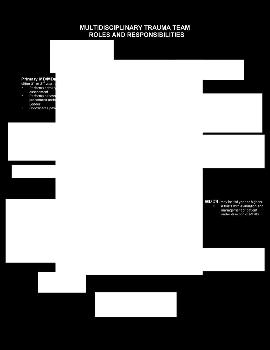
31 UNIVERSITY OF KENTUCKY HOSPITAL NEW DOCUMENT #: CHANDLER MEDICAL CENTER FORMER DOCUMENT#: ISSUED: 6/91 EMERGENCY DEPT. POLICY REVISED: 6/93, 8/94, 7/97, 2/99, 5/00, 4/01, 1/05 & 5/08 SUBJECT: Trauma Resuscitation Roles and Responsibilities PURPOSE: To establish guidelines for staff roles and responsibilities during trauma resuscitation of the critically injured patient. PROCEDURE: Trauma Attending/Trauma Fellow A. Provides guidance and oversees trauma resuscitation. B. Performs and/or assist with procedures. Trauma Team Leader A. Assigned to the Trauma chief resident/emergency Medicine Resident present or EM Attending depending upon assigned rotation schedule. *Trauma Alert Red and Pediatric Trauma Alert Red patients will be managed by the Trauma Attending or Pediatric Surgery Attending with the assistance of Emergency Medicine Service. B. Designated as Trauma Team Leader in charge of trauma resuscitation and decisionmaking under the guidance of the Trauma Attending or EM Attending depending on rotation schedule. C. Responsible for: Overall patient evaluation and management. Ensuring completion of primary and secondary surveys. Communicating assessment findings to recording RN. Determining priority of procedures and necessary tests. Determining need for and timing of operative intervention when indicated and contacting the OR. Determining the need for appropriate consultations. Making physician assignments. Dismissing ancillary personnel when services are no longer needed. Deactivating Trauma Alerts when appropriate. Ensuring traffic control. D. The Trauma Team Leader may assume or delegate the role of evaluation and management depending on patient acuity. The Trauma Team Leader maintains responsibility at all times. The Trauma Team Leaders role may not be delegated to first-year residents. 31
32 Emergency Medicine MD or MD #2 A. Assigned to the EM resident, Anesthesiology Attending or Trauma Resident. B. Responsible for: Primary MD or MD #3 MD #4 Airway management and assuring cervical spine immobilization. Coordinates initial assessment. A. Assigned to second-most Senior Resident, preferably second-year or higher. Responsible for: Assisting Trauma Team Leader in patient evaluation and management. Performs primary and secondary assessment. Performing necessary procedures under the direction of the Trauma Team Leader. Coordinating orders. A. Assigned to third-most Senior Resident, preferably first-year or higher. B. Responsible for: Assisting in patient evaluation and management under the direction of MD #3. Contacting CT Scan and Radiology Special Procedures, as needed, under the direction of the Trauma Team Leader. Coordinating orders and completing Trauma H & Ps under direction of MD #3. Emergency Department Charge Nurse A. Pre-notifies physicians, primary patient care nurse, circulating nurses and financial counselor of impending arrival. B. Responsible for: Primary Patient Care Nurse Delegating responsibility of checking equipment and supplies within the Trauma Resuscitation area. Assisting with traffic control. Acting as family liaison as needed. Disseminating prehospital patient report to appropriate MD s/rn s. A. Prepares Trauma Resuscitation area prior to patient arrival, and ensures that needed equipment/supplies are readily available. B. Responsible for ensuring documentation from admission to time of disposition or report to oncoming RN including: 32
33 Notification and response times of all medical and ancillary personnel. EMS/Air Medical report. Mechanism/time of injury & patient history. Vital signs; cardiac rhythm strips. Ongoing assessment/re-evaluation. Procedures performed by physicians. Nursing process. C. Directs nursing team members as needed and assumes overall nursing responsibility for patient to include: Circulating Nurse/EM Paramedic A. Responsible for: Assists with initial assessment. Confirming labs are obtained and sent. Ensures patient is placed on Cardiac monitor. Monitors VS Gives 5 minute updates. Ensures PIVs are functional & insertion of additional IV s as needed. Obtains Labs from arterial puncture. Accompanying patient outside the ED, as needed. Ensuring initial notification of family and providing ongoing clinical updates. Notifying Capacity Command Center of bed requirement as soon as possible. Assisting with initial assessment. Obtains first manual BP pressure. Assists with medication administration. Assisting physicians with procedures. Assisting in removal of patient clothing. Inserting additional peripheral IV s and drawing labs. Assisting with nursing procedures (e.g. nasogastric tubes, Foley catheters). Assisting with administration of colloids/crystalloids (via fluid warmer, as needed). Paramedics will not administer colloids. Emergency Department Nursing Care Technician A. Responsible for: Assisting in set-up of trauma resuscitation area. Obtaining blood from Blood Bank as instructed. Obtaining IV fluids and blankets from warmer. Assisting in removal of patient clothing. Processing lab specimens. Obtaining additional supplies as directed by nurses. Ensuring patient has an arm/wrist identification band. Processing patient clothing/valuables. Assisting with CPR, as needed. 33
34 Scribe or Documenting Nurse Assisting with patient transport. A. Responsible for: Documents Resuscitation. Assists with crowd control. Assists with procedures as needed. Financial Counselor(s) A. Pre-notifies Radiology, Respiratory Therapy, Operating Room, Blood Bank etc. prior to patient arrival. B. Identifies patient from EMS run sheet, Air Medical records, and/or patient s family and updates computer when possible. C. Prepares lab slips, X-Ray requests, EKG requests and ID bracelets. D. Secures patient valuables in ED safe. Respiratory Therapy A. Assists with airway management. B. Ventilates patient. C. Prepares, monitors and documents all required aspects of mechanical ventilation. D. Assists during patient transport. Radiology Technologist A. Responsible for obtaining radiography studies in a timely manner as ordered. Ultrasound Technologist A. Performing Focus Abdominal Sonogram Test (FAST) as ordered in a timely manner. 34
35
36 UNIVERSITY OF KENTUCKY HOSPITAL POLICY NUMBER: ED08-52CHANDLER MEDICAL CENTER FIRST ISSUED: 06/91 EMERGENCY DEPT. POLICY CURRENT AS OF: 05/08 SUBJECT: PURPOSE: Trauma Labs Describe a standard set of laboratory studies obtained for each trauma patient, and to list specific additional studies for frequently encountered situations. We understand certain cases may require additional studies, to be obtained at the discretion of the treating physicians. PROCEDURE: Lab specimen should be obtained via a femoral arterial stick in patients that are: (a) Unstable, (b) intubated, or (c) have major mechanisms of injury. Venous samples are appropriate for patients that are: (a) Stable, (b) non-intubated, (c) have minor mechanisms of injury, and (d) ALL pediatric patients. Standard Lab Panel for Trauma Alert Red and Trauma Alert Patients * ABG Panel (ABG, Hct, Na, K, ica, Glu) * Type and crossmatch (4 units packed red blood cells) * Coags (PT, PTT, INR) Additional Studies for Altered Mental Status (GCS 14) 1 * Serum alcohol level, urine screen for drugs of abuse Female Trauma Patients of Child Bearing Age 2 * Urine pregnancy test * If positive, Kleihauer-Betke test 1. Altered mental status is defined as GCS 14, not readily explained by pre-hospital medication administration or shock. 2. Female trauma patients without history of prior hysterectomy (Refer to Evaluation and Management of Injury in Pregnancy, located in the protocol manual). Medical Director, Emergency Medicine Trauma Program Director Emergency/Trauma Services Director Formerly
37 UNIVERSITY OF KENTUCKY HOSPITAL NEW DOCUMENT #:08-14 CHANDLER MEDICAL CENTER FORMER DOCUMENT #:08-25 EMERGENCY DEPT. POLICY ISSUED: 9/93 REVISED: 3/00, 4/01, 1/05, 8/08 SUBJECT: SEE ALSO: PURPOSE: Care of Patients in Need of Immediate Surgery When the Operating Room is at Maximum Capacity OR Policy, OR 01-09, OR Access for Trauma. To provide criteria and procedures to manage the needs of patients who require access to the OR at times when the Operating Room is at maximum capacity. CRITERIA: Patients meeting the following criteria may access the Operating Room under this policy: I. Acute Intracranial Bleeding - Rapidly progressing process or lesion leading to herniation or paralysis (ex: acute subdural hematoma, acute epidural hematoma, acute intracerebral hematoma, and/or acute spinal cord compression). II. Exsanguinating Hemorrhage: A. Penetrating neck or chest trauma B. Blunt neck or chest trauma C. Intraabdominal bleeding (ex: penetrating or blunt trauma, ruptured abdominal viscerae, aortic, or iliac aneurysm) III. Potential for Acute Arterial Hemorrhage (established diagnosis) A. Traumatic thoracic aortic injury B. Thoracic aortic dissection or aneurysmal rupture C. Leaking abdominal aorta/iliac artery aneurysm IV. Airway Emergencies Requiring Surgical Intervention A. Traumatic disruption of airway B. Airway compromise due to obstruction C. Intrathoracic and/or lung hemorrhage with respiratory compromise PROCEDURE: In conjunction with the OR response, the staff in the ED will respond in the following manner: I. Trauma Service Responsibilities A. Senior most surgeon present 1. Responsible for determining priorities and management of patient care that necessitates immediate OR access and policy activation. 2. Responsible for notifying OR clerk and charge nurse. 3. Responsible for notifying ED staff. 37
38 Designates trauma team roles as scrub or circulator. B. Remaining surgeons 1. Responsible for retrieving required carts: i.e. craniotomy, airway, chest, abdominal or vascular depending on need. 2. Responsible for opening room, setting up sterile field, serving as scrub and circulating personnel until OR personnel arrive. C. Prior to entering OR room, all staff will apply head covering, mask, and shoe covers. II. Emergency Department Staff A. ED Charge Nurse 1. Responsible for determining need for Trauma Alert Red and for anticipating necessity for accessing the Operating Room. 2. If aware of OR maximum capacity, assess ED staff, assignments and current activities (if time allows). 3. Responsible for reassigning ED patients of primary patient care nurse if patient going to OR (may also send NA if ED staffing allows). 4. Responsible for determining availability and need for additional resources. 5. Communicates need for respiratory therapist to accompany patient to OR. 6. Responsible for ensuring that blood bank delivers blood to OR. B. Patient Care RN 1. Assumes nursing responsibility for the patient; delegates responsibility to others, when available, to accomplish tasks. 2. The ED nurse will prepare the patient for transport and accompany the patient immediately to the OR. NOTE: Patients directly admitted to the OR via Air Medical Services will be accompanied by the Air Medical staff who will continue resuscitation until relieved by the ED nurse or OR personnel (i.e., Anesthesia). 3. The ED nurse will facilitate continued resuscitation in the OR until the call team arrives. The ED nurse will be responsible for the following: a. Monitoring and preservation of airway patency 1. Assisting with intubation or surgical airway as necessary b. Continued volume resuscitation 1. Maintenance/titration of volume infusion rate (crystalloid and colloid) 2. Autotransfusion c. Ongoing hemodynamic monitoring 1. Obtaining specimens for laboratory studies 2. Administration/titration of drugs d. Assisting physicians with procedures e. Documentation on ED Nursing Care Record The ED nurse will remain in the OR until released by the anesthesiologist and/or the arrival of the OR call team. 38
39 Quality Monitoring: **All cases will be reviewed by the Trauma Coordinator and the Multidisciplinary Trauma Committee. Emergency/Trauma Service Director Medical Director, Emergency Medicine REVIEWED: 8/97, 6/00; 8/08 39

40
41 UNIVERSITY OF KENTUCKY HOSPITAL POLICY NUMBER: CHANDLER MEDICAL CENTER FIRST ISSUED: 1/91 Emergency Dept. Policy CURRENT AS OF: 08/08 SUBJECT: SEE ALSO: Nursing Care Record: Trauma/Critical Care 07-12, Nursing Care Record Purpose: Provision of guidelines for use of the trauma/critical care nursing record will allow a standardized method for use of the assessment tool resulting in more effective utilization and accuracy of the tool and the data collected. Rationale: Effective and timely management of the multiple trauma or critically ill patient demands a rapid, organized, systematic approach to assessment, planning, implementation and evaluation. The trauma/critical care nursing record provides the framework for organization of the approach and provides a documentation tool to facilitate establishment of a permanent record of comprehensive baseline and monitoring assessment data as well as facilitating the documentation of the implementation and evaluation phases of the nursing process. The nursing record organizes specific data points to assist in determination of patient status and detection of potential or actual human response to illness. Considerations 1. The trauma/critical care nursing record is included in the patient's permanent medical record. 2. Use of the assessment tool is initiated by the nurse when deemed appropriate. The tool must be used with all critically ill or injured patients who require ongoing monitoring and are classified as acuity Level I or II. 3. As assessments, interventions and/or evaluations are completed the corresponding time and information will be recorded in the appropriate area. 4. Evaluative notes will be recorded under the Nursing Evaluation section of the nursing care record. 5. The record must be signed and initialed by all nurses delivering care to the patient including the recording nurse. Assurance of adequate signatures to document care provision is the responsibility of the patient care nurse. 6. A mechanism for quality assurance will be implemented to audit charts for completeness. Staff will receive feedback surrounding quality assurance audit filters regarding form completion. 41
42 Requisites 1. Black or blue ball point pen essential. 2. Trauma/Critical Care Nursing Record (sample of form attached). Procedure: 1. Legibly print patient name in addressograph area. 2. Record demographic information: a. Classification b. Time of arrival - in military time c. Date d. Age e. Sex f. Weight - in kilograms or pounds g. Mode of arrival: - If Air Med, specify: service. - If Lexington Fire Department, specify unit number: EC-1, EC-5, etc. - If ambulance, specify: Scott County, Jessamine County, etc. h. Recording nurse Specify by name according to i. Patient care nurse role assigned. Record MD j. Circulating nurse names in appropriate areas. k. Physician l. Chief complaint/pre-hospital report - include as many details as are available concerning: 1. Mechanism of injury: driver, passenger, restrained, unrestrained, ejected, rollover, speed traveling, air bag, etc. Description of injury, object (if available) for penetrating trauma - caliber of gun, length or type of knife, removal of impaled objects, etc. 2. Pre-hospital treatment: IV, intubation, immobilization, meds given and fluids infused (type and amount). 3. Chief complaints: particularly important with non-trauma admits - e.g., MI, GI bleeding to include duration of pain - treatment administered. 4. Injury time: Actual time of MVC, GSW, etc. This information is available from patient, pre-hospital personnel, referring hospital, paperwork, etc. 5. Scene flight: Check box if patient arriving via helicopter directly from injury scene. m. Past medical history - check the appropriate box and include any other conditions you identify. 3. Record information in boxes of trauma team response section. a. If trauma activation is called, check the appropriate box for trauma alert, trauma alert red or trauma consult. b. For each team responder (e.g., trauma attending, trauma chief, trauma resident, emergency medicine resident, anesthesia, neurosurgeon, U/S tech, RT, X-ray, etc.) complete time called and time arrived section under each responder. c. Complete name section under each responder by writing in responder's first initial and last name. 4. Initial Assessment
43 a. Complete initial assessment section by documenting the primary and secondary survey findings. Document time assessment of system occurs in space provided. All sections should be completed in full. If you are unable to assess a particular area or it is not applicable document accordingly. b. All trauma patients must have a completed Glasgow coma scores, pupil evaluation & trauma score and on arrival and on discharge from the ED. 5. After completion of the initial assessment, shade injured areas on diagram using legend at bottom of page. 6. Document lab draws with time, site, prep and person obtaining labs. 7. Vital Signs a. Vital signs should have a PTA set documented. Then the frequency of the vital signs should correspond to the acuity of the patient. Vital signs will include Temp, BP, HR, RR, SpO2, GCS and pupils. b. Critical Care monitoring should be documented every hour at minimum e.g. ICP, CPP, CVP, PCWP, etc. c. Intake and Output should be documented at regular interval or as ordered by MD. d. Drips will be documented at regular interval for dosage and rate or when titrated. 8. Ventilator Settings will be completed initially and when changes occur. 9. Intervention a. Procedures - record time of procedures, size of tubes & lines placed along with sites and removal of backboard and/or changing of C-Collar in appropriate spaces. Other procedure data can be entered in the nursing evaluation section. 10. Resuscitation Fluids 1. Record fluids patient received prior to arrival in shaded area. 2. Record all fluids patient receives by using a. Time b. Fluid Type c. Crystalloids - liter number (#) d. IV site e. Y for fluid warmer used f. Colloids document Type & unit number (#) & donor number (#) g. Totals must be documented 11. Rhythm strip must be posted and interpreted for all patients with chest pain or a cardiac event. 12. Medications Document all medication administration by entering time administered, name of med, dose, route, your initials and ordering MD. 13. Nursing Evaluation Documentation of evaluation of patient response to interventions should be documented here. Nurses and NCTs should include their initials after documenting and signature should be legible. If additional space is needed use nursing continuation record. 43
44 14. Transport Patient transports to diagnostics should be documented in this space. 15. Discharge Information Document D/C vital signs, admitting service, total I&Os and disposition of valuables. Transfer form should be completed if patient is transferred to another facility. 16. Notifications All notifications should be documented by checking the box and inserting time notified in space provided. 17. Signatures All nursing staff involved in care of the patient should sign and initial the nursing care record. All signatures should be legible. 18. Disposition Document time report is called and last name of RN receiving report. Time of actual patient movement from the department should be noted as well as destination of patient. If "floor" is checked, enter room number. 19. Place patient stickers in upper right corner of patient chart on white and yellow sheets. The white copy is to accompany the patient to the in-patient area and yellow stays in ED. Formerly ED

45 SECTION 3: ED PROTOCOLS 45
46 TERMINATION OF RESUSCITATION ON THE BASIS OF PREHOSPITAL CRITERIA Introduction: The American College of Surgeons (ACS) position in resuscitative thoracotomy, taught in Advanced Trauma Life Support (ATLS), is simple. In blunt or penetrating thoracic or abdominal trauma, if a patient arrives pulseless and without signs of life, resuscitation ends and the patient should be pronounced DOA (Dead on Arrival). Resuscitative thoracotomy has a poor salvage rate with a high risk of iatrogenic blood borne pathogen exposure and/or injury to health care provides. The patient should be pronounced DOA (Dead on Arrival) when any of the following criteria are met: 1. Blunt Trauma whom arrives pulseless with no signs of life (patients > 15 years old). 2. Penetrating Trauma whom arrives pulseless with no signs of life with prehospital CPR > 15 minutes (patients > 15 years old). Signs of Life: 1. Reactive pupils 2. Spontaneous movement 3. Organized ECG activity Trauma Alert Activation should NOT occur initially for patients meeting the pre-hospital criteria. The Emergency Department faculty will evaluate the patients immediately and determine the following. 1. Pulselessness - if a pulse or any sign of life are detected, Trauma Alert Activation should occur immediately. 2. Normothermic (T > 90 degrees F) If all the above-mentioned physical findings are verified, the patient will be pronounced DOA and no further resuscitation activity should ensue. The ED Faculty should ensure completion of the Trauma Admission Form in its entirety, completing under diagnosis Patient pronounced DOA and noting any injuries diagnosed by gross physical examination (e.g., femur fracture, penetrating head injury, etc.). The pink copy of the Trauma Admission Form will be forwarded to the Trauma Coordinator for entry into the Trauma Registry. The original copy will become part of the patient s permanent medical record. References: American College of Surgeons Committee on Trauma: Advanced Trauma Life Support, ed.8, Chicago, 2008, The College. Committee on Trauma: Resources for Optimal Care of the Injured Patient Chicago, 2006, American College of Surgeons. Revised 11/08 46
47 Revised 11/08 EMERGENCY DEPARTMENT RESUSCITATIVE THORACOTOMY Purpose: Emergency Department resuscitative thoracotomy may be necessary to salvage patients who present in extremis and may otherwise die without aggressive therapy. Emergency Department thoracotomy is not indicated in the resuscitation of all trauma patients who present in extremis. The following protocol is intended to be a guide and is not intended to be all-inclusive or exclusive. Additional patients not covered by this protocol who might benefit from Emergency Department thoracotomy will be rare and case-specific. The procedure is performed in conjunction with other resuscitative efforts and should not be employed in isolation. Under certain conditions, resuscitative efforts might best be accomplished in the Operating Room. An Emergency Department resuscitative thoracotomy should only be performed by general surgery PGY-3, or higher, level residents or attendings. Indications: Definitions: Procedure: 1) Penetrating thoracic trauma that arrive pulseless, with signs of life. 2) Penetrating non-thoracic, non-cranial trauma that arrive pulseless, with signs of life. 3) Cardiac arrest in blunt chest or abdominal trauma after arrival in Emergency Department with an obtainable blood pressure. 4) Suspected systemic air embolism. 1) Signs of life: reactive pupils, spontaneous movement, or organized ECG activity. 2) Aggressive fluid resuscitation: Packed RBC 2 units or Lactated Ringers 2 liters or equivalent volume over 15 minutes. 1) Rapid bilateral antero-lateral Betadine or Chlorahexidine prep while thoracotomy tray opened. Thoracotomy trays are located in Trauma Bay Omni cells. 2) Left antero-lateral thoracotomy incision located beneath nipple in males and in inferior breast fold in females. Incision extends from left sternal border to anterior border of latissimus dorsi and chest entered along the superior aspect of fourth or fifth rib. Care must be taken to avoid injury to heart and lung. A right anterolateral thoracotomy may be preferred for primary right chest wounds. 3) Insert rib spreader with handle located toward table laterally. 4) Examine pericardium, if tense hemopericardium present (pericardium distended with maroon discoloration) then proceed to step 7. 5) If systemic air embolism is suspected or massive hemorrhage from lung parenchyma or hilum is present, then place Satinsky clamp across hilum medially. 6) Retract left lung with left hand. Locate aorta by running right hand medically along posterior chest wall. Aorta located along lateral aspect of vertebral bodies 47
48 and will be postero-lateral to esophagus. Dissect around aorta inferior to pulmonary hilum and apply aortic cross-clamp. 7) Enter pericardium by longitudinally incising pericardium anterior and parallel to phrenic nerve. This is best accomplished by grasping pericardium with forceps and cutting with Metzenbaum scissors. Pericardial incision is carried inferiorly to diaphragmatic reflection and superiorly to level of superior pulmonary hilum. Care must be taken to avoid injury to left atrial appendage and phrenic nerve. This is best accomplished by lifting tip of scissors laterally as incision is made. 8) Manually lift heart from pericardial sac. If hemopericardium present, then examine for cardiac perforation. Teflon pledgetted 3-0 prolene suture on a taper needle is present in thoracotomy suture pack for repair of cardiovascular wounds. If hemopericardium is not present, then begin open cardiac compression. Aortic cross-clamping, if not previously performed, is indicated if no hemodynamic response is noted. 9) Additional exposure may be accomplished by extending thoracotomy incision across sternum into contralateral chest cavity. Reference: American College of Surgeons Committee on Trauma: Advanced Trauma Life Support, ed.8, Chicago, 2008, The College. Powell DW, Moore EE et al. Is emergency department resuscitative thoracotomy futile care for the critically injured patient requiring prehospital cardiopulmonary resuscitation? J Am Coll Surg Aug; 199(2):
49 EMERGENCY DEPARTMENT TRAUMA THORACOTOMY TRAYS TRAY #1 QUANTITY INSTRUMENT 1 - Finochietto Buford Rib Spreader (10 Spread) 1 - Tuffier Rib Spreader 1-6 ¾ Curved Mayo Scissors 2-9 Metzenbaum Scissors 1 - #10 Disposable Blade 2-5 ½ Tissue Forceps 2-9 DeBakey Forceps 2-8 DeBakey Needle holders 4 - Vanderbilt Clamps 1 - Medium Satinsky Clamp 1-9 ½ DeBakey Aortic Clamp 8-5 ¼ Towel Clamps 1 - Liston Bone Cutter Straight 1 - Lebsche Sternal Chisel 1 - Hammer 4 - Surgeons Towels 1 - Teflon Sheet 6 x Laparotomy Pads SUTURE BAG QUANTITY SUTURE PRODUCT NUMBER 2 pkgs 2-0 Silk (SH-CR) 30 Ethicon #C01 6T 1 pkg Cotton Umbilical Tape 30 Ethicon #U-11 T 1 pkg Teflon Felt Pledgets ¼ Deknatel #K pkg Teflon Felt Pledgets Deknatel #L pkgs 3-0 Prolene (SH) 36 Ethicon #8522H 4 pkgs 3-0 Prolene (SH) with Teflon Pledgets 4 pkgs 4-0 Prolene (RB-1) 36 Ethicon #8557H 10 pkgs 2-0 Ticron (T-10) 30 D & G #
50 RSI INTUBATION Julia E. Martin M.D. Assistant Professor Department of Emergency Medicine General information: Airway control is always the most important objective in the initial resuscitation and stabilization. It takes the highest priority in primary assessment. The trauma team must be prepared for any airway emergency. RSI involves the use of neuromuscular blocking agents and sedatives to facilitate endotracheal intubation. Rapid Sequence induction technique is used to prevent regurgitation and aspiration of gastric contents. Requires preoxygenation and denitrogenation by using 100% oxygen via non-rebreather face mask to prevent apnea related hypoxia during the procedure. Once paralytic is on board, mask ventilation is not attempted at this point. During induction, a skilled assistant provides manual in-line axial stabilization of the head while a second assistant presses the cricoid cartilage to prevent gastric aspiration. Cricoid pressure is maintained until the cuff on the ET tube is inflated and tube placement is confirmed. Main disadvantage is once anesthesia has been induced there is no turning back. The only contraindication to RSI intubation is a practitioner who is not skilled in airway management. Indication for surgical airway is the inability to intubate the trachea. In neck trauma, intubation may be difficult or impossible and surgical airway may be required. Short acting agents are used to allow patient to resume spontaneous respirations and to allow close monitoring of neurological status. Oral endotracheal intubation is usually the preferred method. If the head and neck are stabilized by an assistant there is almost no risk of spinal cord injury by oral tracheal intubation. Always anticipate vomiting. Even patients, who otherwise seem relatively unresponsive, may vomit during attempted intubation without RSI. This may result in loss of airway control and aspiration of gastric contents. Struggling patients increase muscle activity making hypoxemia worse and increase ICP. As a general rule, presume all trauma patient s have just eaten. Risk for aspiration is greatest during anesthesia induction and instrumentation of the upper airway. This risk is minimized by applying cricoid pressure. Patients with severe closed head injury are of major concern because intracranial pressure can rise precipitously during intubation. Rapid sequence induction of anesthesia and oral intubation are now recommended for patients with head injuries to minimize the rise in ICP. Remember, rendering patient apneic, when endotracheal intubation is beyond the skill of the operator, may be rapidly fatal. Indication for RSI Endotracheal Intubation of the Acute Trauma Patient: Trauma patients with GCS < 8 Significant facial trauma with poor airway control Airway obstruction Closed head injury or hemorrhagic CVA 50
51 Burn patients with airway involvement and inevitable airway loss Class 3-4 hemorrhagic shock Failure to maintain adequate oxygenation (PaO2 < 60 despite 100% FiO2) Paralysis due to high spinal cord injury Need for positive pressure ventilation Blunt chest trauma with compromised ventilatory effort Mandibular fractures with loss of airway muscular support Evaluation: Talking patient usually indicates airway is patient for the moment. Respiratory distress associated with trauma to the upper airway is frequently made worse by blood or gastric contents in the airway and requires prompt action. These patients are often combative because of hypoxia. When evaluating an awake patient with severe facial trauma ask them if they are getting enough air. If they cannot answer, stick out their tongues fairly easily or are hyperventilating, they should probably be intubated. In unconscious patients, it is probably best to intubate. Tachypnea may be subtle but an early sign of airway or ventilatory compromise. Tachypnea is often also associated with pain and/or anxiety. Agitated and combative patients that are not hypoxic or have a significant head injury are better managed with Haldol 5-10 mg. Signs of Airway Obstruction: Agitation = hypoxia Obtudation = suggests hypercarbia Cyanosis = hypoxia Retractions and use of accessory muscles Snoring, gurgling, stridor = partial obstruction at pharynx Hoarseness = laryngeal obstruction DRUGS: Sedatives: Versed: Benzodiazepine Rapid onset (1-2 min) and short duration (20 min) Amnesic Anticonvulsant Muscle relaxant Slight decrease in blood pressure and increase in pulse rate. No increase in ICP. Dose: 0.1 mg/kg Etomidate: Nonbarbiturate, nonnarcotic sedative-hypnotic induction agent. 51
52 Good agent in multisystem trauma patient because it evokes minimal change in HR and CO compared to Thiopental. (ideal agent in any patient in shock including cardiogenic and septic shock) Decreases ICP and IOP during procedure Rapid onset (<1 min) and short acting (5 min) Vomiting, esp. with combined with a narcotic Dose: 0.3 mg/kg Thiopental: Ultrashort acting barbiturate sedative Dose: 3-5 mg/kg Onset sec Last 10 min. Does not cause increase in ICP but can cause severe hypotension, therefore avoid in multi-traumatized patients. Can also induce bronchospasm. Fentanyl: Narcotic Little or no histamine release Rarely causes hypotension Consider in head-injured patients as a premedication to prevent increase in ICP (blunts pressor response) Rapid injection may cause chest wall rigidity. Dose: 3-5 mcg/kg Onset in 2 min with min duration. Paralytic Agents: Vecuronium: Nondepolarizing agent 1/3 more potent and pancuronium and duration of action is 1/3 to ½ as long (25-40 min vs Pancuronium which last 2-3 hours) Onset 2-3 minutes Dose not cause the degree of tachycardia seen with pancuronium. No histamine release. Defasciculating dose: 0.01 mg/kg Paralytic dose: 0.1 mg/kg Succinylcholine: Depolarizing agent, which causes muscle fasciculations which can be prevented by pretreatment with a non-depolarizing neuromuscular agent. Rapid onset (30-60sec) with short duration of action (5-7 min). Dose: Adult: 1.5 mg/kg Pediatric (<10 y.o): 2.0 mg/kg Contraindications: Burns > 7 days old Extensive crush injuries > 7 days old. Paraplegia > 7 days old. Narrow-angle glaucoma 52
53 Neuromuscular Diseases: Guillain-Barre, myasthenia gravis, Multiple sclerosis, muscular dystrophy, Parkinson s disease. Others susceptible to increased potassium: Renal failure (no real evidence that RSI increases K+) Rhabdomyolysis Rocuronium: Non-depolarizing agent Onset < 1 min. Duration min. Dose: mg/kg Expensive Adjunctive: Atropine: Succinylcholine will cause bradycardia in infants and children therefore they should be premedicated with atropine. Also pretreat any adult who is already bradycardic. Children < 8 y.o. Dose: 0.01 mg/kg up to 0.5 mg (minimum dose of 0.1 mg) Lidocaine: Dose: 1.5 mg/kg Some studies recommend intravenous Lidocaine to blunt the pressor response of increased pulse, increased blood pressure, increased intracranial pressure, and increased intraocular pressure associated with intubation, its usefulness is controversial. However, because a single dose of lidocaine is unlikely to cause harm, it seems reasonable to use in the patient who has a known or suspected head injury. Should be administered 2-3 min prior to intubation. Procedure: The 5 P s of rapid sequence intubation: Preparation Preoxygenation Pretreatment Paralysis (with anesthesia) Placement (of the endotracheal tube) 1. Preoxygenation with 100% oxygen for 3-5 minutes via NRB mask (or 3 vital capacity breaths, avoid BVM if possible). 2. Secure IV s, ECG, pulse oximeter. 3. Prepare intubation equipment: ETT with stylet, suction, BVM, laryngoscope. 4. Premedication: Lidocaine (head injury) 1.5 mg/kg Vecuronium (defasciculating dose) 0.01 mg/kg Versed 0.1 mg/kg Atropine (peds) 0.01 mg/kg Etomidate 0.3 mg/kg 53
54 5. Perform Sellick s maneuver, maintain maneuver until after confirmation of tube placement. 6. Succinylcholine 1.5 mg/kg (Peds: 2.0 mg/kg) 7. Wait sec, place ETT. 8. Confirm ETT placement by: listening for bilateral breath sounds, chest rise and fall, tube fogging, & positive ETCO2. Final confirmation by CXR. 9. Release Sellick maneuver. 10. Secure ETT. Basic RSI Time (min) Preparation and Preoxygenation Administer Vecuronium 0.01 mg/kg IV Lidocaine 1.5 mg/kg IV (Fentanyl 3 mcg/kg IV) (Atropine 0.01 mg/kg in kids < 10) Apply cricoid pressure and in-line cervical stabilization Administer Versed 0.1 mg/kg IV or Etomidate 0.3 mg/kg IV -55 sec Administer Succinylcholine 1.5 mg/kg IV (2mg/kg if <10 y.o.) Intubate RSI without Succinylcholine Time (min) Preparation and Preoxygenation Lidocaine 1.5 mg/kg IV (Fentanyl 3 mcg/kg IV) (Atropine 0.01 mg/kg in kids < 10) Apply cricoid pressure and in-line cervical stabilization Administer Versed 0.1 mg/kg IV or Etomidate 0.3 mg/kg IV Administer Rocuronium mg/kg Intubate 54
55 References: Wilson R, Walt A. Anesthesia for the Trauma Patient. Management of Trauma pitfalls and Practice, 2 nd edition. Baltimore: Williams & Wilkins; 1996: 85-90, American College of Surgeons. Airway Management. Advance Trauma Life Support. American College of Surgeons; 1993: Tintinalli J, Ruiz E, Krome R. Advanced Airway Support. Emergency Medicine a Comprehensive Study Guide, 4 th edition. New York: McGraw-Hill; 1996: Walker L. Using Rapid Sequence Induction to Facilitate Tracheal Intubation. Emergency Medicine Reports. 1993;15: Hardwood-Nuss A. Trauma Airway Management. The Clinical Practice of Emergency Medicine, 2 nd edition. Philadelphia: Lippincott-Raven; 1996:

56 56
57 Initial Management of Head Injuries Brain injury is the most common cause of death in trauma. Brain injury is divided into primary and secondary forms. Primary brain injury, which occurs immediately upon impact, can be reduced only through prevention initiatives. Secondary brain injury, which ensues within hours to days later, results from a cascade of cellular events (intracellular calcium, cell membrane permeability changes, depletion of cellular energy, free radical generation, cell signaling molecules) that harms or even destroys neuronal tissue in and around the site of the primary injury. Secondary injury is exacerbated by 4 major factors: hypoxia, hypotension, hypercarbia and intracranial hypertension. Additional factors which also affect secondary brain injury include: hypocapnea, hyperthermia, glucose imbalance, acute hypo-osmolarity, electrolyte imbalance, anemia, acid-base disorders and coagulopathy. Since secondary brain injury is a major contributor to brain injury mortality and has a negative effect on neurologic outcome, clinicians must work diligently to identify and treat causative factors. Airway maintenance, ventilation, CO 2 control and restoration of circulating volume using isotonic fluid are essential initial treatments. SBP < 90 and SaO 2 < 90% must be strictly avoided in head-injured patients. Intracranial pressure (ICP) elevations cannot be predicted by CT scan. ICP monitoring is indicated in salvageable patients with GCS 8 and an abnormal CT scan. Some evidence also suggests ICP monitoring is indicated in patients with a normal CT if GCS 8 and age > 40, SBP < 90 or bi- or uni-lateral posturing. Though older devices were subarachnoid, subdural or epidurai, modern ICP monitors are intraparenchymal or intraventricular. ICP monitoring guides therapy, and if an intraventricular catheter is in place, permits drainage. ICP monitoring also predicts outcome, as patients who respond the therapy for intracranial hypertension have a more favorable outcome. Normal ICP is 0-15mmHg. Therapy should be initiated to lower ICP when it reaches 20mmHg. More important than ICP is cerebral perfusion pressure (CPP), the difference between MAP and ICP (CPP=MAP-ICP). CPP < 50mmHg must be strictly avoided and recommended CPP is 50-70mmHg, though optimal CPP is unproven. CPP may be increased by lowering ICP or by raising MAP. ICP may be lowered by removal of noxious stimuli and adequate sedation. Morphine is sedative and analgesic without increasing ICP. Midazolam may reduce MAP and raise ICP. Propofol is the recommended sedative agent in brain injured patients because it lowers ICP. MAP can be increased by use of alpha-adrenergic agents. Alternative measures for brain oxygenation include jugular venous oxygen saturation (SjO 2 ) and brain tissue partial pressure of oxygen (PbrO 2 ). If measured, interventions to increase cerebral oxygenation when SjO 2 drops below 50% and/or PbrO 2 drops below 15mmHg. Mannitol is a hyperosmolar plasma expander that also functions as an osmotic diuretic. Mannitol expands plasma volume, reduces blood viscosity, increases cerebral blood flow and oxygen delivery and because of its osmotic effects may reduce brain water and secondary brain injury. Mannitol (0.25g-1g/kg) can be used to lower ICP based upon clinical signs alone (herniation or progressive neurologic decline) or ICP monitoring. Clinicians must maintain adequate intravascular volume in the face of mannitol therapy. 57
58 Hyperventilation has theoretical benefits in lowering pco 2 and thus cerebral blood volume which lowers ICP. Hypocapnea can, however, produce cerebral ischemia and recent data indicate that hypocapnea may be more harmful than hypercapnea. Moreover, prolonged hyperventilation is probably ineffective because adaptation occurs and cerebral blood flow returns to baseline. Current guidelines for CO 2 control are to achieve a pco 2 =35 and avoid hyperventilation to pco 2 < 35 mmhg for the first 24 hours post-injury. Hyperventilation may be used as a temporizing measure only in cases of refractory intracranial hypertension. Additional treatments for severe traumatic brain injury include barbiturates which are recommended for refractory intracranial hypertension in hemodynamically stable patients. Hemodynamic instability and severe ileus are side effects of this therapy. Steroids are contraindicated for the treatment of traumatic brain injury. Anticonvulsants (phenytoin) may be used to prevent early post-traumatic seizures and therapy duration is 7 days. Prophylactic antibiotics are not recommended for indwelling ICP monitors. DVT prophylaxis is recommended in patients with TBI, with compression devices initially and progressing to anticoagulants as neurosurgery indicates is appropriate. Pre-Hospital: Ensure patent airway while maintaining cervical spine precautions (endotracheal intubation using RSI protocol) if necessary. Ventilate patient with 100% oxygen. Keep PaO2 > 60mmHg (Sat >90%). Keep ETCO mmHg during transport. Establish adequate IV access. Maintain mean arterial pressure (MAP) >90. Mannitol may be given with ingoing signs if neurologic deterioration: Motor score < 3 Decreasing motor score Lateralizing motor findings Pupillary changes (dilation or sluggish reactivity) Sedation/Neuromuscular blockade can be useful in optimizing transport of head injured patient. Both treatments interfere with neurologic examination and should be avoided if possible. Reassess neurologic status frequently. Transfer should not be delayed for diagnostic testing. Steroids have not been found to be of benefit to the head-injured patient and are not recommended as therapy for severe head injury. Initial Resuscitation of the Head Injured Patient 58
59 Insure patent airway (endotracheal intubation is indicated in patients with GCS < 8). Maintain cervical spine immobilization. Maintain MAP > 90 throughout patient course in attempt to maintain cerebral perfusion pressure (CPP) 50-70mmHg. Insure adequate volume repletion before adding vasopressors (CVP >12). Maintain adequate circulating volume: o NS preferred o Monitor for signs of Diabetes Insipidus o Place Foley catheter Prophylactic anti-convulsants may be indicated. 59
60 Severe Head Injury Management Algorithm (GCS < 8) Neurologic assessment (GCS < 8) Established definitive airway Obtain ABG BVD 100% O2 or Mechanical Ventilation CT Head (see protocol) Maintain PCO IVF NS (preferred) MAP > 90 Decompress stomach (Place NG/OG) Insert Foley Agitated/ Monitor Urine Output Lateralizing signs PIP > 40 neurologic deterioration Yes No Mannitol ¼ gm 1 gm/kg Consider Sedation/ NMBA Hold sedation Monitor UOP 60
61 FACIAL BONE FRACTURE EMERGENCY DEPARTMENT RADIOLOGIC ALGORITHM Indications: 1. Obvious facial bone fracture or fracture suspected on physical exam. 2. Facial bone fracture detected on head CT. 3. Facial bone fracture detected on radiograph from referring facility. Upper and Midface Lower Face (Mandible) Axial Spiral CT 3mm Slices Coronal Reconstructions* Upper and Mid-face Optional Coronal face, 3mm Coronal orbits, 2mm Lower Face (Mandible) Optional Panorex *Axial spiral CT with 3mm slices provides sufficient detail for diagnosis and treatment planning for facial fractures (forehead to mandible). These films are not often needed urgently and should be obtained at the request or discretion of the maxillofacial trauma consultant. Facial plain films are sometimes helpful for treatment planning. While (formal) coronal CT is usually only needed when coronal reconstructions do not provide adequate detail for surgical treatment planning, an urgent coronal CT may sometimes be needed to resolve an equivocal CT with regards to optic nerve integrity or compression. Mandibular Panorex is a useful study in patients who can sit upright and cooperate with the exam. Thus Panorex is not often logistically obtainable in multi-trauma patients and axial CT with coronal reconstructions, which provides satisfactory detail for treatment planning, is the usual diagnostic of choice. 11/08 61
62 SPINE CLEARANCE Basic Principles General 1. Entire spine is immobilized during primary survey. 2. Radiographic clearance of the spine is not required before emergent surgical procedures. Presence of a spinal column injury is simply assumed until excluded. 3. Secondary and tertiary exams include examination of the spine for tenderness as well as testing all motor roots, sensation and reflexes. 4. Tertiary exams are performed only on alert and unimpaired patient without distracting injuries. 5. If any spine fractures are found, entire spine must be radiographed. 6. For patients with radiographic injury spine consultation requested for focused preoperative evaluation regarding relative instability and severity of injury prior to intubation. 7. Patients remain on spine precautions until spine is cleared. Cervical 1. C-spines are not cleared until after the tertiary exam is completed. 2. Cervical CT scan is the preferred radiographic modality when physical exam is not adequate. 3. With impaired or unconscious patient, rigid collars are taken off within 2 hours and replaced with semi-rigid pressure reducing collar. 4. Enter patients in cervical algorithm for C-Spine clearance. Thoraco-Lumbar 1. CT scan of thoracic and lumbar spines if there are clinical findings on secondary or tertiary exams or an unreliable exam. Multi-detector CT-scan with reformatted axial collimation is superior to plain films. 2. Radiographic Thoraco-Lumbar clearance is not needed prior to OR for non spine surgery. Thoracic & Lumbar clearance may however be required for some non supine positioning in the OR, depending upon acuity and case type. 3. Tertiary exam is necessary to clear thoracic and lumbar spines. 8/08 62

63 63
64 Initial Management of Spinal Cord Injury 1. Priorities: Airway, Breathing & Circulation 2. Maintain complete spine immobilization using: a. Semi-rigid cervical collar b. Modified logroll maintaining spine in neutral position at all times c. Remove patient from long board within 2 hours. 3. *If patient is hypotensive determine cause and treat hypovolemia with fluids and definitive surgical intervention as directed. 4. If hypotension due to Neurogenic Shock confirmed, consider inotropic agents to maintain blood pressure (MAP 60 70mmHg). *Effort must be made to reduce secondary injury. *Methyprednisolone use insufficient evidence to support routine use 5. Patient should be removed from long spine board & placed on pressure reducing surface within 2 hours of trauma room arrival. 6. Radiographic studies to determine location of injury include: a. Plain films b. Spiral CT scan c. MRI 7. Determine if injury is complete or incomplete and fracture is stable or unstable. 8. Fully document complete neurological exam during secondary survey & prior to OR if possible. 9. Document level of sensory and motor function using dermatomes. 10. Obtain Spine Surgery (Orthopedic or Neurosurgery) consult. 11. Place urinary catheter to monitor urinary output & prevent bladder distension. 12. Place gastric tube to prevent gastric distension & aspiration. 13. Physical Medicine and Rehab consult on admission to begin a timely transfer to Acute Spinal Cord Injury Rehabilitation. 8/08 64
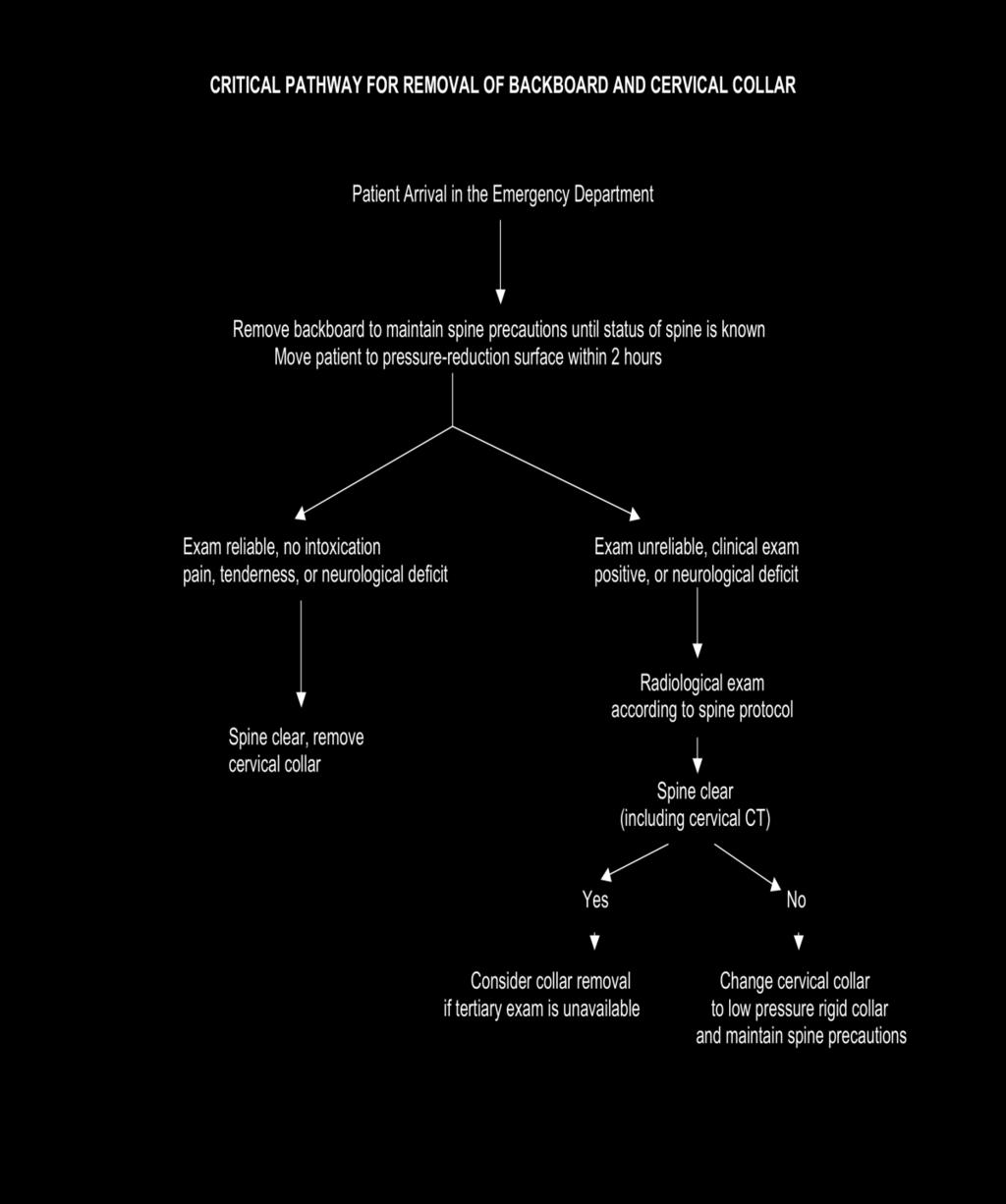
65 65
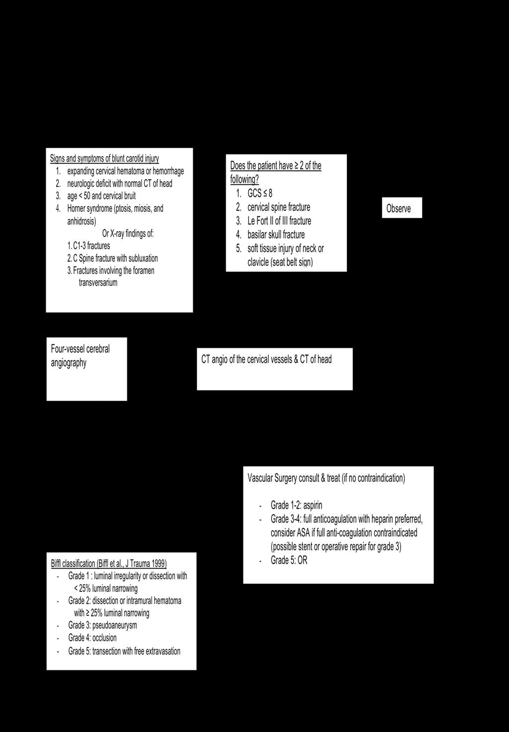
66 66
67 Penetrating Neck Injury Evaluation and Treatment Algorithm University of Kentucky Hospital Trauma Center Overview The neck is divided into zones: Zone 1-Clavicles to cricoid cartilage Zone 2-Cricoid to angles of the mandible Zone 3-Angles of the mandible to skull base Management of patients with penetrating wounds to the neck has historically been determined by zone of injury. Because zones 1 and 3 are challenging to expose surgically, patients with injuries in zones 1 and/or 3 warrant thorough diagnostics because non-therapeutic surgery in these areas is both difficult and morbid. Zones 1 and 3 should be approached surgically only if an injury is felt to be present. However, zone 2 of the neck is easily exposed surgically. Controversy has existed as to whether patients with zone 2 injuries should undergo exhaustive diagnostics to exclude or characterize injuries in this area, or simply undergo neck exploration with limited or no preoperative evaluation of the esophagus and cervical vasculature (esophagography or esophagoscopy or both plus angiography). Recently published studies have changed the management of penetrating neck trauma in 2 important ways. First, evidence suggests that for patients with no clinical evidence of vascular injury (shown on the algorithm as Mandatory Criteria for Neck Exploration ) then physical examination has 100% sensitivity, thus definitively excluding vascular injury without any angiography. This is most true if the injury is to Zone 2. The second major advance in the care of these patients has been the advent of helical CT angiography as an alternative to conventional catheter-based angiography, producing equivalent results. Furthermore, the anatomic detail provided by the neck CT may permit the clinician to exclude injury to the esophagus if the CT clearly shows a missile trajectory remote from the esophagus. However, the precise role that CT will play in excluding injury to the esophagus in these patients remains to be established. If the CT does not conclusively exclude injury to the esophagus, contrast esophagography and/or esophagoscopy (or both) should be performed. Summary 1. CT scan of the neck including CT cervical angiography is the initial diagnostic of choice. 2. Some asymptomatic patients may avoid angiography entirely. 3. Esophageal injury must be definitively excluded, which may require esophagography or esophagoscopy or both. References regarding the utility of CT/CTA for evaluating penetrating neck injury: 1. Gracias VH, Reilly PM, Philpott J, Klein WP, Lee SY, Singer M, Schwab CW. Computed tomography in the evaluation of penetrating neck trauma: a preliminary study. Arch Surg 2001;136: Mazolewski PJ, Curry JD, Browder T, Fildes J. Computed tomographic scan can be used for surgical decision making in zone II penetrating neck injuries. J Trauma 2001;51: Múnera F, Soto JA, Palacio DM, Castañeda J, Morales C, Sanabria A, Gutiérrez JE, García G. Penetrating neck injuries: helical CT angiography for initial evaluation. Radiology 2002; 224:
68 4. Fergusion E, Dennis JW, Vu JH, Frykberg ER. Redefining the role of arterial imaging in the management of penetrating zone 3 neck injuries. Vascular. May-jun 2005;13(3): [Medline]. References demonstrating the diagnostic accuracy of physical exam for vascular injuries in Zone II requiring intervention: 1. Nason RW, Assuras GN, Gray PR, Lipschitz J, Burns CM. Penetrating neck injuries: analysis of experience from a Canadian trauma centre. Can J Surg 2001;44: Sriussadaporn S, Pak-Art R, Tharavej C, Sirichindakul B, Chiamananthapong S. Selective management of penetrating neck injuries based on clinical presentations is safe and practical. Int Surg 2001;86: Azuaje RE, Jacobson LE, Glover J, Gomez GA, Rodman GH Jr, Broadie TA, Simons CJ, Bjerke HS. Reliability of physical examination as a predictor of vascular injury after penetrating neck trauma. Am Surg 2003;69: Osborn TM, Bell RB, Qaisi W, Long WB. Computed tomographic angiography as an aid to clinical decision making in the selective management of penetrating injuries to the nec: a reduction in the need for operative exploration. J Trauma. Jun 2008;64(6): [Medline]. 68
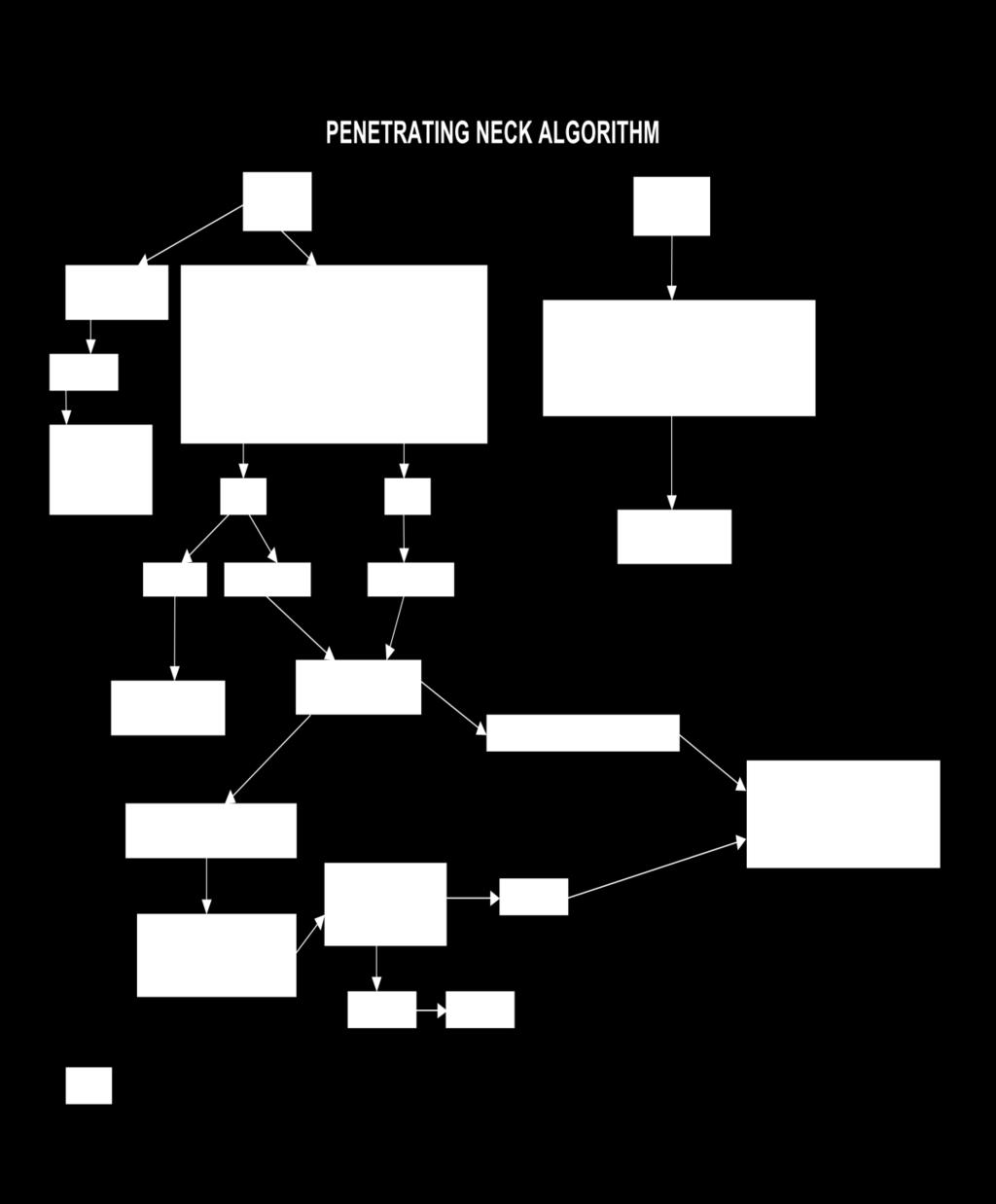
69 69
70 Blunt Cardiac Injury (BCI) Blunt cardiac injury (BCI), formerly called myocardial contusion, encompasses a spectrum of disease ranging from histologic injury to the myocardium without clinical manifestation to blunt cardiac rupture. 1 BCI contributes to 20% of prehospital deaths from blunt trauma. 2 An exact incidence of BCI does not exist because there is no gold standard for diagnosis, i.e., the available data are conflicting with regard to how the diagnosis should be made (ECG, echocardiography, enzyme analysis, etc). This lack of a diagnostic standard makes the literature difficult to interpret and leads to confusion in clinical practice. The major priority is identification of patients at risk for adverse events resulting from BCI and providing appropriate workup, monitoring and treatment. Conversely, patients not at risk can potentially be discharged home. Patients with appropriate mechanism of injury and clinical evidence of cardiac dysfunction (electrophysiologic or mechanical) can be considered to have BCI. BCI results from 5 possible mechanisms: direct pre-cordial impact, crush between sternum and spine, deceleration or torsion causing a tear in the heart at a point of fixation, hydraulic effect resulting in rupture from elevated intra-abdominal and caval pressure, and blast injury. 3 Few clinical signs are diagnostic of BCI. Chest pain is the most common finding, but dyspnea, chest wall ecchymosis and rib fractures may also be present. Associated injuries include hemothorax, sternal fracture and great vessel injury. Clinical signs consistent with BCI include dysrhythmias, cardiac ischemia, low cardiac output and hypotension. 4 Diagnostic tests include ECG, echocardiography, and enzyme analysis. Controversy exists regarding the application of these tests. Frequency of diagnosis of BCI will be proportional to the aggressiveness with which it is sought. Appropriate workup commands achieving a balance between cost-effectiveness and information acquisition with attention to the clinical value of information gained in changing patient management. Guidelines for using diagnostic tests are as follows: A. Level 1 evidence supports obtaining an ECG in the emergency department for at-risk patients (described above). 5 Using any ECG abnormality, including sinus tachycardia, bradycardia conduction delays and PAC s/pvc s, the diagnostic sensitivity of ECG is 100%. 6 B. Echocardiography is not effective as a screening tool and does not identify patients at risk for complications. 7 Transthoracic (TTE) or transesophageal echocardiography (TEE) should be obtained in patients with evidence of hemodynamic instability or in whom coincident coronary ischemia is suspected. C. CK and CKMB fraction analysis is NOT indicated in suspected BCI because associated skeletal and visceral injury creates serum CK abnormalities that contribute to an unacceptable false-positive and negative rate. 8 However, a body of evidence exists suggesting some value to cardiac troponin I (ctni) or troponin T (ctnt). That evidence is as follows: i. Diagnosis of BCI should not rely solely on ctni or ctnt. ECG should be included. ii. Normal ECG and normal ctni is 100% sensitive for BCI. 9 iii. Abnormal ECG and abnormal ctni is 100% specific for BCI. 9 iv. ctni is of little added benefit in patients with a markedly abnormal ECG (diagnosis is already made)
71 v. Though the utility of ctni in patients with normal ECG s is limited, ctni obtained 4-6 hours after the injury in patients with sinus tachycardia or non-specific EKC changes or in older patients may give reassurance that the likelihood of BCIrelated complications is low. 10 References: 1. Mattox KL, Flint LM, Carrico CJ, Grover F, Meredith J, Morris J, et al. Blunt cardiac injury. J Trauma 1992;33(5): Schultz JM, Trunkey DD. Blunt cardiac injury. Crit Care Clin 2004;20: Fulda G, Brathwaite CE, Rodriguez A, Turney SZ, Dunham CM, Cowley RA. Blunt traumatic rupture of the heart and pericardium: a ten-year experience ( ). J Trauma 1991;31(2): Miller FB, Shumate CR, Richardson JD. Myocardial contusion. When can the diagnosis be eliminated? Arch Surg 1989;124(7): Pasquale NK, Clarke J. Screening of blunt cardiac injury The Eastern Association for the Surgery of Trauma. Available: 6. Fabian TC, Cicala RS, Croce MA, Westbrook LL, Coleman PA, Minard G, et al. A prospective evaluation of myocardial contusion: correlation of significant arrhythmias and cardiac output with CPK-MB measurements. J Trauma 1991;31(5): Karalis DG, Victor MF, Davis GA, McAllister MP, Covalesky VA, Ross Jr JJ, et al. The role of echocardiography in blunt chest trauma: a transthoracic and transesophageal echocardiographic study. J Trauma 1994;36(1): Fabian TC, Cicala RS, Croce MA, Westbrook LL, Coleman PA, Minard G, et al. A prospective evaluation of myocardial contusion: correlation of significant arrhythmias and cardiac output with CPK-MB measurements. J Trauma 1991;31(5): Salim A, Velmahos GC, Jindal A, Chan L, Vassiliu P, Belzberg H, et al. Clinically significant blunt cardiac trauma: role of serum Troponin levels combined with electrocardiographic findings. J Trauma 2001;50(2): Collins JN, Cole FJ, Weireter LJ, Riblet JL, Britt LD. The usefulness of serum Troponin levels in evaluating cardiac injury [discussion]. Am Surg 2001;67(9): Revised: 08/08 71
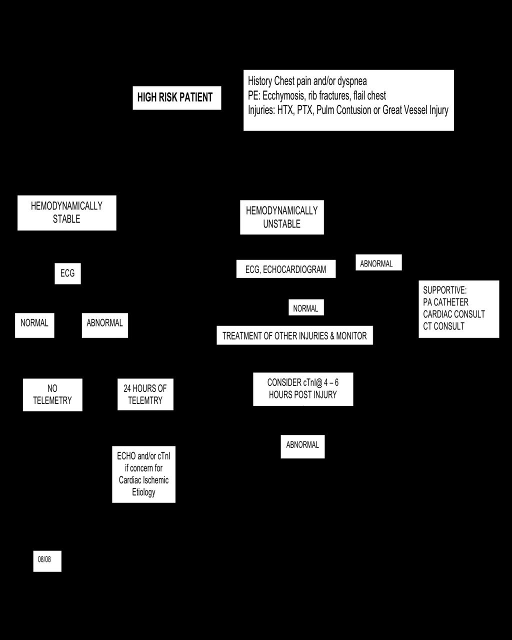
72 72
73 Revised 8/08 THE DIAGNOSIS OF BLUNT INJURY TO THE THORACIC AORTA What diagnostic test is best? Angiography, Transesophageal Echocardiography, or Computerized Axial Tomography Paul A. Kearney, M.D., F.A.C.S. Associate Professor of Surgery Chief, Section of Trauma and Critical Care University of Kentucky Chandler Medical Center Background Blunt chest trauma with resulting aortic injury is a significant cause of death following highspeed motor vehicle collisions. 1-3 The vast majority of these patients (80%-90%) expire at the collision scene. 1-3 For the remaining 10%-20%, the mortality rate is high. Thirty percent expire within 6 hours and 40%-50% within 24 hours of injury. 1 The recent multicenter trial by the American Association for the Surgery of Trauma reported an overall mortality of 31%, with 63% of the deaths attributable to aortic rupture. 4 Expeditious evaluation and timely surgical intervention are essential for patient survival. Screening The goal of screening patients is to attain a zero nontherapeutic surgery rate without overlooking any significant aortic or arch vessel injury. Mechanism of injury, clinical exam, and the initial chest radiograph should reliably select patients who require further diagnostic evaluation. Chest radiographs demonstrating mediastinal hematoma have good sensitivity (93%) for aortic and arch vessel injury. 5-7 When combined with mechanism of injury, sensitivity rises to 98%. 5 More importantly the negative predictive value of a normal upright chest radiograph is almost 100%. A normal chest radiograph virtually excludes aortic and/or arch vessel injury. 5-7 Unfortunately, the specificity of an abnormal chest radiograph is only 10%-45%. Since most mediastinal hematoma originates from thoracic vascular structures other than the aorta, a definitive diagnostic test is required to establish the diagnosis of aortic injury. Computerized axial tomography (CT) of the chest can be used to screen patients for subsequent aortography CT is more sensitive than chest radiograph in detecting mediastinal hematoma. 8,9 Screening thoracic CT is cost-effective and the negative predictive value of a normal study is 100% Consequently, CT can be used to reduce the negative aortogram rate in patients with an abnormal chest radiograph Diagnosis-Aortography Many authors advocate the liberal use of aortography based on mechanism of injury and chest radiographs. 1,3,9,13 Using this approach, aortography yields positive results in only 10% of patients. Although angiography remains the gold standard, the procedure is invasive and time consuming, requires the use of intravenous contrast material and ionizing radiation, and can result in false negative or false positive results. 3,9,13,14 Transporting an injured patient to the angiography suite is not without risk and interrupts the patient s ongoing evaluation, resuscitation, and treatment. On the other hand, when performed properly, aortography has a sensitivity and specificity of 99%. 9 Complications occur in less than 1% of patients. 9 73
74 Aortography remains the only study that provides detailed images of the entire thoracic aorta and the arch vessels. Diagnosis-Transesophageal Echocardiography Transesophageal echocardiography (TEE) provides high-resolution real-time axial and longitudinal images of the aorta TEE can accurately demonstrate injury to the thoracic aorta In our hands, TEE was more sensitive (100%) and specific (98%) than aortography. 14,15 TEE offers a number of advantages over aortography and CT scanning. The study can be performed at the bedside, eliminating transport risks. Concomitant diagnostic and therapeutic procedures can continue unhindered. TEE provides real-time images so that areas of interest can be examined repeatedly in different planes. Simultaneous evaluation of cardiac pathology and function can also be obtained. If urgent surgical intervention is indicated for other injuries, TEE can be performed in the operating room without delaying TEE or the surgical intervention. The study can be performed more rapidly than aortography making TEE ideal for the evaluation of the unstable trauma patient with a number of diagnostic and treatment priorities. Unfortunately, TEE requires the expertise of a well-trained and interested echocardiographer. Airway compromise, esophageal pathology, and unstable cervical spine fractures are contraindications to TEE. The depth and extent of injury are frequently difficult to determine particularly when atherosclerotic disease diminishes the sensitivity of the examination. In our experience, this has resulted in nontherapeutic thoracotomy. 14 There are blind spots related to the tracheal air column and the arch vessels simply cannot be imaged. Diagnosis-Computerized Axial Tomography (CT) CT scanning technology is advancing rapidly. Perhaps the most promising technology for a fast, accurate, and less-invasive diagnostic test for detecting injuries to the thoracic aorta and arch vessels is the newer generation helical CT scanner. 9,19-21 Rather than providing indirect evidence of aortic injury (detection of mediastinal hematoma), the helical scanners can provide direct evidence of aortic injury obviating the need for confirmatory tests. Sensitivity, specificity, and accuracy depend heavily on the technical skill of CT performance and the interpretative expertise. 9 Confirmatory angiography or TEE must be performed for indeterminate or equivocal CT scan results. Although CT can be performed more rapidly than angiography, transport of the injured patient to CT is not without risk and interrupts the patient s ongoing evaluation, resuscitation, and treatment. Summary Patients with suspected blunt injury to the thoracic aorta are a challenge for the trauma surgeon. Multisystem trauma, critical illness, and hemodynamic instability in this patient group result in diagnostic and treatment dilemmas. We employ a practical, evidence-based algorithm for the screening and diagnosis of injury to the thoracic aorta. Both mechanism of injury and an abnormal mediastinum on chest radiograph are required to trigger a diagnostic evaluation. Every attempt is made to obtain an upright AP chest radiograph to minimize distortion and magnification. However, this is not possible in all patients. Widening of the mediastinum alone is neither sensitive nor specific for mediastinal hematoma. 5,6,7 Instead, we employ the criteria for mediastinal hematoma (abnormal mediastinal silhouette) as defined by Mirvis and Ayella. 6,7 No further diagnostic evaluation is undertaken in patients with a normal chest radiograph, unless there are compelling physical findings that suggest aortic or arch vessel injury (i.e. Pulse deficit, 74
75 unequal blood pressure/pulse measurement, unexplained hemodynamic instability, unexplained neurologic deficit). Thoracic CT scan using the aortic protocol is performed. 9,19 A negative scan yields observation. A scan positive for aortic injury prompts surgical intervention or appropriate non-operative management. Patients with indeterminate scans undergo angiography or TEE to establish or exclude the diagnosis. REFERENCES: 1. Parmley LF, Mattingly TW, Manion WC, et al. Nonpenetrating traumatic injury of the aorta. Circulation 1958;17: Greendyke RM. Traumatic rupture of the aorta: special reference to automobile accidents. JAMA 1966;195: Culliford AT: Traumatic aortic rupture. In Hood RM, Boyd AD. Culliford AT (eds): Thoracic Trauma. Philadelphia, WB Saunders, 1989, pp Fabian TC, Richardson JD, Croce MA, et al. Prospective study of blunt aortic injury: multicenter trial of the American Association of the Surgery of Trauma. J Trauma 1997; 42: Woodring JH. The normal mediastinum in blunt traumatic rupture of the thoracic aorta and brachiocephalic arteries. J Emerg Med 1990;8: Mirvis SE, Bidwell JK, Buddemeyer EU, et al. Imaging diagnosis of traumatic aortic rupture: A review and experience at a major trauma center. Invest Radiol 1987;22: Ayella RJ, Hankins JR, Turney SZ, et al. Ruptured thoracic aorta due to blunt trauma. J Trauma 1977;17: Harris JH, Horowitz DR, Zelitt DL. Unenhanced dynamic mediastinal computed tomography in the selection of patients requiring aortography for the detection of acute traumatic aortic injury. Emerg Radiol 1995;2: Patel NH, Stephens KE, Mirvis SE, et al. Imaging of acute thoracic aortic injury due to blunt trauma: A review. Radiology 1998;209: Mirvis SE, Shanmuganathan K, Miller BH, et al. Traumatic aortic injury: Diagnosis with contrast-enhanced thoracic CT. Five year experience in a major trauma center. Radiology 1996;200: Demetriades D, Gomez H, Velmahos GC, et al. Routine helical computed tomographic evaluation of the mediastinum in high-risk blunt trauma patients. Arch Surg 1998;133: Dyer DS, Moore EE, Ilke DN, et al. Thoracic aortic injury: How predictive is mechanism and is chest computed tomography a reliable screening tool? A prospective study of 1,561 patients. J of Trauma. 2000;48: Kirsh MM, Behrendt DM, Orringer MB, et al. The treatment of acute traumatic rupture of the aorta: A 10-year experience. Ann Surg 1976; 184: Smith MD, Cassidy JM, Souther S, et al. Transesophageal echocardiography in the diagnosis of traumatic rupture of the aorta. N Engl J Med 1995;332: Buckmaster MJ, Kearney PA, Johnson SB, et al. Further experience with transesophageal echocardiography in the evaluation of thoracic aortic injury. J of Trauma 1994;37: Vignon P, Gueret P, Vedrinne JM, et al. Role of transesophageal echocardiography in the diagnosis and management of traumatic aortic disruption. Circulation 1995;92: Goarin JP, Catoire P, Jacquens Y, et al. Use of transesophageal echocardiography for diagnosis of traumatic aortic injury. Chest 1997;112: Mollod M, Felner JM. Transesophageal echocardiography in the evaluation of cardiothoracic trauma. Am Heart J 1996;132:
76 19. Mirvis SE, Shannuganathan K, Buell J, Rodriquez A. Use of spiral computed tomography for the assessment of blunt trauma patients with potential aortic injury. J of Trauma 1998;45: Gavant M. Helical CT grading of traumatic aortic injuries. Impact on clinical guidelines for medical and surgical management. Rad Clinics of North America 1999;37: Wicky S, Capasso P, Meuli R, et al. Spiral CT aortography: an efficient technique for the diagnosis of traumatic aortic injury. European Radiology 1998;8:
77 BLUNT THORACIC INJURY WITH SUSPECTED INJURY TO THE THORACIC AORTA OR ARCH VESSELS Patients who sustain blunt thoracic trauma are at risk for injury to the heart and great vessels. Patients should be selected for additional diagnostic studies based on mechanism of injury and evidence of mediastinal hematoma on chest radiograph. Every attempt is made to obtain an upright AP chest radiograph to minimize distortion and magnification. However, this is not possible in all patients. Widening of the mediastinum alone is neither sensitive nor specific for mediastinal hematoma (5,6,7). Instead, we employ the criteria for mediastinal hematoma (abnormal mediastinal silhouette) as defined by Mirvis and Ayella (6,7). No further diagnostic evaluation is undertaken in patients with a normal chest radiograph, unless there are compelling physical findings that suggest aortic or arch vessel injury (i.e. Pulse deficit, unequal blood pressure/pulse measurement, unexplained hemodynamic instability, unexplained neurologic deficit). MECHANISM OF INJURY (History of significant deceleration) 1. High-speed MVC (>30-40mph) 2. Substantial vehicle deformity or associated fatalities 3. Unrestrained and/or ejection from vehicle 4. Pedestrian struck by vehicle 5. Falls > 10 feet 6. Hemodynamic instability INJURY 1. Sternal and/or scapular fracture 2. Multiple rib fractures and/or flail chest CHEST RADIOGRAPH (One or more of the following) 1. Upper mediastinal widening 2. Indistinct aortic contour 3. Obscuration of the aortopulmonary window 4. Widened left paraspinal stripe 5. Deviation of the NG tube or trachea to the right 6. Depression of the left mainstem bronchus 7. Left apical cap (apical capping) * Isolated fractures of the first and second ribs without evidence of mediastinal hematoma do not correlate with aortic or arch vessel injury and are not an indication for further imaging. References: 1. Mirvis SE, Bidwell JK, Buddemeyer EU, et al. Imaging diagnosis of traumatic aortic rupture: A review and experience at a major trauma center. Invest Radiol 1987;22: Ayella RJ, Hankins JR, Turney SZ, et al. Ruptured thoracic aorta due to blunt trauma. J Trauma 1977;17:
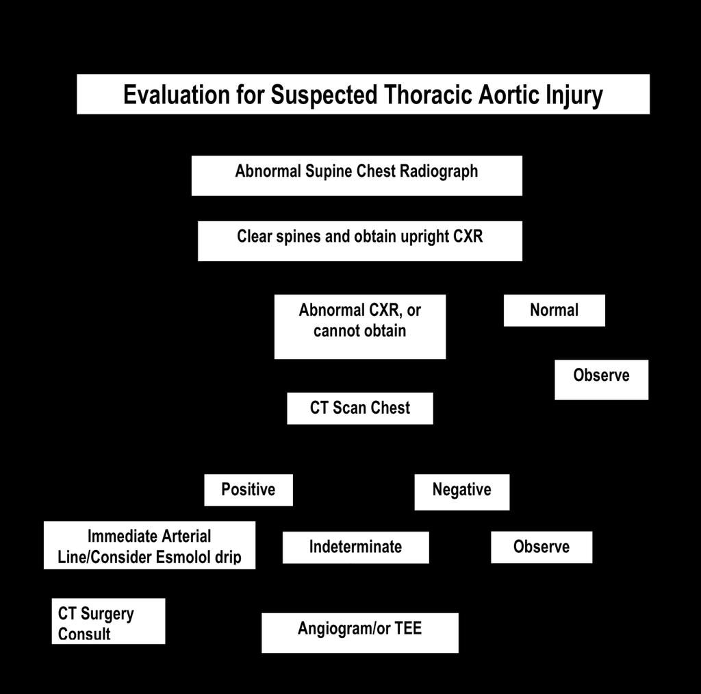
78 78
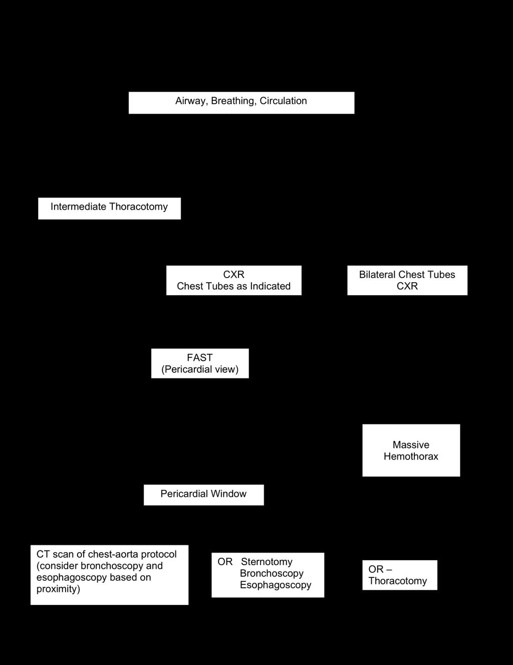
79 79
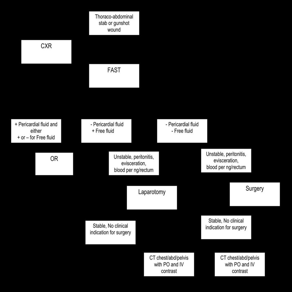
80 80
81 DIAGNOSTICS: ROLE OF DPL, CT, FAST (ultrasound) 05/08 The goal of the abdominal evaluation of multi-system trauma patients is the safe, accurate, and timely determination of the presence or absence of intra-abdominal injury, particularly those requiring surgical intervention. Physical examination can be unreliable in polytrauma patients, particularly when the abdominal examination or the level of consciousness has been altered by alcohol, drugs, central system trauma, or distracting pain. Diagnostic peritoneal lavage (DPL), CT scan, and abdominal sonography (FAST) are the most frequently employed diagnostic studies used for the abdominal examination of multi-system trauma patients. Each study has advantages and disadvantages. The goal of this session is to integrate these complementary studies into a rational diagnostic algorithm for the evaluation of the abdomen. Introduced by Root et al. in 1965, DPL (Diagnostic Peritoneal Lavage) was the time-honored standard for evaluation of the abdomen for many years. DPL is rapid (< 30 minutes) and safe (< 2% complication rate) with well-established standards for surgical intervention on the basis of cell counts or the aspiration of free blood after lavage. Overall sensitivity is 98%, with a specificity of 98% and a diagnostic accuracy approaching 100%. The procedure is inexpensive and can be performed at the bedside while other diagnostic and therapeutic interventions proceed. The procedure is invasive. Previous abdominal surgery makes the procedure more difficult and may enhance complications and diminish specificity. Pelvic fractures also diminish specificity. DPL cannot evaluate extraperitoneal structures (thorax, retroperitoneum, and pelvis) and must be supplemented with other diagnostic procedures. DPL is not organ specific. Therefore, a positive study may lead to a non-therapeutic laparotomy for a trivial injury. In recent years, DPL has been replaced by FAST in the unstable patient and has a limited role in the stable patient. Abdominopelvic CT scanning for blunt abdominal trauma was introduced by Federle et al. in the early 1980s and has gained wide popularity in the United States. CT scanning has a sensitivity of 96%, a specificity of 98%, and an accuracy of 97%. CT is organ specific, allowing the identification and grading of injured organs and the quantification of intraperitoneal fluid or blood. This allows for non-operative management of stable patients, thereby reducing the rate of nontherapeutic laparotomy. Extraperitoneal injuries (thorax, retroperitoneum, and pelvis) can be identified and graded. CT scanning is relatively expensive. Sensitivity and specificity depend on quality of the scan and skill of the interpreter. Contrast aspiration and allergy may occur. Bowel and pancreatic injuries may be missed. Even with the new, more rapid scanners, CT is time consuming, and transport to the scanner interrupts other diagnostic and therapeutic interventions. Consequently, CT is limited to hemodynamically stable patients. CT scan is the mainstay in the abdominal evaluation of the stable blunt trauma victim. CT now has a role to play in the evaluation of penetrating torso injury. The use of ultrasound or FAST (Focused Assessment with Sonography in Trauma) in abdominal trauma first evolved in Germany. FAST has been employed successfully at trauma centers in the United States for over a decade. US is noninvasive, rapid (2 to 4 minutes), and relatively inexpensive. The examination can be performed at the bedside, does not interfere with other diagnostic and therapeutic interventions, and can be repeated as needed. The primary goal of FAST is to detect free fluid in Morison s Pouch, the pelvis, peri-splenic region and the pericardium. The sensitivity of FAST ranges from 80% to 100%; the specificity, from 89% to 100%; and the accuracy, from 86% to 99%. Extraperitoneal structures can be imaged (thorax, pericardium, retroperitoneum). The technique can be easily learned and performed by the 81
82 treating surgeon. Sensitivity, specificity, and accuracy of US clearly improve with experience. Although extremely sensitive for peritoneal fluid, US is much less sensitive for specific organ injury (liver, spleen, etc.). As is true of CT, US can miss bowel injuries. However, repeat examinations mitigate the latter weakness, and organ-specific diagnostic sensitivity improves with the experience of the examiner. Sensitivity, specificity, and accuracy also depend on image quality. Obesity, bowel gas, and subcutaneous emphysema interfere with imaging. References 1. Root HD, Hauser CW, McKinley CR, et al. Diagnostic peritoneal lavage. Surgery 1965;57: Federle MP, Goldberg HI, Kaiser JA, et al. Evaluation of abdominal trauma by computed tomography. Radiology 1981;138: Kearney PA, Vahey T, Burney RE, et al. Computed tomography and diagnostic peritoneal lavage in blunt abdominal trauma. Their combined role. Arch Surg 1989;124: Hoffman R, Nerlich M, Muggia-Sullam M, et al. Blunt abdominal trauma in cases of multiple trauma evaluated by ultrasonography: a prospective analysis of 291 patients. J Trauma 1992;22: Goletti O, Ghiselli G, Lippolis PV, et al. The role of ultrasonography in blunt abdominal trauma: results in 250 consecutive cases. J Trauma 1994;36: Forster R, Pillasch J, Zielke A, et al. Ultrasonography in blunt abdominal trauma: influence of the investigator experience. J Trauma 1992;34: Liu M, Lee CH, P eng FK. Prospective comparison of diagnostic peritoneal lavage, computed tomographic scanning, and ultrasonography for the diagnosis of blunt abdominal trauma. J Trauma 1993;35: Rozycki GS, Ochsner MG, Jaffin JH, et al. Prospective evaluation of surgeons use of ultrasound in the evaluation of trauma patients. J Trauma 1993;34: McKenney M, Lentz K, Nunez D, et al. Can ultrasound replace diagnostic peritoneal lavage in the assessment of blunt trauma? J Trauma 1994;37:
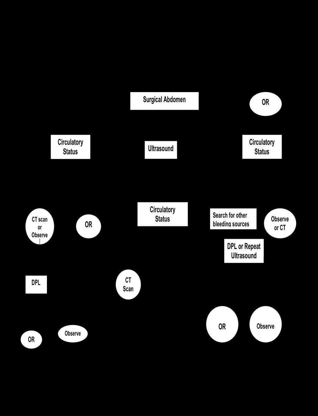
83 83

84 84
85 Genitourinary Trauma 5/08 Trauma to the GU tract is present in approximately 10% of all injuries. The following discussion pertains to the evaluation and management of the stable patient. Work-up of GU trauma begins with assessment of the urethra. Injuries of the female urethra are rare. Urethral injuries are usually due to blunt trauma associated with pelvic fracture or straddle-type injuries. The primary posterior site of urethral injury is the prostatomembranous junction. The primary anterior urethral injuries are most commonly caused by straddle-type injuries or perineal trauma. The location of injury is typically the bulbar urethra. Physical finding of blood at the meatus, perineal hematoma or extensive laceration, a high riding prostate, or a large hematoma found on rectal exam mandates a retrograde urethrogram prior to insertion of a Foley catheter. Management requires suprapubic urinary diversion or endoscopic assisted placement of Foley catheter. Once urethral injury is ruled out, a Foley catheter should be inserted. It is essential to document the color of the urine, as gross hematuria mandates further workup of the GU tract. Evidence suggests the microscopic hematuria is not diagnostic for GU trauma and as such a urinalysis should not be part of the workup. If there is gross hematuria, evaluation of the remainder of the GU tract kidneys, ureters (in penetrating trauma), and bladder needs to be performed. This can be accomplished by various imaging techniques. However, with the advancement of the CT technology, it has evolved to become the standard of care. Therefore, for gross hematuria, a CT scan of the abdomen and pelvis to evaluate the kidneys, as well as CT cystogram should be obtained. The kidneys are the most commonly injured organs in the GU tract. Management depends on hemodynamic stability and the grade of injury by CT scan. Most renal injuries do not require an operation in a stable patient. However, incidental intra-operative finding of perinephric hematoma should be explored if the mechanism is penetrating trauma. A blunt ureteral injury is a case report! Most ureteral injuries are penetrating. They require surgical intervention. Basic principles of ureteral reconstruction include debridement of devitalized tissue, followed by tension-free anastomosis with absorbable suture in a spatulated fashion over a double J stent. Bladder ruptures can result from penetrating or blunt trauma. Extraperitoneal bladder rupture is commonly associated with pelvic fracture; and intraperitoneal bladder rupture is a result of blunt lower abdominal force on a full bladder. Classic physical findings of bladder rupture include suprapubic pain, hematuria, and inability to void. A CT cystogram should be obtained. Extraperitoneal bladder rupture is generally managed with Foley catheter drainage, and intraperitoneal bladder rupture requires immediate surgical intervention. 85
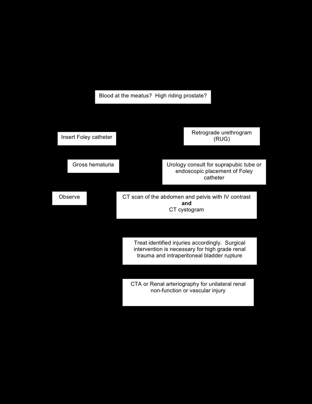
86 86
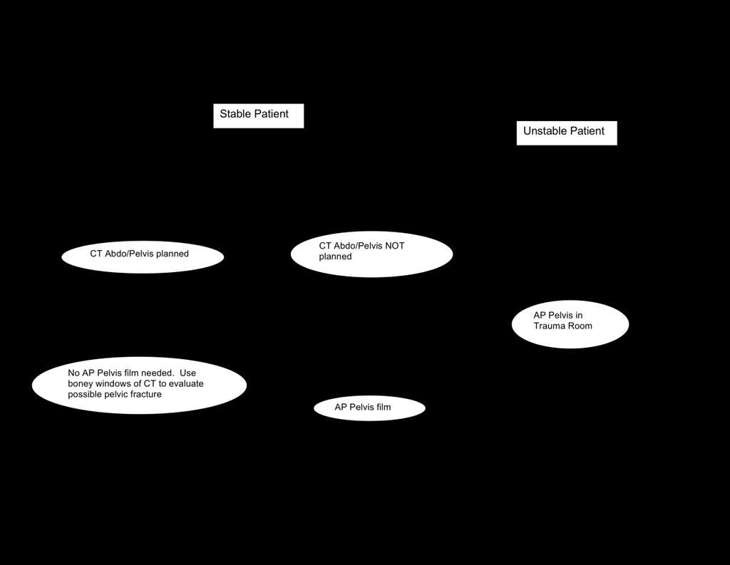
87 87
88 88
89 Traumatic Peripheral Vascular Injury Any injured extremity should be thoroughly evaluated for a possible vascular injury. The presence of obvious arterial injury from a blunt and/or penetrating mechanism rarely requires imaging and should not delay emergent operative exploration. The presence of hard signs strongly supports vascular injury and typically necessitates emergent repair. These hard signs are: 1. Bruit/Thrill 2. Active/Pulsatile hemorrhage 3. Pulsatile/Expanding hematoma 4. Signs of limb ischemia and or compartment syndrome including the 5 "P's" - pallor, paresthesias, pulse deficit, paralysis, and pain on passive extension of the compartment (pain on passive extension is the earliest and most sensitive physical finding) 5. Diminished or absent pulses with + Doppler signals (this is not a sensitive prognostic finding, as up to 30% of patients with major vascular injuries requiring repair have normal pulses or Doppler signals distal to the injury due to collateral flow) [1] The Arterial Perfusion Index, API, is a validated tool for screening for peripheral vascular injury[2]. This is performed by placing a blood pressure cuff above the ankle or on the bicep of the limb of concern. The systolic pressure is determined with a Doppler probe at the dorsalis pedis or brachial artery. Repeat this procedure on the ipsilateral uninjured limb. The API is calculated by dividing the systolic pressure in the injured limb by the systolic pressure in the uninjured limb. An API < 0.9 has a sensitivity of 95% and specificity of 97% for a major arterial extremity injury. In a study on blunt orthopedic extremity injuries the negative predictive value is 100% for an API > 0.9 to exclude an arterial injury.[3-5] The purpose of these algorithms is to diagnose the occult injury early before irreversible tissue ischemia is present. In patients where the hard signs are NOT present it is imperative to maintain a high suspicion of peripheral vascular injury in the injured extremity [2, 6, 7]. If hard signs are not present but peripheral vascular injury is suspected then expedient consultation with Vascular Surgery is indicated and the use of imaging, per Vascular Surgery, should be liberal to avoid missed injuries. References: 1. Drapanas, T., et al., Civilian vascular injuries: a critical appraisal of three decades of management. Ann Surg, (3): p Bravman, J.T., et al., Vascular injuries after minor blunt upper extremity trauma - pitfalls in the recognition and diagnosis of potential "near miss" injuries. Scand J Trauma Resusc Emerg Med, (1): p Mills, W.J., D.P. Barei, and P. McNair, The value of the ankle-brachial index for diagnosing arterial injury after knee dislocation: a prospective study. J Trauma, (6): p Lynch, K. and K. Johansen, Can Doppler pressure measurement replace "exclusion" arteriography in the diagnosis of occult extremity arterial trauma? Ann Surg, (6): p Johansen, K., et al., Non-invasive vascular tests reliably exclude occult arterial trauma in injured extremities. J Trauma, (4): p ; discussion Dennis, J.W., et al., Validation of nonoperative management of occult vascular injuries and accuracy of physical examination alone in penetrating extremity trauma: 5- to 10- year follow-up. J Trauma, (2): p ; discussion
90 7. Gelberman, R.H., J. Menon, and A. Fronek, The peripheral pulse following arterial injury. J Trauma, (11): p
91 91
92 Open Long Bone Extremity Fracture Protocol Open long bone extremity fractures are associated with significant trauma. The expedient management of these injuries ensures the best possible fracture treatment outcome. The determination of the grade of open fracture is the responsibility of the orthopedic trauma service. The extent of the type of open fractures often requires intra-operative evaluation. Antimicrobial Treatment for Open Fractures Depending on Type Open Fracture Type Type I Type II Type III Type A, B, & C Antibiotic Treatment Kefzol 1 gm IV q8h x 24h Kefzol 1 gm IV q8h x 24h + Gentamicin 4mg/kg/d x 48h Kefzol 1 gm IV q8h x 24h + Gentamicin 4mg/kg/d x 48h + PCN 4 million U q4h x 48h Antibiotic Treatment if PCN Allergic Clindamycin 600 mg IV q6 x 24h Clindamycin 600 mg IV q6 x 24h + Gentamicin 4mg/kg/d x 48h Clindamycin 600 mg IV q6 x 48 + Gentamicin 4mg/kg/d x 48h If patient exposed to barn or farm wound contamination then add high dose PCN x 24 hours (Type I and II) or 48 hours in Type III. If patient is has pen-allergy consider flagyl. Open fracture management and evaluation, including antibiotics should be initiated as soon as possible from the timing of wounding, not based on arrival to ER. If a patient gets an operation on this open fracture after completion of the above duration of antibiotics is given they should get only a perioperative dose. Wound care for open long bone extremity fracture requires coverage of the wound with a Hibiclens or Betadine soaked gauze. Mandatory documentation of neurovascular exam is required in all extremity injuries pre and post extremity fracture care. Frequent neurovascular examinations are required before and after fracture management to detect extremity compartment syndrome. The LAST clinical finding lost in developing compartment syndrome is the pulse. The body has evolved to perfuse cells until the very end so it makes sense that the pulse is the last clinical finding to be lost in developing compartment syndrome. * All open fractures must be evaluated by the Ortho Trauma Service for proper management (stabilization, wound care, further fracture grading, and definitive fracture management) 92
93 UNIVERSITY OF KENTUCKY HOSPITAL POLICY NUMBER: 08 CHANDLER MEDICAL CENTER FIRST ISSUED: 05/07 Emergency Dept. Policy CURRENT AS OF: 08/08 Subject: Management of Injury in Pregnancy Purpose: To establish guidelines for rapid assessment and treatment of critically injured pregnant patients. PROCEDURE: To effectively manage trauma patients that are pregnant, the Trauma Team must mobilize the resources essential to diagnosis and treat both mother and fetus. For purposes of this policy, a potentially viable fetus is one at 24 weeks gestation, although some exceptions may exist. I. Mechanisms of Injury Motor Vehicle Accident Assault Domestic Violence Other trauma, such as gunshot wound II. Initial Evaluation and Management 1. If 24 weeks gestation, sustaining a traumatic injury, activate Trauma Alert 2. Page OB chief Resident 3. Place roll under the torso to give a gentle right-side-up position (10-15 degrees) 4. Early intubation when indicated 5. Chest x-ray and FAST completed during the primary survey 6. Consider avoiding the plain pelvic radiograph if: a. Hemodynamically normal patient b. No gross instability or tenderness on physical exam c. Planned CT scanning would cover imaging of the pelvis 7. Simultaneous fetal assessment should be completed by the OB team 8. In the case of maternal shock with a positive FAST, where emergent laparotomy is indicated, the fetal assessment can be completed in the OR 9. If distress is identified in a potentially viable fetus, every effort should be made to expedite transfer to the OR for emergent cesarean section via a midline incision 10. When significant mechanism or concern for associated intraabdominal injury are present, exploratory celiotomy at the time of C-section is indicated III. Diagnostic Studies 1. Lab studies: a. Urine pregnancy or serum beta-hcg, especially with a questionable history b. Clotting factors and plasma fibrinogen c. Kleihauer-Betke test, especially when blunt uterine trauma is suspected 93
94 2. Imaging a. Avoid duplicating films b. Shield the fetus whenever possible c. See Appendix 1 for estimated fetal exposure Appendix 1: Estimated Fetal Exposure for Various Radiographic Studies Plain Films: dose per exam (rad) Estimated Fetal Lateral Cervical Spine Chest Pelvis CT Scans Head <0.050 Chest <0.100 Abdomen/Pelvis 2.6 Summary Focus all initial evaluation and resuscitation efforts on the mother. 1. Complete a primary survey before moving the mother from the trauma bay for any reason. 2. Secondary survey should include simultaneous fetal evaluation. 3. Plan radiographic studies and avoid duplication. Shield the fetus whenever possible. 4. Emergent cesarean sections should be performed through a midline incision. Medical Director, Emergency Medicine Trauma Program Director 94
95 UNIVERERSITY OF KENTUCKY HOSPITAL POLICY NUMBER: 08-55a CHANDLER MEDICAL CENTER FIRST ISSUED: 05/07 Emergency Dept. Policy CURRENT AS OF: 08/08 Initial E&M of the Pregnant Trauma Patient Traumatic injury in female with reported pregnancy at or beyond 24 weeks: Primary Survey Roll backboard R-side up degrees PCXR FAST Stable Complete Secondary Survey Fetal evaluation in ED Unstable- No clear source Complete Secondary Survey Fetal evaluation in ED Plan additional imaging, lab studies Unstable w/ Positive FAST Proceed emergently to MAIN OR Fetal evaluation in OR *FETUS IN DISTRESS* Emergent C-section via midline incision (MAIN OR) Trauma present for laparotomy after delivery *NORMAL FETUS* Additional work-up as indicated Fetal monitoring for at least 6 hours (or as directed by OB) Maternal death/traumatic arrest Initiate perimortem C-section ideally within 4 minutes of maternal arrest (If gestational age likely >24 weeks) 95

96 EMERGENCY DEPARTMENT EVALUATION OF BURN PATIENTS Stabilization A. Maintain airway - the supraglottic airway is extremely susceptible to obstruction from edema as a result of exposure to superheated air. Assess for clinical signs of inhalation injury: - Facial burns/singeing of the eyebrows and nasal hairs - Carbonaceous sputum and acute inflammatory changes in the oropharynx, raspy voice - History of impaired mentation and/or confinement in a burning environment - Assess for toxic inhalation, or carbon monoxide poisoning B. Circulation 1. Replace volume based on Rule of Nines calculation. Follow the Parkland Formula (2-4cc x % TBSA burned x weight/kg = total to be administered in first 24 hours. Half should be given in first 8 hours from time of burn and second half over next 16 hours) for administration of fluid. The amount of fluid given should be adjusted according to individual patient's response: a. Adult UO: ml/kg/hour b. Child <30 KG: UO 1.0 ml of urine/kg/hour. It is necessary to administer Maintenance IV fluid containing glucose in addition to burn formula 3. Initiate two large bore IVs; overlying burns shouldn't prevent IV placement (upper extremities preferred) Assessment 96
97 A. Assess for associated injuries B. History - time and mechanism of the injury; enclosed fire, or if toxic chemicals involved C. Assess burn 1. First degree: (Sunburns): characterized by erythema, pain, and the absence of blisters NOT COUNTED IN TBSA 2. Second degree: (Superficial Partial Thickness or Deep partial thickness): characterized by a red or mottled appearance with swelling and blister formation. The surface may have a weeping, wet appearance and is painfully hypersensitive 3. Third degree: (full thickness): skin appears dark and leathery; may also appear translucent, mottled, or waxy white; the surface is painless and generally dry, but may also be moist D. Circumferential extremity burns: 1. Remove rings and bracelets and Assess distal circulation 2. Check pulses with a Doppler (absent pulse may indicate inadequate fluid resuscitation) d. Observe for cyanosis, impaired capillary refill, or progressive neurological signs (i.e., paresthesia and deep tissue pain) E. Limb Escharotomy Relieve compromised distal circulation in a circumferentially burned limb by escharotomy, which can be done without anesthesia, due to the insensitive full - thickness burn 1. The incision must extend across the entire length of the eschar in the lateral and/or medial line of the limb including the fingers and joints 2. The incision should be deep enough to allow the cut edges of the eschar to separate F. Thoracic Escharotomy: Circumferential burns of the thorax occasionally impair respiratory excursion. Bilateral, mid-axillary escharotomy incisions should be considered Special Burn Requirements A. Chemical burns 1. Flush burns for at least 20 to 30 minutes; alkali burns require longer irrigation 2. Brush dry powder off before irrigation 3. Alkali burns to the eye require continuous irrigation during the first eight hours B. Electrical burns - frequently more serious than they appear on the surface 1. Initial care as above 2. Full spinal immobilization 3. EKG monitoring 4. Urinary catheter a. Observe for myoglobinurea (due to rhabdomyolysis) b. Increase IV rate fluid to ensure UO of at least 100 ml/hour c. Consult Medical Control for Mannitol 25 GM IVP and IV infusion with 12.5 GMs of mannitol/1000 cc NS to maintain the diuresis C. Explosive Injuries 97
98 Any patient involved in an explosion should be considered as having a mechanism for traumatic injuries. Even if the patient states they were NOT thrown a distance. The force of a flash flame explosion is enough energy to cause concussive type of injuries. 98
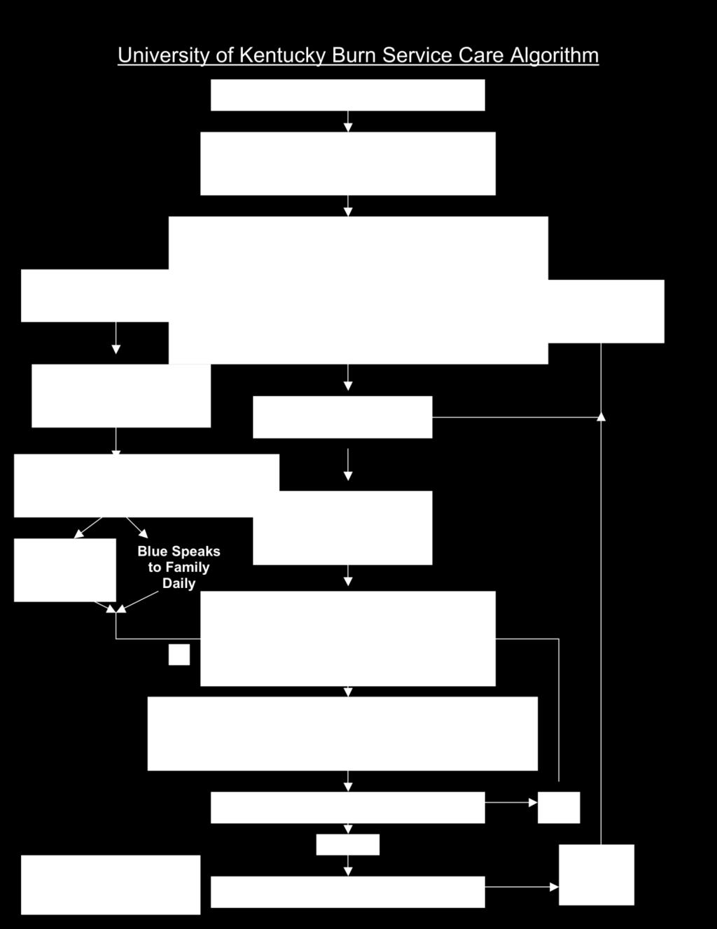
99 99
100 100
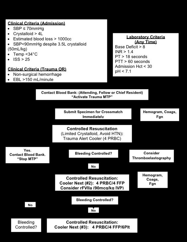
101 101
102 102
103 The Anticoagulated Trauma Patient Warfarin use has been shown to increase the severity of head injury and a higher mortality rate. Mortality of trauma patients with head injury while on warfarin ranges from 33% to 50%. i Furthermore, it has been reported that the head injured patients on warfarin have an increased risk of mortality from 2-fold to 4-fold, when compared with non-anti-coagulated patients with similar degrees of head injury. ii As part of the trauma workup, one should always obtain an adequate history, which includes a list of current home medication. Early identification of warfarin use has been shown to reduce mortality on patients with intracranial hemorrhage from 48% to 9%. iii In the same study, mean time to warfarin reversal (normal coagulation profile) was 1.7 hours in the early identification group compared to 4.3 hours. Another important aspect of the anti-coagulated patient is the decreased reliability of their neurological exam. It has been shown that GCS of 15 and no loss of consciousness does not reliably rule out intracranial pathology after trauma. Indeed, one study reported two anticoagulated patients with no loss of consciousness that eventually died from consequence of intracranial hemorrhage. 2 Therefore, all patients with known warfarin use should have a CT scan of the head as part of their trauma workup regardless of their mental status. 1 Lavoie A, Ratte S, Clas D, Demers J, Moore L, Martin M, Bergeron E. Preinjury warfarin use among elderly patients with closed head injuries in a trauma center. J Trauma Apr;56(4): Mina AA, Bair HA, Howells GA, Bendick PJ. Complications of preinjury warfarin use in the trauma patient. Trauma May;54(5): Janczyk et al. Rapid warfarin reversal in anticoagulated trauma patients with intracranial hemorrhage reduces hemorrhage progression and mortality. Abstract presented at AAST annual meeting, September 30, Phillip Chang, M.D. Revised: 08/08 CT of head neg Check PT/PTT pos yes Known Warfarin use Transfuse two units of FFP & give 5mg of Vitamin K IV before results are available Transfuse as needed no Check PT/PTT Warfarin reversal based on lab values. If there is evidence of warfain use, Ct of head should be obtained Close observation Neurosurgery consult & check fibrinogen level 103
104 SECTION 4: ICU SPECIFIC PROTOCOLS Revised: 08/08 University of Kentucky Medical Center Acute Care Surgery (Blue) Service Intensive Care Unit Mechanical Weaning/Extubation Guidelines Critically ill trauma/surgical patients frequently require mechanical ventilation to maintain adequate tissue oxygenation until they are able to take on the work of breathing without assistance. Discontinuing ventilator support should be done at the earliest possible time after the disease process has been adequately reversed. In the vast majority of cases, prolonged or gradual weaning is unnecessary. Rather, patients should be assessed for eligibility for separation from the ventilator and if appropriate, separated. The UK Ventilator Separation Protocol is included in this manual for reference and should be ordered for all mechanically ventilated patients. Weaning is used to describe gradual removal of ventilator support and is reserved for patient who cannot be discontinued from ventilator support without gradual maneuvers. The weaning process must be tailored to meet each patients needs. There are no dogmatic rules or hard and fast guidelines regarding the timing or duration of the process. These are basic guidelines that should be followed. *Nursing Guidelines: Pre Extubation: Extubation: 1. Adequate pain control [remember to decrease and/or withdrawal pain/sedation medications to level of adequate pain control without over sedation]. 2. Ensure complete reversal of NMBA if applicable [4/4 in TOF] 3. Assess for hemodynamic stability 4. Patient should be awake, alert and ideally follows commands 5. Correct acid/base and electrolyte abnormalities 6. Assess nutritional status [Is patient s caloric need being met with dietary regimen] 7. Assess characteristics of secretions 8. Breathing through an endotracheal tube requires more respiratory muscle work than atmospheric breathing following extubation, therefore limit CPAP trial to 30 minutes. 1. Separation criteria met prior to extubation 2. Cool humidified oxygen set up at the bedside 3. Suction out endotracheal tube and mouth prior to extubation 4. Deflate endotracheal tube cuff just prior to extubation 5. Racemic epinephrine available Post Extubation: 1. Assess breath sounds q 4 hours and PRN 2. Assess for any signs of respiratory distress 3. Monitor vital signs and SaO2 per unit guidelines 4. Elevate head of bed or reverse Trendelenburg 30 degrees 5. Encourage incentive spirometry, deep breathing and coughing per unit guidelines 104
105 Post Extubation Therapy: Breathing Exercises: The most effective method of preventing post-extubation pulmonary complications is to encourage maximal voluntary deep breathing and coughing. Incentive spirometry is perused to augment voluntary efforts. Aerosol Therapy: epinephrine Stridor - Upper airway stridor is best treated with nebulized racemic Bronchoconstriction - Bronchodilators delivered via nebulized aerosol effectively reverse airway constriction. Specific drugs have varying B1 and B2 effects that will influence the choice of a particular medication. Patients that do not exhibit clinical evidence of bronchoconstriction will not benefit from this therapy. Secretions: Thick, tenacious, inspissated secretions can be effectively treated with heated aerosol. Some patients may require the addition of a mucolytic agent [Mucomyst] delivered per hand held nebulizer. A mucolytic agent is an irritant that can cause bronchospasm and should be discontinued after 48 hours due to bronchorrhea. Percussion and Postural Drainage: The indication for therapy is very narrow and specific. A. Increased secretions or infiltrate on Chest radiograph B. Atelectasis not responding to breathing exercises C. Collapse of lung, lobe or segment. In order to achieve maximum benefit, the patient must be able to cooperate with the respiratory therapist. A large number of contraindications to this form of therapy, some of which are listed, limit its use in the critically ill patient. Contraindications: A. Unstable cardiac rate or rhythm B. Congestive heart failure C. Unstable neurologic status (aneurysm, increased ICP) D. Abdominal distention and or obesity E. Fractured ribs or flail chest F. Bleeding diathesis *Refer to Mechanical Ventilation by B. Boulanger, M.D., P. Kearney, M.D., G. Hale, R.R.T., J. Coughenour, M.D., for more detailed information on mechanical ventilation. References Boulanger BR, Kearney PA, Hale G, Coughenour JC. Mechanical Ventilation: Division of Surgery, Division of Trauma/Critical Care and the Department of Respiratory Therapy, 4 th Ed,
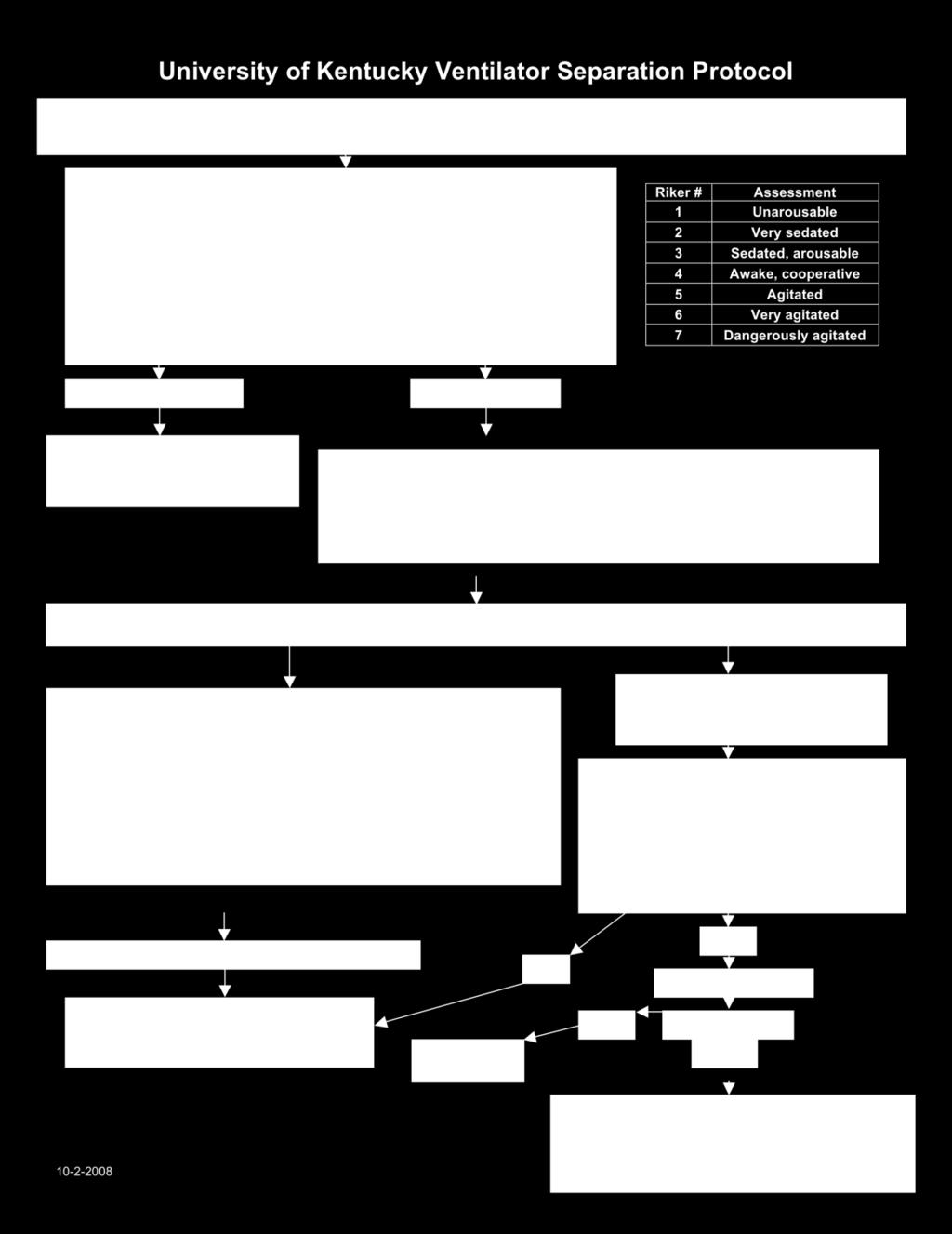
106 106
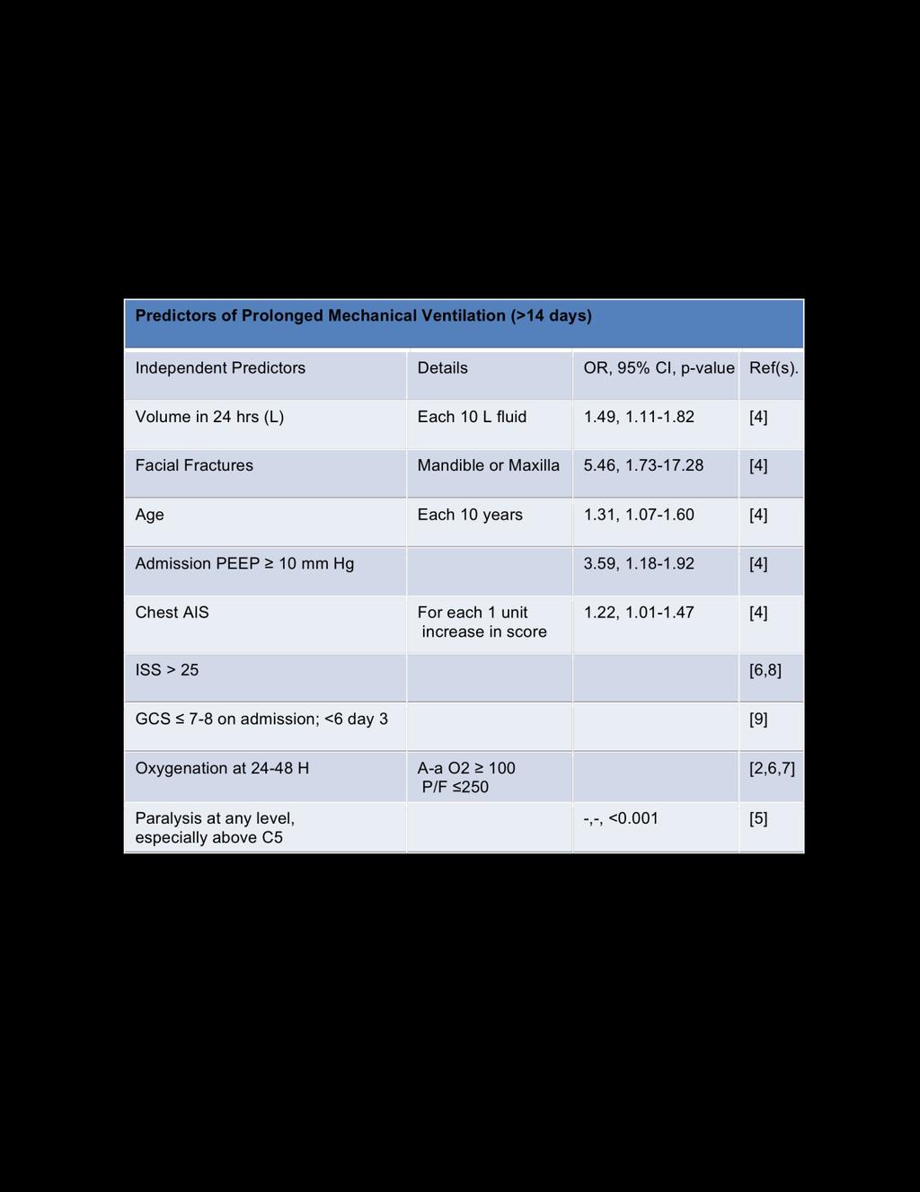
107 107
108 Nosocomial Pneumonia The incidence of nosocomial pneumonia is high in mechanically ventilated patients. National data indicates that critically ill patient intubated for greater than 24 hours are at 6 to 21 times the risk of developing pneumonia (VAP). This risk increases by 1% every ventilator day. (Tablan et al, 1994) Morbidity and mortality associated with the development of VAP is high, adding 5-16 hospital days and increased health care cost. (Kolef, 1993, Rello et al, 2002) The Ventilator Bundle was developed to prevent adverse events that prolong the amount of time a patient requires mechanical ventilation. The Ventilator Bundle consists of stress ulcer prophylaxis, DVT prophylaxis, Elevation of the head of the bed (>30 degrees), antiseptic oral care, as well as Sedation Weaning. This has been incorporated into the Trauma ICU Admission Order Set. VAP is suspected by: 1. Fever 2. Leukocytosis 3. Change in character and volume of sputum 4. New infiltrate on CXR 5. Increasing ventilator and/or oxygen requirements. The diagnosis is confirmed by Protected Alveolar Lavage (PAL). Protected Alveolar Lavage (PAL) Guidelines The procedure is performed by trained respiratory technician, nurse and/or physician. Technique: 1. Confirm physician order. 2. Gather necessary items/equipment. 3. Prepare the patient for the procedure by: a. Adequate pre-oxygenation with 100 % FiO2. b. Ensure patient is adequately sedated. 4. Using aseptic technique (mask, gloves, cap), advance the catheter out of the protective sheath at the opened end and gently insert protected catheter through the endotracheal tube or tracheostomy cannula into the pulmonary tract until resistance is felt. 5. Pull the set back 3-4 cm, remove the plastic spacer, and advance inner catheter to fully expel the distal polyethylene glycol plug. 6. Instill 20 cc of non-bacteriostatic saline. 7. Aspirate lavage sample from the airway while maintaining catheter position 8. Remove catheter from the endotracheal tube with the syringe still attached. 9. Place sample in to specimen container without contamination. 10. Send specimen to lab without delay. 11. Return to previous ventilator settings. 108
109 References Tablan, O.C., Anderson, L. J., Arden N.H., et al. (1994). Guidelines for prevention of nosocomial pneumonia. Hospital Infection Control Practices Advisory Committee. American Journal of Infection Control, 22, Kollef, M., (1993). Ventilator-associated pneumonia: a multivariate analysis. JAMA, 270, Rello, J., Ollendorf, D.A., Oster, G., et al. (2002). Epidemiology and outcomes of ventilatorassociated pneumonia in a large US database. Chest, 122 (6), Attia, J., Roy, J.G., Cook, D.J., et al. (2001). Deep vein thrombosis and its prevention in critically ill adults. Archives of Internal Medicine, 161, Mahul P., Auboyer, C., Jospe, R., et al. (1992). Prevention of nosocomial pneumonia in intubated patients in respective role of mechanical subglottic secretion drainage and stress ulcer prophylaxis. Internal Care Medience, 18, Drakulovic, M., Torres, A., Bauer, T.T., et al. (1999). Supine body position as a risk factor for nosocomial pneumonia in mechanically ventilated patients: a randomized trial. Lancet, 354, Kress, J., Pohlman, A.S., O Connor, M. F., et al. (2000). Daily interruption of sedative infusions in critically ill patients undergoing mechanical ventilation. New England Journal of Medicine, 342, Schleder, B., et al. (2002). The effect of a comprehensive oral care protocol on patients at risk for ventilator-associated pneumonia. Journal of Advocate Health Care, 4(1), Scannapieco, F.A. (1999). Role of oral bacteria in respiratory infection. Journal of Periodontology, 70(7), Reviewed: 03/05 109
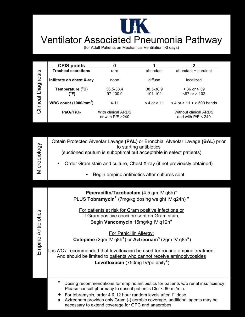
110 110
111 111
112 Reviewed 10/08 University of Kentucky Medical Center Trauma Intensive Care Unit Analgesic and Sedation Protocol Introduction: Injuries, surgical wounds, and the attendant critical illness are associated with moderate to severe pain, as well as anxiety and agitation. Intense sympathetic and parasympathetic responses can and do occur with pain and anxiety. When pain and anxiety are not promptly and adequately relieved, excessive adrenergic activity and hormonal changes indicative of stress occur. These physiologic stress reactions produce a marked increase in metabolism, cardiac workload, and oxygen consumption that may compromise tissue oxygenation in the critically ill patient. The goal is to provide effective, sustained pain relief and sedation with minimal side effects. Analgesia: Morphine is the analgesic drug of choice and may be used alone or in conjunction with a sedative. Morphine may be given intermittently or as a continuous infusion. Continuous intravenous infusion of morphine is preferable because it avoids peaks and troughs in blood levels leading to over and under sedation. When pain control cannot be achieved with morphine, fentanyl is an effective, inexpensive alternative. Agitation: Haloperidol is preferred for agitation. Frequently, escalating doses are required to reach therapeutic levels and clinical effect. Agitation can occur from inadequate analgesia. Therefore, make sure analgesia is adequate before treating agitation. Although rare, haloperidol can produce extrapyramidal symptoms, neuroleptic malignant syndrome, and cardiac events, such as prolongation of QT interval and torsade de pointes. Anxiety: Benzodiazepines are the principle class of drugs for treatment of anxiety. Short acting agents may be used for patients undergoing ICU procedures (central venous access, intubation, bronchoscopy, chest tube placement, tracheotomy, etc.). Benzodiazepines should also be used to insure deep sedation for patients requiring neuromuscular blocking agents. Benzodiazepines should not be used as first line drugs for treating agitation. However, agitation can result from inadequate analgesia or severe anxiety. Benzodiazepines may be used for patients who do not respond to haloperidol or for those patients in whom haloperidol is contraindicated. This protocol is designed to provide effective and consistent pain management as well as effective treatment for agitation and sedation. 112
113 113
114 Nursing Care: 1. Assess patients for subjective and objective signs of pain every 4 hours and PRN. 2. Utilize pain, sedation and anxiety scale as shown above. a. Decrease pain/sedation medication rate and/or frequency as patient requirements decrease. b. AVOID OVER SEDATION 3. Document patient response to medication. 4. Use comfort measure in addition to medication regimen. 5. Always assess vital signs prior to medication administration. a. Use extreme caution with borderline MAP and respiratory rate. 6. It is essential to taper high dose analgesic and sedation medications when a. weaning patient for extubation. 7. Obtain new pain/ sedation orders after patient is weaned, extubated and no longer a. receiving mechanical ventilation. 8. Monitor for extrapyramidal symptoms if patient is receiving haloperidol 9. Administer intermittent haloperidol slowly over 5 minutes if SBP is < 100. Weaning guidelines for continuous infusion narcotic analgesics: 1. Weaning guidelines for Fentanyl: > 2 weeks duration: 100 mcg/hr decreased twice daily > 3 weeks duration: 50 mcg/hr decreased twice daily 2. Weaning guidelines for Morphine: > 2 weeks duration: 2 mg/hr decreased every 6 hours > 3 weeks duration: 2 mg/hr decreased every 12 hours The weaning guidelines are to serve as parameters for weaning narcotics when patients have been receiving narcotics for more than 2 weeks. These are not iron clad rules. Weaning from narcotic analgesics may be individualized for the patient. Listed below are signs and symptoms of withdrawal from narcotics that the critical care clinician should monitor for when weaning the patient from narcotic analgesics. Signs and symptoms of withdrawal: agitation tachycardia tachypnea tremors 114
115 fever increased lacrimation diarrhea nausea and vomiting Daily Awakening Procedure: For all patients meeting inclusion criteria, continuous infusion sedation medications will be interrupted every day at Infusions are restarted at half the previous rates. Infusions will be held until: o Patient is awake and can follow commands (SAS = 4) with medical indication to resume infusion o Patient becomes agitated (SAS = 5-7) and requires resumption of sedation. Inclusion Criteria for Daily Awakening: o Patients initiated on continuous sedation > 48 hours o Patients not concurrently receiving neuromuscular blocking agents o Patients with stable hemodynamic/ventilatory status including: No evidence of ARDS (i.e. P/F ratio > 250) PEEP < 10 mmhg and pco 2 < 50 mm Hg MAP > 60 mmhg without use of vasopressor agents Patient not requiring use of rotobed Exclusion Criteria for Daily Awakening: o Patients initiated on continuous sedation < 48 hours o Patients concurrently receiving neuromuscular blocking agents o Patients with unstable hemodynamic/ventilatory status including: No evidence of ARDS (i.e. P/F ratio <250) PEEP > 10 mmhg and pco 2 < 50 mm Hg MAP < 60 mmhg without use of vasopressor agents Patient requiring use of rotobed 115
116 References 7 Dangerous Agitation Riker Sedation-Agitation Scale SAS Pulling at ETT, trying to remove catheters, climbing over bedrail, striking staff, thrashing side to side 6 Very Agitated Does not calm despite frequent verbal reminding of limits, requires physical restraints, biting ETT 5 Agitated Anxious or mildly agitated, attempting to sit up, calms down to verbal instructions 4 Calm and Calm, awakens easily, follows commands Cooperative 3 Sedated Difficult to arouse, awakens to verbal stimuli or gentle shaking but drifts off again, follows simple commands 2 Very Sedated Arouses to physical stimuli but does not communicate or follow commands, may move occasionally 1 Unarousable Minimal or no response to noxious stimuli, does not communicate or follow commands Jacobi, Judith, et al. Clinical practice guidelines for the sustained use of sedatives and analgesics in the critically ill adult. Critical Care Medicine. 2002;30(1): 119 Riker, RR, et al. Prospective evaluation of the sedation-agitation scale for adult critically ill patients. Critical Care Medicine Jul;27(7): Shapiro BA, Warren J, Egol AB, et al. Practice parameters for intravenous analgesia and sedation for adult patients in the intensive care unit: An executive summary. Critical Care Medicine. 1995; 23: Fox CA, Mansour A, Watson SJ. The effects of haloperidol on Dopamine Receptor Gene Expression. Experimental Neurology. 1994; 130: Riker RR, Fraser GL, Cox PM. Continuous infusion of haloperidol controls agitation in critically ill patients. Critical Care Medicine. 1994; 22: Guinard JP, Carpenter RL, Chassot PG. Epidural and intravenous fentanyl produce equivalent effects during major surgery. Anesthesiology. 1995; 82: George KA, Wright PM, Chisakuta AM. Thoracic epidural analgesia compared with patient controlled intravenous morphine after upper abdominal surgery. Acta Anesthesiologica Scandinavia. 1994; 38:
117 Ready LB, Oden R, Chadwick HS, et al. Development of an anesthesiology based acute postoperative pain management service. Anesthesiology. 1988; 68: Uzbay T, Akarsu ES, Kayaalp, SO. Effects of bromocriptine and haloperidol on ethanol withdrawal syndrome in rats. Pharmacology Biochemistry and Behavior. 1994; 49(4): Sun K, Quinn T, Weissman C. Patterns of sedation and analgesia in the postoperative ICU patient. Chest. 1992; 101: Malacrida R, Fritz ME, Suter PM, et al. Pharmacokinetics of midazolam administered by continuous intravenous infusion to intensive care patients. Critical Care Medicine. 1992; 20(8): Singer M, Noonan KR. Continuous intravenous infusion of fentanyl: Case reports of use in patients with advanced cancer and intractable pain. Journal of Pain and Symptom Management. 1993; 8(4):
118 Revised: 11/08 University of Kentucky Hospital Trauma Intensive Care Unit Neuromuscular Blockade Protocol The use of neuromuscular blocking agents in the treatment of critically ill patients has increased steadily over the past two decades. Increased use is largely due to advancements in medical technology and a better understanding of the treatment of critical illness. Up to 8% of intensive care patients who require mechanical ventilation may require neuromuscular blocking agents. The decision to paralyze a patient should not be taken lightly. The benefits, however, outweigh risks when consideration is given to the efficacy of short-term use of NMBA s. Neuromuscular blocking agents are indicated for the treatment of patients with severe acute respiratory failure (ARDS), marginal tissue oxygenation ( VO 2 or DO 2 ), and severe agitation which compromises patient safety. To assess the depth of neuromuscular blockade, a peripheral nerve stimulator using Train-of- Four stimulation will be utilized. Routine twitch monitoring is essential to ensure a consistent and safe level of blockade while preventing prolonged neuromuscular blockade. Clinical indicators should be utilized to assess effectiveness of blockade (e.g. PIP < 40, improved DO2, ICP<15). Titrate to lowest dose of NMBA to achieve defined parameters. Since pain and anxiety cannot be assessed easily, patients must receive scheduled sedation and analgesia. The neuromuscular blockade will be discontinued every day to determine if the patient is receiving adequate analgesic and sedation, and to evaluate if continued paralysis is needed. The following protocol was developed to assist the physicians and nurses in critical care to achieve optimal management of patients requiring NMBA s. 118
119 NON DEPOLARAZING NEUROMUSCULAR BLOCKING AGENTS Intubated/Ventilator patients Only I. Primary indications for the use of continuous infusion non-depolarizing neuromuscular blocking agents (NMBA). A. To enhance oxygenation during intermittent positive pressure ventilation. Useful in patients with poor lung compliance (i.e., ARDS) or those who compromise ventilation by using forceful expiratory musculature. B. Hypermetabolic patients with marginal oxygen delivery and increased oxygen consumption. High fever (>40C), shivering and extreme agitation. C. Where control of continuous skeletal muscle fasciculation is unresponsive to conventional therapy (i.e., tetanus). D. Intracranial hypertension unresponsive to conventional therapy or when conventional therapy is contraindicated. II. Precautions and contraindications for the use of continuous infusion of NMBA: A. Caution when used in patients with neuromuscular diseases (i.e., myasthenia gravis, Eaton-Lambert syndrome), severe acidosis and hypothermia. B. Caution when used concurrently with aminoglycosides, tetracyclines, clindamycin, metronidazole, cyclosporin, vancomycin, steroids, calcium channel blocking agents, magnesium salts, procainamide and quinidine due to their potentiating effects. [See Section VIII] C. Caution when used in patients with renal, hepatic or pulmonary impairment, and in geriatric or debilitated patients. D. Avoid use in patients who have either experienced or have a family history of malignant hyperthermia. III. Vecuronium should be used as the drug of choice if neuromuscular blockade is indicated. Cisatracurium is available as alternative agent, but must be ordered by an attending physician because of status on formulary. Administer agents by controlled infusion. A. Vecuronium: mix 50 mg in 50 ml NS Concentration: 1 mg/ml Load: mg/kg [use dry weight] Infusion: mg/kg/hr to maintain one detectable twitch 119
120 by train of four monitor [1/4] Start 0.04 mg/kg/hr B. Cisatracurium: Mix 200 mg in 250 D5W or NS Concentration: 800 mcg/ml Load: 0.2 mg/kg [use dry weight] Infusion: 1-10 mcg/kg/min to maintain one detectable twitch by train of four monitor [1/4] Start 1 mcg/kg/min [please note that infusion is in mcg/kg/min.] IV. Nondepolarizing neuromuscular blockade can be reversed if clinically required if spontaneous reversal of paralytic agent does not occur after agent is discontinued. A. Neostigmine mg plus glycopyrrolate 0.5 mg B. Edrophonium mg plus atropine 0.5 mg If above regimens fail to reverse blockade, the following considerations must be addressed minutes is required to antagonize block. 2. Blockade may be too intense to be antagonized. 3. Respiratory acidosis prevents antagonism. 4. Possible drug interactions. 5. Organ system failure may decrease excretion of NMBA V. Sedatives and or analgesics must be used concurrently with NMBA s. Refer to Pain/Sedation Protocol VI. Adverse effects associated with NMBA. A. Reactions associated with histamine release: 1. Bronchospasm 2. Flushing 3. Erythema 4. Hypotension 5. Tachycardia 6. Pruritis 7. Urticaria 8. Wheal formation B. Cardiovascular effects including changes in: 1. Heart rate 2. Cardiac output 3. Cardiac filling pressure 120
121 4. Mean systolic blood pressure 5. Mean arterial pressure 6. Systemic vascular resistance C. Malignant hyperthermia VII. Physiologic conditions that affect the amount of NMBA needed. Requires Less NMBA Acidosis Hypothermia Hypocalcemia Hypokalemia Neuromuscular Disease Requires More NMBA Alkalosis Hyperthermia Hypercalcemia Hyperkalemia Burns VIII. Pharmacological agents that affect the amount of NMBA needed. Requires Less NMBA [potentate blockade] Aminoglycosides Beta-blocking agents Clindamycin Midazolam Polymyxin Procainamide Quinidine Tetracyclines Vancomycin Requires More NMBA [antagonizes blockade] Azathioprine Carbamazepine Methylxanthines Phenytoin IX. The efficacy of NMBA s for altering oxygen consumption can be measured by monitoring the following parameters: 1. Cardiac output/index 2. Venous oxygen saturation 3. Arterial oxygen saturation 4. Oxygen consumption X. Depth of neuromuscular blockade should be monitored with a peripheral nerve stimulator using the Train of Four (TOF) mode. An appropriate blockade is defined 1-2 twitches in the TOF. The following guidelines should be followed when initiating and titrating for appropriate blockade: 1. Establish baseline by determining the lowest current that evokes a 4/4 response. Monitor TOF at 10 MA above baseline. 121
122 2. Ulnar nerve is preferred site for assessing TOF. Facial nerve and Superficial Peroneal nerve are alternative sites if both upper extremities are not accessible. 3. If no response is elicited follow the trouble shooting algorithm. If medication is excessive, the infusion should be held until one twitch is detected. Restart the drip at the lowest dose per kg and titrate to 1/4 or 2/4 twitches. 4. Initially a TOF should be determined every hour until three consecutive TOF elicit the same response. A TOF can be determined every 4 hours thereafter. If the patient is demonstrating renal or hepatic insufficiency, the TOF should be utilized every 2 hours Hz should not be used unless attempting to determine level of excessive neuromuscular blockade or sustained tetanus. 6. Refer to the Trouble Shooting Algorithm with 0/4 twitches. 7. Brow diaphoresis is indicative of inadequate blockade. Nursing Care: 1. Lacrilube and artificial tears q 2 hours and PRN 2. Ensure complete eyelid closure to prevent scleral abrasions (use paper tape to tape eyelid closed). 3. Oral Care q 2 hours 4. PT Consult for splints per physician s orders 5. Consult PT when NMBA discontinued 6. Monitor skin integrity q 4 hours 7. Consider Kinetic Therapy 8. Deep vein thrombosis prophylaxis 9. Reposition q 2 hours 10. Passive range of motion q 4 hours 11. Change electrode placement q day and PRN to avoid skin irritation 12. Place new battery in TOF monitor prior to use. 13. Please evaluate the patient for adequate pain and sedation before increasing the NMBA. The goal of therapy is to provide optimum ventilation and if this can be achieved by increasing the amount of analgesia and/or sedative this should be your first priority. Refer to Trouble Shooting Algorithm 122
123 Non Depolarizing Neuromuscular Blocking Agents Intubated/Ventilator Patients Only Severe acute respiratory failure, marginal tissue oxygenation, Hyperthermia/shivering, severe agitation, and increased ICP Appropriate and Adequate Pain/Sedation (Imperative that pain/sedation be addressed 1 st ) Patient Responds Increase in Sa0 2 and/or Sv0 2, Decreased ICP YES NO Maintain Adequate Pain and Sedation Initiate Neuromuscular Blockade Obtain Baseline TOF Patients w/o organ Renal Failure Hepatic Failure dysfunction Vecuronium Vecuronium Cisatracurium Titrate to 1/4 or 2/4 twitches [75% to 90% blockade] (If the patient shows any S & S of pain/agitation (dilated pupils, etc), consider increasing pain/sedation medication before increasing or restarting the NMBA. Assessment for pain/sedation cannot be over emphasized and should be ongoing) Administer Benzodiazepines per protocol Administer Narcotic Analgesic per protocol Stop NMBA q 0800 to assess adequacy of sedation/analgesic determine if continued paralysis is needed. and to Restart drip at previous rate if patient exhibits signs and symptoms of inadequate oxygenation. Notify H.O. if the patient is severely compromised & requires a bolus. 123
124 124
125 Trouble Shooting Guidelines for Neuromuscular Blockade If unable to obtain twitch[0/4]: check electrodes. Good contact? NO YES Change electrodes Good Lead Placement NO YES Change Lead Placement Site Edematous NO YES Check Battery Use Alternate Site [refer to diagram] Battery Good NO YES Change Battery Patient Dose Excessive Stop NMBA and Check TOF in 1 hour 125
126 References Jacobi, Judith, et al. Clinical practice guidelines for the sustained use of sedatives and analgesics in the critcally ill adult. Critical Care Medicine. 2002; 30(1): 119 Riker, RR, et al. Prospective evaluation of the sedation-agitation scale for adilt critically ill patients. Critical Care Medicine July; 27(7): Shariro BA, Warren J, Egol AB, Greenbaum DM, et al. Practice parameters for sustained neuromuscular blockade in the adult critically ill patient: An executive summary. Critical Care Medicine. 1995; 23: Coursin DB, Meyer DA, Prielipp RC. Doxacurium infusion in critically ill patients with atracurium tachyphlaxix. American Journal of Health-System Pharmacy. 1995; 52: McCoy EP, Mirakhur RK, Connolly M, et al. The influence of the duration of control stimulation on the onset and recovery of neuromuscular block. Anesth Analg. 1995; 80: Kirkegaard-Nielsen H, Helbo-Hansen H, Severinsen IK, et al. Comparison of tactile and mechanomyographical assessment of response to double burst and train-of-four stimulation during moderate and profound neuromuscular blockade. Canadian Journal of Anesthesia. 1995; 42(1): Gooch JL, Suchyta MR, Balbierz JM, et al. Prolonged paralysis after treatment with neuromuscular junction blocking agents. Critical Care Medicine. 1991; 19(9): Helbo-Hansen HS, Bang U, Nielsen HK, et al. The accuracy of Train-of-four monitoring at varying stimulating currents. Anesthesiology. 1992; 76(2): Jarpe MB. Nursing care of patients receiving long-term infusion of neuromuscular blocking agents. Critical Care Nurse. 1992; 10: Davidson JE. Neuromuscular blockade. Focus on Critical Care. 1991; 18(6): Clarens DM, Kelly KJ, Gilliland SS, et al. A retrospective analysis of long-term use of nondepolarizing neuromuscular blocking agents in the intensive care unit, and guidelines for drug selection. Pharmacotherapy. 1993; 13(6):
127 Intra-Abdominal Hypertension and Abdominal Compartment Syndrome Assessment and Monitoring Guidelines The abdominal cavity can be considered a single cavity and change in the volume of contents will elevate abdominal pressures. Abdominal Compartment Syndrome (ACS) is a condition in which the increased pressure in the anatomic space results in organ dysfunction. Undetected increases in intra-abdominal pressure (IAP) can be life threatening. Identification of patients at risk is essential to prevent hemodynamic and respiratory compromise from undetected ACS. ACS is preceded by intra-abdominal hypertension (IAH) and organ dysfunction may precede development of ACS. Definition of ACS: Intra-abdominal pressure (IAP) > 20 mmhg (with or without an APP < 60 mmhg) in a minimum of three standardized measurements taken four to six hours apart plus at least one new end-organ failure. Definition of IAH: IAP > 12 mmhg. I. Etiology of Increased IAP Acute A. Intra-abdominal Hemorrhage Post resuscitation visceral hemorrhage Hypothermic or consumptive coagulopathic bleeding Rupture of abdominal aortic or visceral artery aneurysm Post traumatic intra-abdominal hemorrhage B. Retroperitoneal Hemorrhage Blunt trauma (i.e., pelvic fracture, kidney laceration) Hemorrhagic Pancreatitis Ruptured abdominal aortic aneurysm C. Accumulation of Fluid/Visceral Swelling Septic shock Peritonitis (i.e., perforated viscus, postoperative abscess) Paralytic ileus Bowel obstruction Mesenteric venous thrombosis Mesenteric ischemia/reperfusion Pancreatitis D. Other Tension pneumoperitoneum Intra-abdominal packing Chronic A. Ascites B. Pregnancy C. Large abdominal tumor/ovarian mass 127
128 II. Physiologic Consequences Cardiopulmonary Effects Increased IAP increases intra-thoracic pressure (ITP) which impedes venous return and causes a number of physiologic derangements. A. Pulmonary Decreased compliance (see higher peak airway pressures) Increased inspiratory pressure Hypercarbia (decreased ventilation) Hypovolemia (compresses SVC, decreasing preload = decreased CO) Respiratory Acidosis (decreased FRC and TV = decreased ventilation) Increased pulmonary vascular resistance (increases pulmonary shunt and increases work on heart to generate same CO) B. Cardiac Decreased ventricular compliance (requires increased preload for same CO) Increased CVP, PWP, PAP (Falsely elevated when euvolemic!) Diminished venous return (compresses SVC/IVC) Tachycardia (decreased preload, need increased HR to keep same CO) Decreased cardiac output (seen when compensatory mechanisms fail) Increased SVR * Venous stasis may increase risk of DVT/PE Renal Effects Increased IAP compresses the inferior vena cava and renal veins. Direct extrinsic pressure on the kidney creates a circumferential constriction. The combination of direct trauma, hypoperfusion and venous backpressure can create an intra-renal compartment syndrome. As a consequence, urine output diminishes. Neurological Effects By increasing ITP, increased IAP impedes venous outflow from the cerebral circulation. ICP CPP 128
129 Gastrointestinal/Hepatic/Wound Healing Increased abdominal pressure reduces blood flow to the abdominal viscera. celiac and portal blood flow mucosal blood flow fascial blood flow- (increases risk of wound infection and dehiscence) bacterial translocation Multiple Compartment Syndrome IAP -> ITP -> CVP -> ICP -> CPP When ICP remains elevated despite maximal medical maneuvers, give consideration to MCS which may require decompressive laparotomy and/or decompressive craniotomy. Grades of Intra-abdominal Hypertension (mmhg) Normal = 0 11 Grade I = (IAH) Grade II = Grade III = Grade IV = >25 Please notify physician if IAP is > 12, or per physician s orders. Profound physiologic derangements can occur with IAH reforcing the need to recognize and treat IAH early before ACS develops. Abdominal Perfusion Pressure (APP) = MAP IAP Goal APP > 60 mmhg III. Procedure for Measuring Abdominal Pressures A. Supplies Needed ml Normal Saline 2. Transducer/pressure tubing ml syringe gauge needle 5. Providone-iodine swab 6. Alcohol Swab 7. Kelly 8. 4 x 4 gauze 129
130 B. Guidelines 1. Place patient in supine position 2. Clamp tubing of the indwelling urinary catheter distal to sampling port with Kelly clamp, using 4 x 4 gauze to protect tubing. 3. Clean sampling port with providone-iodine/alcohol 4. Attach IAP monitoring device to foley 5. Insert 18-gauge needle into sampling port of bladder drainage system. 6. Attach 60-ml syringe to 3-way stopcock and withdraw 60 ml of normal saline from the flush system. 7. Instill 60 ml of normal saline into the bladder 8. Using the proximal stopcock, level and zero the transducer at the symphysis pubis. Please mark position if you are going to be doing more intra-abdominal pressure measurements. 9. Measure abdominal pressure at end expiration (please note that the intraabdominal pressure waveform is a relatively flat line with excursion that corresponds to respiratory cycle. 10. Record reading on flow sheet 11. Subtract NS instilled into the bladder from urine output 12. Label and date flush bag/transducer used for intra-abdominal pressure readings if repeated measurements are ordered. Please ensure that the flush bag/transducer is labeled for intra-abdominal pressure readings only. References Scalea TM, et al. Increased Intra-abdominal, Intra-thoracic, and intra-cranial pressure after severe rain injury: multiple compartment syndrome. J. Trauma 62(3): , March Malbrain ML, et al. Results from the International Conference of Experts on Intra-abdominal Hypertension and Abdominal Compartment Syndrome. Intensive Care Medicine 2006;32(11): & 2007;33(6): Sugrue M. Abdominal compartment syndrome. Curr Opinion in Critical Care 2005;11: Watson, RA, Howdieshell, RT. Abdominal Compartment Syndrome. Southern Medical Journal. 1998; 91(4): Harrahill M. Intra-abdominal pressure monitoring. Journal of Emergency Nursing. 1998;24(5): Iberti, TJ, Kelly KM, Gentili, DR, et al. A simple technique to accurately determine Intraabdominal pressure. Critical Care Medicine. 1987;5(12): Iberti, TJ, Lieber CE, Benjamin, E. Determination of Intra-abdominal Pressure Using a Transurethral Bladder Catheter: Clinical Validation of the Technique. Anesthesiology. 1989;70(1):47-50 Gallagher, John J. Ask the Experts. Critical Care Nurse. 2000;20(1): Schein, M, Wittmann, DH. The Abdominal Compartment Syndrome Following Peritonitis, Abdominal Trauma, and Operations. Complications in Surgery 1996; 15(5) 130
131 Guidelines for Operative Procedures in the Intensive Care Unit I. Operative Procedures A. Non-Emergent Procedures 1. Percutaneous Tracheostomy 2. Percutaneous Endoscopic Gastrostomy 3. Planned relaparotomy for the management of intra-abdominal sepsis 4. Planned relaparotomy for pack removal B. Emergent Procedures Note: Indicated operative procedure to sustain life when patient condition or time precludes transport to the operating room. 1. Compartment Fasciotomy 2. Bedside Decompressive Laparotomy II. Staffing Needs A. Non-Emergent Procedures 1. Surgical Attending 2. Surgical Resident 3. RN 4. Other necessary staff based on procedure and need for anesthesia: Percutaneous Tracheostomy Respiratory Therapist Anesthesia to be administered Anesthesiologist ICU tech to assist in obtaining supplies B. Emergent Procedures 1. Surgical Attending 2. Surgical Resident 3. OR Safari team 4. Anesthesia resident 5. ICU RN III. Monitoring Requirements A. Non-Emergent and Emergent Procedures 1. Electrocardiogram 2. Pulse Oximetry 3. Arterial Blood Pressure If indicated: 4. Train of Four twitch assessment 5. End-tidal CO2 Note: Adherence to strict nursing policies and procedures for monitoring will be maintained at all times. Refer to Appendix A and Appendix B. 131
132 IV. Administration of Medications A. Non-Emergent and Emergent Cases 1. Surgical Attending 2. Surgical resident 3. Anesthesiologist 4. RN Note: If anesthesia is to be administered, the RN will not administer. RN will follow guidelines set forth for conscious sedation See Appendix A and Appendix B V. Supplies Needed A. Non-Emergent Procedures 1. See Appendix C for Percutaneous Tracheostomy 2. See Appendix D for Percutaneous Endoscopy Gastrostomy B. Emergent Procedures 1. Overhead OR light 2. See Appendix G for Compartment Fasciotomy 3. See Appendix I for Bedside Decompressive Laparotomy 4. See Appendix H for Minor OR Tray Supplies Note: Supplies provided by OR Sterile per Safari Team VI. Guidelines during procedure A. Non-Emergent Procedures 1. Minimal traffic in and out of room other than indicated staff 2. Unit visiting may be closed as per Surgical Attending discretion B. Emergent Procedures 1. Unit closed to visitors during operative procedure References Mayberry, J.C. (200) Bedside Open Abdominal Surgery. Utility and Wound Management. Critical Care Clinics, 16 (1), 200, Porter, J.M., Ivatury, R. R., Kavarana, M. & Verrier, R. (1999). The Surgical Intensive Care Unit as a Cost-Efficient Substitute for an Operating Room at a Level I Trauma Center. The American Surgeon, 64, Waydhas, C., Schneck, G., & Duswald, K. H. (1995). Deterioration of respiratory function after intra-hospital transport of critically ill surgical patients. Intensive Care Medicine. 21,
133 Smith, I. Flemming, S., & Cernaiano, A. (1990) Mishaps during transport from the intensive care unit. Critical Care Medicine, 18, Indeck, M., Peterson, S., Smith, S., et al. (1988). Risk cost and benefit of transporting ICU patients for special studies. Journal of Trauma, 28, Stiegmann, G., Goff, J., Van Way, C., et al. (1988). Operative versus endoscopic gastrostomy in the intensive care patient. Critical Care Medicine, 16, Toursark, B., Zweng, T. N., Kearney, P.A., et al. (1994). Percutaneous Dilatational tracheostomy: Report of 141 cases. Annals of Thoracic Surgery, 57, Hill, B. B., Zweng, T.N., Maley, R.H., et al. (1996). Percutaneous dilatational tracheostomy: Report of 356 cases. Journal of Trauma. 32, Hwang, T.L., Chiu, C.T., Chen, H.M., et al. (1995). Surgical results for severe acute pancreatitis: Comparison of the different surgical procedures. Hepatogastroenterology, 42, Tsiotos, G. G., Smith, C. D., & Sarr, M. G., (1995). Incidence and management of pancreatic and enteric fustulas after surgical management of severe necrotizing pancreatitis. Archives of Surgery, 130, Brant, C. P. Prieve, P. P., Eckhauser, M. L. (1993). Diagnostic laparoscopy in the intensive care patient. Avoiding the nontherapeutic laparotomy. Surgical Endoscopy, 7,
134 HIGH DOSE VITAMIN C (Ascorbic Acid) Protocol PLA Burn ICU admission order set. IV Meds If > 30% TBSA burn (? depth) - High dose Vitamin C 66mg/kg/hr x 24 in LR Calculation box 66mg/kg/hr X INSERT KG -> XX mg/hr/25mg/ml = Administer over 24hours then discontinue infusion Rate ml/hr PHARMACY INSTRUCTIONS 1. 66mg/kg/hr x -> XX mg/hr/25mg/ml = KG Rate ml/hr x 24hr L/1000ml = Rate ml/hr Total Dose = L/dose 2. Remove 50ml of LR from 1Liter bag 3. Add 50ml (25Gram vial) Vitamin C to 1 Liter of Lactate Ringers 4. Prepare entire total dose (24hr. infusion) 5. Cover each bag with an amber sleeve 6. Deliver entire total dose to the Burn ICU Ex. 80kg pt. x 66mg/kg/hr = 5280mg/hr /25mg/ml = 211.2ml/hr Total dose 5.069L/24hr period 134
135 ICU Anemia Management Protocol (Adults) Patients commonly suffer from anemia on admission to the intensive care unit (ICU). Additionally, anemia frequently develops or worsens during the course of a patient s ICU stay. Anemia can cause prolonged ICU stays, increase the costs of healthcare, and lead to other negative patient outcomes. Anemia is usually treated in these patients but not always in a cost-effective and resource-sensitive manner. In recognition of the ongoing shortage in the nation s blood supply, the cost of agents used to treat anemia, the potential harmful effects of transfused blood products and the need to improve patient outcomes, the Pharmacy and Therapeutics Committee recently approved the Adult ICU Anemia Management Protocol. Prevention Minimize phlebotomy (avoid daily lab draws, obtain all blood needed at once, use specimen tubes that can be used for multiple labs, add tests to blood already in the lab when appropriate) Adequate nutritional support Transfusion of PRBCs The protocol defines guidelines for blood transfusions. To receive PRBCs, patients must meet one of three criteria presented in the Table. After the transfusion of one unit, the average 70 kg adult will experience an increase in the Hgb by 1 g/dl. The goal after receiving a transfusion is a Hgb 7 g/dl (Hct 21). If clinically indicated to monitor response and further bleeding, blood counts should be collected 30 minutes after the end of the infusion. Routine post-transfusion hematocrits are not mandatory. The most bloodeconomical manner of monitoring hematocrit is with blood gas panels, which require only 1cc of blood (venous or arterial), are cost neutral compared to specific hemograms or hematocrits and for which results are obtained more quickly by virtue of processing on the blood gas analyzer. Since 2001, almost all red blood cell units prepared and stored in the United States are leukocyte-reduced. Table. Indications for PRBC Transfusion Variable Transfusion Trigger g/l*, Transfusion Goal g/l, %HCT %HCT Critical Illness (no active 70, , hemorrhage) Critical Illness & septic 70, , shock (>6h) Critical Illness & septic , , 30 shock (<6h) Critical Illness with chronic 70, , cardiac disease Critical Illness with acute cardiac disease , ,
136 If the decision to transfuse PRBCs is made then 1 U should be given first to monitor for a response as measured by hemodynamic status and measurement of Hg/HCT for appropriate transfusion response. If an appropriate clinical response is not seen then increments of 1 U of transfusion with monitoring for clinical/laboratory response. Adapted from, Hébert PC et al. Controversies in RBC Transfusion in the Critically Ill. Chest May;131(5): Epoetin Alpha (EPO) EPO administration has demonstrated only a slight decrease in PRBC transfusion in critical illness. This decrease was only appreciated in liberal transfusion models which are not applicable today. In studies with strict transfusion models the decrease in the number of PRBCs transfused was not statistically significant. EPO has been demonstrated to reduce mortality in trauma patients however this inference is from largely retrospective data. EPO was found to increase thromboembolic complications in trauma patients, however, these findings occurred in patients that were not on medical thromboembolic prophylaxis. EPO did show a mortality benefit with no increase in thromboembolic events in those patients on medical thromboembolic prophylaxis. Further studies are needed to determine the clinical benefit in trauma patients. Adjunctive Medications It is important for anemic patients to be placed on the appropriate adjunctive agents. Patients being treated for anemia should also receive: Folic acid 1 mg orally/per tube daily; Cyanocobalamin (vitamin B 12 ) 500 mcg orally/ per tube daily for five days, then weekly; Ferrous sulfate 325mg orally/per tube daily unless contraindicated. Beta blockade to keep the heart rate less than 90 should be considered in patients with known or suspected heart disease. The preferred agent is metoprolol starting at 12.5 mg orally/per tube twice daily, and titrated as tolerated to a maximum dose of 100 mg twice daily. The patient must be adequately resuscitated and have appropriate pain control before altering their physiology with beta blockade. Indicators of adequate resuscitation are clearance of acidosis (normal lactate, base deficit and serum HCO 3 - ), normotension, adequate urinary output, and ScVO 2, ScVO 2 or StO 2 ). Revised 11/08 136
137 SECTION 5: ICU and FLOOR PROTOCOLS FIFTH FLOOR QUALITY IMPROVEMENT PROJECT The goal of the Trauma/Emergency Surgery service is to provide cost-effective, high-quality patient care. In order to achieve this goal a customer service model will be implemented. The physician/nurse team will make excellent patient care the focus of their effort. This will be accomplished by the development of best practice models using evidence based clinical data. This will allow the physician/nurse team to direct ancillary services in the most cost-effective manner for excellent patient care. General Guidelines for Patient Care The very best way to care for patients is to understand physiology and pathophysiology. This allows the clinician to understand the treatment being rendered. In general, if it sounds stupid it is stupid! Remember that every beneficial intervention also has the potential to cause harm. When a treatment or intervention is no longer required discontinue the intervention/treatment. Nasogastric tubes are useful for gastric decompression and to prevent emesis associated with bowel obstruction, On the other hand the tubes are uncomfortable and predispose the patient to sinusitis, GERD, and aspiration. Proton pump inhibitors and H2 antagonists reduce the incidence of stress ulceration. However, they are associated bacterial overgrowth in the stomach that may lead to nosocomial pneumonia. IV catheters and fluids can be life saving but they are also associated with thrombophlebitis and infection. Foley catheters may be needed for urinary retention or monitoring of intake and output. However, they are associated with urethral strictures and infections. Phlebotomy for blood tests is a necessary part of patient care. However, excess phlebotomy can and does result in anemia that can be harmful to patients. Medications are extremely important for patient care but drug interactions and adverse reactions can be harmful to patients. These are a few examples of the myriad of rather simple interventions and therapies that can and do harm patients. Modern medical care is exceedingly expensive. Ask yourself why a particular intervention, test, or medication is being used. Is this needed? How does this benefit the patient? What is the harm? How much is this going to cost? What information will I gain? Through rigorous self-examination the answer for many of these questions is easier than you think. Mobilization There is clear, irrefutable evidence that extended bed rest is harmful to patients. Supine position and lack of mobilization leads to reductions in pulmonary functional residual capacity, atelectasis, diminished cough, and accumulation of dependent lung water. This makes the patient more prone to pulmonary complications which are the most frequent cause of admission or readmission to the ICU in the Medical Center. Bed rest also leads to rapid loss of muscle mass leading to de-conditioning which may increase hospital length of stay, lengthen rehabilitation, or create the need for rehabilitation that would have not been otherwise needed. Patients at bed rest are more prone to pressure ulcers, bowel dysfunction, and venous thromboembolic disease. A primary focus of patient care should be early mobilization of the patient. Weight-bearing status should be determined within 24 hours of admission to the hospital. Ambulation of the patient should be an immediate goal. If there are limitations on weight-bearing these should be identified and appropriate resources (physical therapy, equipment, etc) should be applied immediately. At the very least, patients should be out of bed and placed in a chair. This does not mean bringing the bed into a sitting position but actually getting the patients out of the bed into a chair. If traction or hemorrhage risk mandates bed rest, the patients should be nursed with the head of the bed elevated at least 30 degrees. When spine precautions are in place the patient should be placed in reverse trendelenburg. 137
138 Respiratory Function Respiratory therapy is essential for acutely ill and injured patients. Many of our patients have comorbid lung disease and/or a history of heavy tobacco use. Lung function is further compromised by bed rest, obesity, chest wall trauma, pain, surgical incisions, chest tubes, or via the use of cervical collars, back braces, and/or traction. It is unrealistic to expect respiratory therapy service to assume this task for most patients. Pulmonary toilet should be a major goal for excellent nursing care. Prevention is the key element here. Apply vigorous pulmonary toilet early! Use the respiratory therapist wisely. It is far easier to prevent respiratory failure than to treat the consequences. Patients also require education as to their role in pulmonary toilet. Patients often equate pain with further damage. They need to understand that pulmonary toilet may cause pain but not harm. The simplest and best pulmonary toilet is to mobilize the patient. This requires patient effort resulting in work of breathing that maintains respiratory muscle function. Mobilization also shifts lung water, increases lung functional residual capacity, and reduces atelectasis. Mobilization should be supplemented with education on maximum voluntary ventilation, as well as coughing and deep breathing. Inspirometers and blow bottles can and should be used to supplement coughing a deep breathing. Some patients may require bronchodilators and/or chest physiotherapy. Occasionally expectorants are also required. Some patients may require nasotracheal suctioning. Pain control is essential. Patients should be comfortable but not so somnolent that they can t actively participate in pulmonary toilet. A word about supplemental oxygen Supplemental Oxygen is expensive and unnecessary for most patients. Oxygen does not improve respiratory failure! In fact, supplemental oxygen usually masks worsening respiratory function that would respond to pulmonary toilet. Virtually all human beings have arterial desaturation when recumbent or sleeping. Spot pulse oximetry in these circumstances is of no clinical value and frequently results in unnecessary application of supplemental oxygen. Respiratory rate and effort combined with auscultory findings, radiograph, and subjective patient complaints are much better determinants of respiratory failure than arterial desaturation. In the latter circumstance, supplemental oxygen should be used along with pulmonary toilet. Central nervous system injury Patients with head and/or spinal cord injury do not have normal pulmonary toilet. These patients rely heavily on positioning, mobilization, and active pulmonary toilet to avert respiratory failure. Nasotracheal suctioning along with chest physiotherapy, bronchodilators, and breathing exercises should be used liberally in this patient group. Tracheostomy Tracheostomy is frequently required for the management of airway, pulmonary toilet, and/or respiratory failure in patients with complex critical illness and/or injury. The vast majority of these are performed percutaneously although some are still placed using the older, but still reliable, open surgical technique. Regardless of placement technique, complications, daily tracheostomy tube/site care, and downsizing/decannulation are the same. Complications Dislodgement: The most common early and lethal complication of tracheostomy is tube dislodgement. In order to prevent this complication the tracheostomy tube is secured to the skin with sutures and fixed in place with a secure neck strap. Sutures often give a false sense of security and will not in and of themselves eliminate dislodgement. The most important defense against early dislodgement is a secure neck strap and strict avoidance of undue tension and/or torque on the fresh tracheostomy. It takes about four days after insertion to develop a mature tract. After four days, dislodgement rarely 138
139 results airway compromise and the tube can simply be reinserted through the neck. Prior to four days, airway loss is likely and the patient may require endotracheal intubation to reestablish the airway. This is a true emergency that requires the immediate attention of the surgical house staff. The patient can be temporized with 100% oxygen delivered via bag valve mask and gentle occlusion of the tracheostomy site. Plugging: The most common intermediate complication of tracheostomy is mucous plugging. This will be the most frequent problem encountered with ward patients. Secretions build up in the tracheostomy tube lumen leading to sudden occlusion and airway compromise. Virtually all of the tracheostomy tubes in use at UKMC have a removable inner cannula. The routine practice of maintaining cuff deflation and routine inspection/cleaning of the inner cannula will prevent this complication. Stenosis: Tracheal stenosis is a late complication of tracheostomy. Virtually all tracheostomies (85%) are associated with some degree of tracheal stenosis. Only 1-2% of patients develop critical stenosis that compromises the airway and produces clinical symptoms. The duration of endotracheal intubation (> 11 days) prior to tracheostomy, technique used (open > percutaneous), higher placement (between rings 1-2 vs. 2 or lower), and smaller airway (children, females) all increase the rate of clinically significant tracheal stenosis. Almost all clinically significant stenosis occur within twelve weeks of tracheostomy. The hallmark of clinically significant tracheal stenosis is respiratory distress and/or stridor and wheezing when the tracheostomy tube is plugged or removed. If plug removal or tracheostomy tube reinsertion alleviates symptoms the tube should remain in place until a diagnostic evaluation can be performed. Tracheal-arterial fistula: Small amounts of bleeding may occur simply from the irritation of suctioning, site care and/or the tube itself. A rare but lethal complication of tracheostomy is tracheal-arterial fistula that occurs from erosion of the tip of the tracheostomy tube into the great vessels of the upper thorax. The hallmark of this complication is a herald bleed defined as a moderate to large amount of bleeding that stops spontaneously. If bleeding persists or produces airway compromise, the endotracheal tube cuff can be inflated to tamponade bleeding and maintain the airway. Herald bleeding requires immediate investigation! Infection: Tracheostomy site infection is exceedingly rare. The presence of purulent, foul smelling secretions accompanied by an expanding zone of erythema establishes the diagnosis. Site care and appropriate antimicrobial therapy are effective in controlling this complication. Tracheocutaneous fistula: This is a rare complication of tracheostomy that is defined as a persistent air leak present for more than one week after decannulation. While a small percentage of these will close after 7 days most will require a surgical intervention to achieve closure. Bronchorrea: Copious secretions usually indicate a residual or recurrent pulmonary problem. Occasionally, these secretions are due to the endotracheal tube itself. The tube can and does irritate the upper airway producing excessive secretions. Decannulation is the treatment of choice. If the patient still requires a tracheostomy (coma), a drying agent such as robinol can be used. Tracheostomy tube/site care The tracheostomy tube and site should be inspected at least once daily. The site itself should be inspected for purulence and erythema. Gentle cleansing with a small amount of soap and water followed by a dry dressing provides ample site care. The inner cannula should be removed, inspected, and cleaned as necessary to remove build up of dried secretions. The tracheostomy tape should be snug enough to prevent excess movement of the tube but not so tight as to produce skin breakdown/ulceration. The balloon should be deflated on cuffed tubes. Sutures, if present, can be 139
140 removed after six days. Tracheostomy bypasses the normal humidification provided by the oro/nasopharynx so patients are prone to evaporative water loss and desiccation of the airway mucosa. Humidified air or oxygen (if required) should be used at all times to prevent the latter complications. Downsizing and decannulation The general practice of downsizing a tracheostomy tube prior to decannulation is controversial. Be that as it may, downsizing the tracheostomy tube prior to decannulation is the routine practice at UKMC. The stated advantages are a reduction in size of the tract to reduce tracheocutaneous fistulas and to detect tracheal stenosis prior to decannulation. After four days a well-established tract exists between the surface and the trachea. Downsizing can proceed safely at this point. Decannulation should not proceed until the patient is clinically stable. Respiratory failure should be stable or improving and suctioning requirements should be nominal (>2-4 hours). The patient should have a vigorous cough and be able to handle their secretions. There should be no residual airway issues. As a first step, the plastic tracheostomy tubes (Usually Shiley occasional Portex or Bivona) are removed and replaced with a 6mm metal tube (Jackson). These can then be plugged. If the patient tolerates plugging (see complications: tracheal stenosis), has a good cough and minimal secretions, decannulation can proceed. The tracheostomy site should be covered with an occlusive dressing. The patient should be monitored closely over the next 24 hours for any evidence of respiratory distress. Closure of the tract usually requires hours. Comatose patients can be downsized but should not be decannulated. This group of patients rarely has a good cough and cannot protect their airway. Consequently, they remain at risk for aspiration and/or respiratory compromise. Dysphagia and aspiration following tracheostomy Dysphagia and/or aspiration following tracheostomy are very common. If managed correctly, this is rarely a clinical problem and oral feeding can be safely resumed without an elaborate ritual. The dysphagia team is seldom, if ever required. Almost 30% percent of normal individuals will have some amount of aspiration on barium swallow. Contrast studies often overestimate the problem and lead to unnecessary, complicated, and expensive solutions to a simple problem. Most patients who require a tracheostomy have not had oral intake for some time. This is complicated by the fact that the tracheostomy itself may interfere with swallowing. This is simply a problem of training/initiating a swallowing bolus! To initiate oral intake, cuffed tracheostomy tubes should be deflated or downsized to a smaller metal tracheostomy. Oral intake should never begin with liquids. It is much harder to develop a swallowing bolus with liquids. The initial oral feeding challenge should be with thickened liquids and/or solids. Most importantly, the patient should be sitting upright. Why don t you try to swallow liquids in a supine position! If the patient doesn t succeed try, try again. Give the patient good instructions and several attempts before conceding failure. Decannulation, if indicated, can also be accomplished prior to another try. Only an occasional patient will fail and require an alternative feeding access. Intravenous Access Three quarters (75%) of all hospital bactermia events are associated with intravenous catheters! There is a general hospital wide practice to heparin lock and keep both peripheral and central venous catheters. Keeping multiple IV sites is simply not a good practice. Each and every IV site represents a potential nosocomial infection site for patients. Both insertion technique and indwell time influence subsequent thrombophlebitis. Many catheters are placed under less than ideal conditions and should be removed as soon as possible. In all but the most unusual circumstances, a patient will require a single functioning IV access site. Proper inspection and site care should be used to maintain function and sterility. All other intravenous access sites should be removed. 140
141 Wound Care Wound care is much easier than most physicians and nurses think. The process has become unnecessarily complicated, confusing, and expensive. The simple answer is soap, water, and gentle handling of tissues. Astringents (peroxide, betadine, acetic acid, alcohol, Daken s, etc.) should rarely if ever be used in a wound. There is a widely held misconception that more frequent wound care somehow makes wounds heal faster. This is simply not the case. Keeping wounds clean, moist, and covered allows the body to heal the wounds considerably faster. Astringents, frequent dressing care, and overzealous packing are more often responsible for delays in healing. Wound Classification Clean Wounds - These are surgical incisions that follow an elective surgical procedure that does not involve the aerodigestive tract. Examples would be neurosurgical procedures, vascular procedures, hernias, most elective orthopedic procedures. Clean contaminated wounds These are surgical incisions that follow an elective surgical procedure that crosses the aerodigestive tract. Examples would be most ENT procedures, operations on the gastrointestinal tract, or operation on the lung. Contaminated wounds These are incisions that follow an emergent surgical procedure where there is obvious or potential infection. Examples would be perforations of the GI tract, strangulated hernias, complicated soft tissue infections, open fractures. Dirty wounds This is really a matter of degree. The difference between contaminated and dirty wounds is really the degree of contamination. Complex wounds with large devitalized areas, gross fecal contamination, large amounts of purulent material, dirt, foreign bodies etc. are usually classified as dirty. Important Definition Dehiscence refers to separation of the wound edges. Dehiscence can further defined as involving the skin and subcutaneous tissue (superficial) or extending to the deeper layers (fascial dehiscence). Evisceration refers to the protrusion of visceral contents through the wound. Not all dehiscence has evisceration but by definition all eviscerations have dehiscence. Wound management Wounds are managed in one of three ways: 1. Primary closure of the skin and subcutaneous tissues (most wounds) 2. Delayed primary closure. Wounds are left open initially. The skin is then closed primarily between day 3 and 4. Bacterial counts are lowest in the wound at this point and delayed primary closure has the greatest success. 3. Healing by secondary intention. This technique applies for most open wounds. Closed wounds Wounds that have been closed primarily will seal within 36 hours. After that point it is very unlikely that environmental contamination would compromise the wound. The general rule is to leave the surgical dressing on for 24 hours. After that point the wounds can be covered with a light dry dressing to absorb minor drainage, prevent irritation and for patient comfort. These wounds should be carefully inspected at least once a day for signs of infection (redness, swelling, excessive tenderness, purulent drainage). The subjective patient complaint of wound pain (fever may or may not be present) that increases or is out proportion to wound size is often the earliest sign of surgical wound infection. Open wounds Contaminated or dirty wounds are often packed and left open. The main clinical reason for this practice is the high incidence of wound infection if the wounds are closed. The wounds are generally packed tightly to achieve hemostasis after the initial operative procedure. Unless there is a planned return to 141
142 the operating room for exam under anesthesia, further debridement, and irrigation, these dressings should be taken down and the wound examined at 24 hours. The first dressing change can be quite painful and provisions should be made for adequate analgesia prior to proceeding. Most of the pain emanates from the densely innervated wound edge and care should be taken in this area. If the dressing is adherent gentle wetting with saline will facilitate removal and reduce discomfort. Clean wound care rather than sterile technique should be the standard practice. At the first dressing change the decision can be made to initiate saline wet to dry dressing or application of a vacuum dressing. At this point a decision can also be made about washing/showering the wound with soap and water. Dressing changes need only be once or twice a day and packing of the wound should be gentle. Saline irrigation of the wound base is permissible. As mentioned previously, astringents should be avoided and every effort should be made to avoid wound dessication by irrigating the wound in between dressing changes if necessary. Regarding dressing fixation, the skin should be protected from tape adhesives by using duoderm and Montgomery straps or by application of Bandnet dressing (preferred). Absolutely every effort should be made to simplify wound care prior to discharge. Patients should be given clear instructions on clean wound care, showering should be encouraged. When possible, wounds can/should be dressed with tap water rather than sterile saline which is considerably more expensive and unnecessary for most wounds. Open wounds and evaporative water loss. Open wounds can and do result in significant fluid & electrolyte disturbances. Dehydration from evaporative water loss and malnutrition from protein loss are significant problems with large wounds. This can be amplified if the wound is associated with an enterocutaneous fistula. The patients should be assessed for signs of volume depletion such as excessive thirst, diminished skin turgor, and or low urine output. Keeping the wounds covered and moist reduces evaporative water loss and may reduce protein loss as well. Wound infection The earliest and most frequent sign of wound infection is excessive wound pain and tenderness. Low grade fever, wound redness, and drainage often appear later and can be easily seen with a good exam and dressing change. Wounds should be opened in the affected area to allow drainage, irrigation, and gentle packing just like in open wounds. Wound culture and antibiotics are totally unnecessary except in rare circumstances such as when patients exhibit signs of systemic illness and/or there is prosthetic material in the wound. WARNING! When dealing with abdominal wall wounds, drainage may indicate deep wound problems such as fascial failure and/or evisceration. Gastrointestinal Tract There is a widely held misconception that the gastrointestinal tract in quiescent following illness, injury, and/or surgery. The gut plays an active role in overall host defenses, gastrointestinal stress ulceration, and systemic inflammation. The historical term attached to the clinical problem of post operative gut dysfunction was paralytic ileus. This has been shortened in modern medical terminology to ileus. The traditional clinical practice is to withhold oral intake and maintain nasogastric decompression until there was clinical evidence indicating return of bowel function (passage of flatus, bowel movement, or audible bowel sounds). This practice is outdated and not consistent with what is currently known about bowel function in illness. The stomach and small bowel function very well following illness, injury, and /or operative intervention unless there has been mesenteric ischemia or long standing obstruction. The actual root cause of the clinical entity referred to as ileus is delayed return of colonic function. Although ingrained in our medical terminology, ileus is a misnomer and the proper term to use is colonic pseudo-obstruction. There are a number of clinical practices that either exacerbate or contribute to colonic pseudoobstruction. Chief among these are bedrest, narcotic administration (particularly epidural catheters), as 142
143 well as fluid & electrolyte abnormalities. Depending on the clinical circumstances, the clinician also needs to consider other contributing factors such as fecal impaction, resolving peritonitis, intraabdominal abscess, pneumonia, wound infection, retroperitoneal hematoma, and pseudomembranous colitis. Mesenteric ischemia and early mechanical bowel obstruction, although rare, must also be considered in the differential diagnosis. For most patients, early mobilization, judicious use of narcotics, as well as attention to fluid & electrolytes can mitigate or prevent pseudo-obstruction. Routine use of an effective bowel regimen and/or early enteral nutrition is also effective depending on the patient and clinical circumstances. The main risk to the patient with pseudo-obstruction is colonic ischemia and/or perforation which are dependent upon the degree of colonic distention. Perforation/ischemia is much more likely when colonic/cecal diameter is > 11cm. Under these circumstances, more aggressive management is warranted. For most patients, the treatment of pseudo-obstruction is relatively straightforward. Bowel rest, hydration, and correction of electrolytes are essential. Narcotics should be reduced as much as possible. Depending on the clinical situation, other treatable contributing factors need to be rectified or excluded. Nasogastric tubes are completely unnecessary for the vast majority of patients because they are not effective in reducing colonic distention. NG tubes should be withheld unless the patient is vomiting and/or has evidence of gastric distention on X-ray. A combination of stool softeners and cathartics accompanied by a prokinetic agent (metaclopramide) are usually effective. Cathartics and prokinetic agents are more effective when given orally but other routes of administration may be necessary depending on the clinical situation. Rectal stimulation with a suppository and/or enema may also produce results. For refractory patients or those with significant colonic distension, a parasympathomimetic agent (neostigmine) can be administered IV with excellent results. Routine use of neostigmine is precluded by side effects such as bradyarrythmias, bronchorrea, and diaphoresis. Ideally, patients should be monitored during drug administration particularly if they have known cardiac disease. Decompressive colonoscopy which is both diagnostic and therapeutic may be required for patients with significant colonic distention. Enteral Nutrition Not all patients require early enteral nutrition. Well nourished patients who sustain mild to moderate injury or those undergoing elective operations tolerate up to seven days of fasting with little or no adverse consequences. However, patients with documented pre-injury or pre-operative malnutrition as well as those patients with complex critical illness/injury clearly benefit from early enteral nutrition. In fact, the evidence is clear and irrefutable. Infectious complications are significantly reduced in patients who receive early enteral nutrition. Early enteral nutrition also maintains gut integrity, reduces the risk of gastrointestinal stress ulceration, and increases the rate of wound healing. Access routes The main difficulty with early enteral nutrition is achieving and maintaining a reliable feeding access. Gastric feeding is well tolerated and most patients can be fed in the stomach. Unfortunately tolerance is an issue for some patients, monitoring may be difficult and the aspiration risk is higher than that for post-pyloric tubes. As the patient improves clinically, aspiration risk declines and the need for postpyloric access diminishes. Three approaches are used to establish feeding access; nasoenteral feeding tube, surgical jejunostomy, and percutaneous endoscopic gastrostomy (PEG). For the vast majority of patients, a nasoenteral feeding tube is a safe, temporary access. These can be placed blindly, via endoscopy, fluoroscopy, or at the time of surgical intervention. Since these tubes frequently become dislodged they are often secured in place with a bridle. Nasoenteral feeding tubes are not a reliable long term access and should be replaced with a jejunostomy or gastrostomy tube. When a patient has significant foregut pathology, a surgical jejunostomy can be placed. This allows enteral feeding to 143
144 proceed in the absence of an intact/functioning foregut. The most frequently utilized long term feeding access is the PEG. This is a safe, effective way of delivering enteral nutrition for most patients. Assessing enteral feeding tolerance Continuous feeding is the only method used for post-pyloric nasoenteral and jejunostomy feeding tubes. The small bowel will not tolerate bolus feedings. Continuous feeding is preferred for PEG feedings but can be changed over to bolus feedings over time. Feeding intolerance manifests clinically in a variety of ways. The key to delivering effective enteral nutrition is to be aware of the clinical manifestations of feeding intolerance and to realize that signs of intolerance vary depending on the feeding access used. Tube feeding reflux, high gastric residuals, vomiting, aspiration, abdominal distention, and diarrhea are all signs of feeding intolerance. Tube feeding reflux In patients with a post-pyloric nasoenteral tube and a nasogastric tube, the first sign of intolerance can be tube feeding reflux in the nasogastric aspirate. The first maneuver should be to confirm tube positions with a radiograph. NG tubes can migrate distally and feeding tubes can be dislodged. Tubes should be repositioned if necessary. Once tube position has been confirmed, then a downward adjustment in rate and/or the addition of a prokinetic agent may be required. If reflux is significant and accompanied by abdominal distention, the best course of action is to hold tube feedings for hours and reassess the patient. Sudden abdominal distention and reflux in a patient previously tolerating tube feeds is a very worrisome finding that warrants further investigation. High gastric residuals In patients receiving continuous feeding via a PEG elevated gastric residuals are the first sign of intolerance. Residuals should be monitored every 4-6 hours and should not exceed the sum total of the tube feeding over that time period. When the residuals are elevated, feeding should be withheld and rechecked after a period of rest. A prokinetic agent can be added with success in some patients. Again, elevated residuals and abdominal distention in a patient that previously tolerated feeds should alert the clinician to a change in clinical status that warrants investigation. Occasionally, patients will be fed into the stomach using a small bore nasoenteral feeding tube. Residuals cannot be checked via these tubes and no attempt should be made to do so. Vomiting/Aspiration Vomiting and/or aspiration may be the first sign of feeding intolerance. In the awake patient, complaints of nausea will precede the event, so don t ignore this complaint. This manifestation is more likely in patients being fed in the stomach via PEG or nasoenteral feeding tube. Remember that post-pyloric feeding reduces but does not eliminate vomiting/aspiration risk. The most prudent course of action is to hold feedings. Depending on the clinical suspicion for aspiration, an evaluation by a physician is warranted. Vomiting will usually dislodge a nasoenteral feeding tube, so replacement and/or verification of position is warranted. Abdominal distention Frequently overlooked, abdominal distention and bloating are the earliest and most reliable signs of intolerance. All patients receiving enteral nutrition should be evaluated daily. Not all patients require and intervention but this should be noted and brought to the attention of the physicians caring for the patient. Acute and/or significant distention may indicate mesenteric ischemia or colonic pseudoobstruction. 144
145 Diarrhea Diarrhea is probably the most frequent complication of enteral nutrition. Oddly enough tube feeding is usually not the cause. Sorbitol containing medications are frequently to blame so a review of the medication record is warranted. If indicated, clostridium difficile colitis should be excluded. Higher tube feeding rates may produce diarrhea so a change in rate may be warranted. Anti-diarrhea agents can be utilized if the problem persists. Changes in formula can be made. If the volume of stool exceeds cc per day then holding the feeds may be necessary. Tube maintenance Enteral access tubes are expensive and vital to patient care. Every effort should be made to maintain patency and protect against dislodgement. Small bore feeding tubes Standard nursing guidelines for small bore feeding tubes should be rigorously followed to prevent clogging. The most effective way to maintain patency is to flush with tubes frequently with warm water and to avoid medication known to clog these tubes. Whenever tube feedings are interrupted or medications are administered, the tubes should be flushed with warm water. PEG These tubes can and do become clogged and or dislodged. One of the most important aspects of daily PEG care is to assess the tube site and determine tube depth. Nurses should pay close attention to the insertion site for redness, swelling, and/or tube feeding reflux. Following placement, PEG tubes are secured in place using a silastic bumper. These bumpers are applied loosely to maintain the PEG at the original depth of insertion which is charted in the endoscopic procedure note. Each PEG tube has a centimeter marker on the side. The general depth for most patients is between 3-6 centimeters. The depth at insertion should be recorded on the nursing assessment. If the depth marker is <3cm or > 6cm or there is tube feedings refluxing through the insertion site, feedings should be held and the physician notified immediately. Remember, a sudden change in PEG feeding tolerance accompanied by abdominal distention can indicate PEG tube migration or dislodgement. Diarrhea Not all liquid stools constitute diarrhea. Diarrhea is defined as frequent loose stools exceeding 1000cc per day and/or producing fluid/electrolyte abnormalities. The clinical objective is to identify and remove treatable causes of diarrhea. Medications are probably the most frequent cause of diarrhea. Drugs that produce diarrhea such as prokinetic agents, oral macrolides, cathartics, and sorbitol containing elixirs should be eliminated when possible. Adjustments in tube feeding rate and/or formula change may be required. Clostridium difficile colitis should be excluded or diagnosed and treated. Diarrhea may accompany a high-grade fecal impaction. Diarrhea may also be the only manifestation of intraabdominal infection. Surgical resection of the small or large bowel (ileocecal valve in particular) may produce post-operative diarrhea. Diarrhea may follow resolution of pseudo-obstruction or surgical relief of a mechanical small bowel obstruction. In general, treatment is defined by the cause. Medications should be changed/eliminated. Fecal impaction should be cleared. With the exception of C. difficile colitis, symptomatic relief can be provided with anti-diarrhea agents such as combinations of lomotil, Imodium, paregoric, and narcotics. Clostridium difficile (Pseudomembranous) Colitis The Gram negative bacterium Clostridium difficile is part of the normal colonic flora in roughly 20-25% of patients. The bacteria exist in small numbers in balance with other colonic flora and do not cause any problems. Pseudomembranous colitis develops as a consequence of an overgrowth of C. difficile allowing for increased toxin production and mucosal damage. The major mechanism is antibiotic administration that alters/reduces colonic flora allowing room for increased growth of the drug resistant 145
146 C. difficile bacterium. The disease can be produced by as little as one dose of antibiotics and most often follows single dose antimicrobial administration for perioperative prophylaxis. There is some data to suggest that mechanical bowel preparation/cleansing may produce the disease as well. The most frequent offending antimicrobials are cephalosporin (Rocephin), Clindamycin, ampicillin, and fluroquinolones such as levaquin. The primary manifestation of the disease is diarrhea which may occur up to 14 days after the last antibiotic administration. Occasionally the patients will have abdominal pain and distention. Rarely they will present with or have constitutional symptoms such as fever and systemic toxicity that accompany the diarrhea and abdominal pain. The diarrhea associated with the disease is quite distinct. The frequent passage of small amounts of foul smelling liquid stools should raise clinical suspicion. The diagnosis is easily established by sending a stool specimen for toxin assay. Cultures are of no value because the bacteria are normal resident flora in many patients. First line therapy is metronidazole (flagyl) administered 7-10 days via the enteral route. Intravenous flagyl is effective for patients who will not tolerate the oral route. Vancomycin given enterally is reserved for patients who present with severe disease and/or fail on flagyl. Empiric therapy is appropriate after stool cultures have been obtained and can be stopped if the toxin assay is negative. Endoscopy to identify pseudomembranes is occasionally required to establish the diagnosis. Rarely, a patient will develop toxic megacolon and require emergent surgical intervention. Constipation There is overlap between constipation and colonic pseudo-obstruction. Abdominal pain, distension, nausea, and vomiting accompanied by absence of a bowel movement for more that several days are the most common symptoms. Unfortunately these symptoms are identical to colonic pseudo-obstruction so the diagnosis is difficult in the hospitalized patient. Narcotics, bedrest, dietary changes, fluid & electrolyte abnormalities, particularly dehydration all predispose the patient to constipation. The key is prevention. Early mobilization of the patient and adequate hydration are essential. Patients who require pain medications should receive stool softeners as a routine. Once constipation develops attention should be turned to correcting the problem. Fecal impaction should be excluded by rectal exam. Remember that diarrhea may be a manifestation of fecal impaction. Stool softeners alone are usually not enough. Unless the patient is vomiting, oral cathartics should be tried initially. Oral dulcolax tablets and/or milk of magnesia (MOM) can be administered along with oral metaclopramide (reglan) 10-20mg. Osmotic agents such as sorbitol or phosphate of soda are also effective particularly if administered with dulcolax. These regimens can be repeated. If the patient is vomiting or does not respond to oral therapy, digital rectal stimulation with a dulcolax suppository with or without enemas can be employed. 146
147 5 th Floor Practice Standards Mobilization Standards: Patient will be out of bed to chair three times a day. Patient will also ambulate in hall three times a day if patient is ambulatory. 1. For non-ambulatory patients: OOB three times a day. Bending the bed is not equivalent to getting them out of bed. Getting them out of bed requires patient effort and therefore respiratory and other muscle work. For bedridden patients, active/passive ROM will be performed every 12 hours along with turning every 2 hours to decrease soft tissue injury. Every patient should have incentive Spirometer when admitted unless specifically contraindicated. Incentive Spirometer will be included in admission kit by nursing tech. Incentive Spirometer will be used by every patient 10 times every hour while awake. Turn/cough/deep breathe will be done for all non-ambulatory patients unless contraindicated every 2 hours. The surgical literature clearly shows that pulmonary complications are reduced by any method of pulmonary care that leads to maximum voluntary ventilation on the part of the patient. Coughing, deep breathing, exertion, and incentive Spirometer use should be encouraged and stressed. IV Standards: Nurse will have MD order prior to IV initiation. 1. Unless an emergency situation arises. IV s will not be heparin-locked for procedures. 1. Unless specifically ordered by MD Each patient will have only one IV access. 1. Unless specifically ordered by MD. 2. Unless additional IV sites are needed for blood transfusions or non-compatible drugs. RN will attempt two IV sticks, if unsuccessful; will ask for assistance from one other RN. If IV access is not acquired, RN will notify MD and suggest PICC or deep line placement. IV s and IV tubing will be changed every three days and more often as needed by night shift RN s. RN will assess IV for patency, infiltration, infection every 8 hours and with medication administration. Patient may be heparin-locked with adequate PO intake, and IV will be d/c d unless patient has medication requiring IV, if so the IV will be heparin locked. PICC line Standards: PICC line therapy will be considered when IV initiation is difficult or impossible or when long-term IV therapy is anticipated. RN will acquire MD order prior to PICC line initiation. PICC line placement will be performed by vascular access RN. PICC line dressing will be changed 24 hours after PICC line insertion. Dressing will not have gauze over insertion site so that it can be assessed for infection. After initial change, PICC line dressing will be changed weekly using aseptic technique. PICC line should be assessed every 8 hours. Always use a 10 cc syringe for flushing a PICC line. 1. If patient is receiving infusion through PICC line, flushing every 12 hours is not necessary. 147
148 Flush with 10cc of NS and 2cc of heparin in a 10 cc syringe every 12 hours. After blood draw, lipid administration, blood transfusion, PICC line will be flushed with 20cc of NS. RN is able to discontinue PICC line: pull line, measure length, assess condition (tip of catheter) and document findings. Diet Standards: Diet will be as tolerated. (Progressing diet will be discerned by RN). 1. Unless specific diet is ordered by MD. When a procedure is ordered, MD will specify post-procedure diet or NPO status. Bath/Shower Standards: Baths will be divided between days/evenings/nights. 1. Baths that are appropriate for night shift will be determined by the RN. Appropriate patients will have a bath/shower every day. 1. If patient refuses bath, RN will be notified by NCT. Oral care must be offered by NCT 3x s/day (with am care, after lunch and after supper). Oral care on NPO patients must be done at least 3x/day and more as needed (every 8 hours). 1. If patient refuses, RN will be notified by NCT. Hair care will be done when blood or other debris is in hair. Hair care will be offered by NCT when appropriate. Male patients will be shaved daily unless patient has facial hair that they desire to leave unshaved. Denture care will be offered in morning and at night by NCT. Oxygen saturations Standards: Oxygen saturation will not be measured with vital signs except: 1. Unless specifically ordered by MD. 2. If O2 saturations ordered by MD to remain at a certain level, patient should be on continuous pulse oximetry. 3. If patient returns to the floor from surgery on oxygen, aggressive pulmonary toilet should be implemented. Patient should be kept on continuous pulse oximetry until O2 sats remain above 92% on room air. 4. If patient symptomatic (SOB, chest pain, tachypnea) RN to obtain O2 saturation and notify MD. Remember the most reliable indicator of respiratory distress is patient rate and effort (are they using accessory muscles, are they tachycardic, are they having trouble speaking, are they obtunded). Intakes and Outputs Standards: I & O s will not be measured unless specifically ordered by MD except for: 1. I & O s will be measured every 8 hours on each patient for the first 24 hours to establish base line (per nursing practice guidelines). 2. I & O s will be measured when a patient is on IV therapy. 3. I & O s will be measured after discontinuing a foley catheter until patient voids. 4. I & O s will be measured on patients that are on tube feedings. 148
149 Foley Catheter Standards: RN must have MD order for foley catheter to be initiated. Foley catheter will be assessed for kinks and emptied every 4 hours. Foley catheter will be discontinued the night before discharge. 1. Unless patient is to be discharged with foley catheter in place. For total joint patients, foley catheter will be discontinued 2 days post-operative. In and Out catheterization may be carried out by RN if patient has not voided 6-8 hours after foley catheter has been discontinued. RN may repeat in and out catheterization a second time. If patient has 400cc s or more of urine out with catheterization, RN will place catheter and notify MD. Foley catheter care will be completed with bath every day and more often as needed. Nasogastric Tube Standards: RN may place NG tube if a patient has vomited 1000cc s or more in 8 hours. NG tube drainage should be measured every 8 hours (per nursing practice guidelines). Assess nose/skin for breakdown around NG tube every 12 hours. Chest tube Standards: Chest tubes (site, tubing, drainage chamber) will be assessed every 12 hours. Chest tube dressings will be changed every three days and more often as needed. Chest tube should never be clamped (unless changing the collection chamber). Chest tube cannot be disconnected from suction. 1. Unless specifically ordered by MD Medication Administration Standards: Medication will be administered by RN with MD order in accurate and timely manner. RN must witness patient taking medication (do not leave medication at bedside). Lidocaine (1cc-1.5cc) may be added to runs of potassium (10mEq/100cc). 1. Unless specifically contraindicated by MD. These medications may be ordered by RN as needed under attending MD: A. Bacitracin: apply to affected area BID and PRN. B. Chloraseptic (for irritated/sore throats): 1-2 sprays every 2 hours PRN. C. Tums (for heartburn/indigestion): 2 tabs every 6 hours PRN. D. Benadryl (for insomnia and itching): 25-50mg PO every 6 hours PRN. E. Tylenol (for headache): mg PO every 4 hours PRN. 1. Unless specifically contraindicated (patient allergic or intolerant of medication) 2. Unless specifically contraindicated by MD. Admission Standards: New patients will be admitted during the shift on which they arrive. 1. Unless patient arrives to floor 30 minutes prior to end of shift (7am/3pm/7pm/11pm). 2. If patient arrives to floor one hour prior to end of shift (6am/2pm/6pm/10pm), patient will be settled into room, vital signs assessed and admission paperwork initiated; admission paperwork will be completed by next shift. Patients should be reassigned when there are multiple discharges on one wing so that admissions are evenly divided between RN s. 149
150 Discharge Standards: Discharge will be completed in a timely manner upon completion of all discharge orders and upon pending discharge being entered into the computer by MD or Patient care facilitator. Foley catheter/iv/pca will be discontinued the night before discharge. 1. If discharge orders and pending discharge are not completed 30 minutes prior to end of shift, RN will be responsible for ensuring that IV is discontinued, patient has transportation and belongings are collected. RN will initiate paperwork when possible. Discharge paperwork and process is to be completed by next shift. Transfer Standards: Transfer paperwork of patients that are being transferred to another facility by 11am will be completed by night shift RN. Report will be called by transferring RN. Night shift RN will do as much preparation for transfer as possible (bath, dressing change, IV discontinued). RN should also verify pending transfer order in computer/md chart and that discharge summary has been requested. Report Standards: Report MUST start at 7am/3pm/7pm/11pm. This is the beginning of the shift! RN must pull up E-MAR and address any overdue tasks and both RN s will check orders in SCM to assure that they are up to date. This is the beginning of the shift! Patient assignment will be done by the off going shift. (Night nurses will divide patient for the day shift nurses and day shift nurses will divide patient assignment for night nurses.) Patient assignment will be made with consideration of who had patient during the previous shift, acuity level, # of patients being discharged). Assessment Standards: 1. Nursing assessments will be done at the beginning of every shift Except for patients needing more frequent assessment (per RN s discretion) 2. Patients will be assessed upon arrival to floor from procedure, surgery or transfer from ICU. Vital Signs Standards: Vital signs will be measured per routine. Routine vital signs are as follows: upon arrival to floor vital signs will be measured every 4 hours times 24 hours Every shift. (6a-2p-10p) 1. Unless specifically ordered by MD to be measured more often. 2. Unless patient has IV/epidural PCA. 3. If patient is transferred from another med/surg floor (7 th or 8 th ) vital signs will be measured as they were on transferring floor. 4. Vital signs will be measured more often as needed per RN s discretion. Blood Glucose Monitoring Standards: Blood glucose will be measured on known diabetic patients as per protocol orders not to do blood glucose monitoring Unless MD 150
151 Logroll Guidelines General rules: 1. Patients are transported to UK Hospital immobilized so must consider pre-hospital board times (don t forget referring hospital pre-transfer time as well), thus you may receive patient that has had extended length of time on a board. 2. Evaluate your patient for risk factors associated with skin breakdown such as poor nutritional status, circulatory impairment from cardiac or vascular disease, diabetes, lack of adipose tissue, etc. 3. Hardboards should be utilized in the ICU/Floor for patient transfer and obtaining films only and must be discontinued as soon as possible to prevent breakdown. Do not keep patient on a board for any longer than necessary (2 hours is maximum time on board). 4. Document off/on board times. 5. Don t use slider board to transfer the patient. Sliders are flexible devices that do not offer appropriate spine immobilization. 6. Must reassess sensory/motor function with every turn, transfer and prn. I. Pre-Log Roll Assessment: A. Review Medical Diagnosis (Know your patient) 1. Clearance of spines per MD by radiologic evaluation 2. Level of SCI/stability of spine fractures 3. Other injuries B. Review Medical Order for Activity 1. Spinal Precautions until spines clear a. HOB flat or Reverse Trendelenburg (if not contraindicated) 2. Log Roll 3. Log Roll with cervical spine precautions C. Determine number of staff required to perform logroll 1. Leader positioned at HOB 2. Assistants (1-2) for placement on hardboard, wound/skin assessment, linen change 3. Assistants (3-4) positioned for turning a. Additional staff may be required depending on patient size and/or injuries b. 1 st torso c. 2 nd hips d. 3 rd legs D. HOB flat 1. maintained at all times 2. Reverse Trendelenburg may be used to elevate patient head after logroll procedure completed 151
152 E. Inspect Cervical Collar 1. Correct size? 2. Appropriately applied/positioned? F. Inspect cervical traction (if indicated) 1. Are weights secure and hanging freely? II. Prepare for Log Roll : A. Prepare patient for log roll turn 1. Explain procedure 2. Instruct patient to lay still, not to assist with turn 3. Ensure patient is in proper alignment prior to turn 4. Raise bed to approximate waist level of all participants III. Log Roll Procedure: A. Leader takes position at patient s head 1. Position hands on each side of patient s head 2. Place thumbs at the mandible bilaterally 3. Place fingers behind head at occipital ridge 4. Maintain firm, gently stabilization of neck throughout procedure B. Leader to assess current motor and sensory function of patient C. Leader directs assistants to turn patient (in unison on count of 3 ) toward them onto patient s side 1. Leader monitors alignment (nose & umbilicus) continuously D. Leader directs assistants on opposite side to proceed with turn 1. Placement of rigid backboard a. position rigid backboard for contact with patient s back b. assess skin integrity while patient is on his/her side c. change linen E. Leader directs return to supine position on count of 3 1. Patient should be gently rolled as a unit maintaining spinal alignment F. Continue with patient care 1. Rigid backboard: *Patient should be centered on board If not centered, then: a. Leader maintains cervical alignment as described b. Equal number of assistants on either side of patient c. On count, patient should be repositioned to center of rigid backboard 2. Linen change: a. Leader maintains cervical alignment as described and assess spinal alignment b. Assistants move to opposite side of bed c. Repeat log roll procedure in opposite direction 152
153 G. Leader re-assesses sensory/motor function after all logroll procedures IV. Transfer/Transport Guidelines : A. Log Roll procedure is used at all times until spines are cleared by MD order B. A rigid backboard is used at all times for transfers from one surface to another until spines are cleared by MD order 1. Stretcher to stretcher 2. Stretcher to procedure/diagnostic table 3. Stretcher to bed 4. Bed to procedure/diagnostic table 5. Bed to bed References C. Slider boards must not be used to LIFT or TRANSFER patient. Slider is not a rigid surface, thus not a suitable lifting or transfer device. Exception: slider may be used under hardboard (not next to patient) to reduce friction associated with movement from surface to surface. D. Portable Diagnostic X-rays 1. Place patient on rigid backboard per log roll procedure 2. Leader and assistants lift patient on rigid hardboard in unison count Pancake x-ray board is placed between bed and patient on rigid backboard 4. Count in unison to lower patient/hardboard onto pancake board 5. Notify radiology that patient is ready for films 6. Patient remains on hardboard and pancake board until radiology approves quality of films obtained Patient should not remain on hardboard > 2 hours!! 7. Remove patient from pancake board a. Leader and assistants lift patient on rigid hardboard. b. Pancake board is removed c. Leader and assistants lower patient in unison (still on hardboard) to bed surface d. Remove patient from rigid backboard using log roll procedure Groeneveld, A, et al. Logrolling: establishing consistent practice. Orthopedic Nursing. 2001, Mar-Apr;(20):45-9. Hadley, MN. Cervical spine immobilization before admission to the hospital. Neurosurgery. 2002, Mar;(50):S7-S17. Monhadevan, SV, et al. The evaluation and clearance of the cervical spine in adult trauma patients: clinical concepts, controversies, and advancement, part 1. Trauma Reports. 2004, July. Emergency Nurses Association (2000). Trauma Nursing Core Curriculum:Provider Manual (5 th ed). Park Ridge IL:
154 University of Kentucky Medical Center Trauma Intensive Care Unit Fever Protocol Disordered temperature regulation is a common manifestation of critical illness. Hyperthermia occurs secondary to heat generated by hypermetabolism which accompanies critical illness. This is associated with altered central nervous system thermoregulation resulting in an upward adjustment of the hypothalamic thermostat. Fever has both beneficial and harmful effects. Fever is a beneficial survival mechanism that enables the body to combat infection. Endogenous pyrogens stimulate production of T & B lymphocytes which increases the body s antibody production twenty-fold. The harmful effect of fever is a result of increased metabolic activity which in turn increases oxygen consumption. The goal of therapy should be aimed at allowing the body s defense mechanism to help combat the infectious organism without compromising tissue oxygenation. If the fever is not compromising tissue oxygenation it is not necessary to treat fever. Diagnosis and source of infection are often difficult to identify in the critically ill patient. Also, there are many clinical conditions which produce hyperthermia that should be considered when attempting to identify the source of fever. The following algorithm was designed to assist the resident and the intensive care unit nurse in clinical decision making for appropriate intervention and treatment of the critically ill febrile patient Protocol for Fever 154
155 Fever Evaluation Temperature > 38.9 C [102 F.] or > 38.3 C [101 F] if patient is neutropenic Greater than 48 hours post op YES NO Call House officer 1. Known source of temperature elevation 2. Antibiotic therapy initiated 3. Patient responding to antibiotic therapy YES NO Monitor and Notify House Officer on Rounds Pan cultures in Past 96 Hours YES NO Blood Cultures * Pan culture** (if not one in the past 24 hours) Consider Other Sources*** Intravenous Lines in Place > 72 hours? Wounds? If site is obviously infected change the line to a new site. Send intracutaneous portion for a culture. Obtain a wound specimen and send for C & S ****Ibuprofen mg po/per tube Q 4-6 Hours PRN if Temperature > 39.0 C [102.2 F.] Temperature > 40 C. [104 F.] Receiving NMBA YES NO External Cooling Call House Officer 155
156 Cannot use external cooling if not receiving NMBA or not properly sedated. Cooling may cause shivering, peripheral vasoconstriction driving deep thermal temperature higher and a marked increase in oxygen consumption if not paralyzed or adequately sedated. *Blood cultures should be obtained from 3-4 different sites ~20-30ml. (No data to support waiting between draws) **Pan cultures defined as sputum, blood and urine cultures. ***Special Considerations: Also, consider sinusitis or central nervous system infections [Refer to common source of infections in ICU patient]. ****Acetaminophen 650mg po/pr q4-6 hours PRN should be used for patients with renal insufficiency (Cr>1.5), thrombocytopenia (<50,0000), a known bleeding diathesis, gastrointestinal stress ulceration, or a hypersensitivity to NSAIDS. Other etiologies of fever should also be investigated. Consider: Alcohol withdrawal Atelectasis Deep venous thrombosis Drug withdrawal Drugs * Hematoma Neuroleptic hyperthermia Pancreatitis Subarachnoid hemorrhage SIRS (secondary to shock, trauma) Tissue necrosis Transfusions *Drugs: Allopurinol Antibiotics (penicillin, sulfonamide, cephalosporins) Antihistamines Barbiturates Dobutamine Hydralazine Methyldopa Procainamide Phenytoin Quinidine 156
157 Possible Sites of Infection Seen in the ICU Patient Pneumonia: (The most common nosocomial infection in the ICU). Risk factors include prolonged intubation, chest trauma and ARDS. Pathogens commonly found include: gram negative enteric organisms (Hemophilus, Pseudomonas, and Enterobacter) and/or gram positive organisms (Enterococcus, other strep species and Staphylococcus aureus). Urinary Tract Infection: Pathogens commonly found include: E. coli, Enterococcus, Klebsiella, Pseudomonas, Enterobacter, Proteus and Candida species. Wound Infection: The wound may be erythematous with or without purulent drainage, or subcutaneous crepitus. A surgical wound infection may not be clinically apparent until 5 to 7 days post-operatively. Vascular Catheter Related Infection: The risk of line infection increases with the length of time the vascular cannula has been in place. Sinusitis: The risk factors include: nasogastric tube, nasotracheal tube, nasal packing, facial fractures, recumbent positions, and high dose steroids. Intraabdominal Infection: The risk factors include: peritoneal contamination by GI contents, ascites, or presence of intraabdominal hematoma. If a patient develops bacteremia with Klebsiella, Enterobacter, E. coli, B fragilis, or Enterococci species, an intraabdominal source should be considered and investigated. Acalulous Cholecystitis: Any critically ill patient is at risk. Contributing factors include: opiates, fasting, TPN and shock. Empyema: The risk factors include: pneumothorax, hemothorax, penetrating chest trauma, unrecognized diaphragmatic perforation and pneumonia. Tracheitis: Usually associated with tracheal intubation. Manifestations may include foul smelling purulent tracheal secretions. Fungal Infection: This is usually seen in immunocompromised patients or in patients who have been critically ill for a prolonged period of time and have been on extended courses of broad spectrum antibiotics. Vascular Grafts: Manifestations of vascular graft related infections include: wound drainage, wound infection, graft thrombosis, septic emboli and pseudoaneurysm. Endocarditis: Central venous catheters can be an etiologic factor. Common pathogens include: Staphylococcus and Streptococcus. Mediastinitis: Can be seen after surgical procedures performed through a median sternotomy and with injuries to the aerodigestive tract. Central Nervous System Infection: The risk factors include CSF leak (following craniotomy or basilar skull fracture), craniotomy, intraventricular catheter or penetrating spinal cord injury. 157
158 Nursing Guidelines The goal of therapy is to allow temperature elevation considering the possible benefits of immune functioning, but be aware of the harmful effects that require immediate interventions. 1. Tissue oxygenation: Keep SvO 2 > 60, SaO 2 > 90, in the absence of shivering 2. Hydration: Keep PCWP > 10, CVP > 8, UOP > 30cc/hr 3. Nutrition: Consult R.D. to ensure metabolic needs are being met with current feeding regimen. 4. Pain/Sedation/Monitor for signs and symptoms of pain and assess need for sedation. Adequate sedation will help control shivering and if unsuccessful will need NMBA. 5. External cooling should only be used if temperature is > 40 C. AND the patient is receiving a NMBA or is properly sedated. 6. Antipyretics may be given if patient temperature is < 39.0 C. and adequate tissue oxygenation cannot be achieved. 7. Rectal temperatures correlate most closely with core temperatures and should be used if the patient does not have a pulmonary artery catheter, unless contraindicated or temperature sensing Foley catheter. External Cooling: Hyperthermia is a natural adaptive mechanism in critical illness. Hypothalamic temperature regulation is adjusted upward to accommodate the hyperthermia associated with hypermetabolism and infection. Under these circumstances, attempts to lower temperature to normal can be harmful because CNS autoregulation has been reset at a higher core temperature. External cooling will produce increases in sympathetic tone that markedly increase oxygen consumption. The body will attempt to restore temperature, during external cooling by stimulating skeletal muscle, producing shivering, will increase tissue oxygen consumption. External cooling will cause peripheral vasoconstriction. This will shunt heat deeper and make it more difficult to cool. Recognizing the role of hyperthermia in critical illness, a more permissive attitude is taken towards temperature elevation. Modest rise in core temperature is monitored without treatment, and moderate temperature elevation (>102.2 ) is treated with antipyretics. External cooling is reserved for extreme temperature elevation (>104 ) when compromise of tissue oxygenation and/or direct tissue damage may occur. More aggressive temperature control can and should be employed when marginal tissue oxygenation occurs with lower temperatures. 158
159 References Rosenthal KE, Joshi, M. Evaluating the septic-appearing multiple trauma patient. Critical Care Report. 1990; 1: Bruce JL, Grove SK. Fever: pathology and treatment. Critical Care Nurse. 1992; 12(1): Wasserman MR, Keller EL. Fever, white blood cell count, and culture and sensitivity: their value in the evaluation of the emergency patient. Topics in Emergency Medicine. 1989; 10(4): Robinson-Strane SR, Bublick JS. Dobutamine induced fever. Annals of Pharmacotherapy.1992; 26: S, Livingston DH, Elcavage J, et al. The utility of routine daily chest radiography in the surgical intensive care unit. The Journal of Trauma. 1993;35(4): Larach MG, Localio AR, Allen GC. et al. A clinical grading scale to predict malignant hyperthermia susceptibility. Anesthesiology. 1994; 80(4): Haynes GR, Gottesman J, Dorman BH, et al. Postoperative hyperthermia in a patient having critical brain resection. Southern Medical Journal. 1994; 87(3): Schmitz T, Bair N, Falk M, et al. A comparison of five methods of temperature Measurement in febrile intensive care patients. American Journal of Critical Care. 1995; 4(4): Bessey PQ. Metabolic response to critical illness. Scientific American Medicine Revised: 12/08 159
160 Guideline for Rib Fracture Management Rib fracture repair has been selectively performed for more than 50 years; however, clear operative indications have not been established yet. The long-term outcome of a strictly nonoperative approach to flail chest, flail sternum, and rib series fracture may not be optimal with the development of chronic pain, chest wall deformity, reduced chest wall compliance and rib fracture nonunion. In order to improve patient s morbidity and mortality, indications for rib fracture repair should be considered in patients with flail chest, flail sternum and painful movable rib fractures refractory to conventional pain management 1-7. When considering rib fracture repair, anterior plating with bicortical locking screws or locked intramedullary nails should be used. The anterior approach, which can be performed in a minimal invasive technique, does not violate the pleural space. Further locking screws, where the screw is threaded in to the plate improves fixation stability especially in thin and osteoporotic bone 2,4,7. Conservative rib fracture management should be adequate with the use of PO/IV pain medication as well as intercostal pain pumps 6,7. ORIF of rib fractures does not normally require an OnQ pump for post operative pain management. PO/IV medication or IV PCA should be considered first for pain management. Rib series: > 6 fractured ribs. Consider ORIF repair for pain management only if fractures occur at rib levels 3 7 with a failure of conservative pain management. Flail chest: 4 consecutive unilateral ribs fractured in 2 or more places. Sternal flail: sternum is dissociated from the hemi-thoraces of bilateral, multiple, and anterior cartilage or rib fractures. 160
161 References: 1. Management of flail chest injury: Internal fixation versus endotracheal intubation and ventilation. Ahmed et al. Journal of Thoracic and Cardiovascular Surgery, Operative chest wall stabilization in flail chest-outcomes of patients with or without pulmonary contusion. Voggenreiter et al. J Am. Coll Surg Pulmonary function testing after operative stabilization of the chest wall for Flail chest. Lardinois et al. European Journal of Cardio-thoracic Surgery Surgical Stabilization of Internal Pneumatic Stabilization? A Prospective Randomized Study of Management of Severe Flail Chest Patients. Tanaka et al. Journal of Trauma, Continuous Thoracic Paravertebral Infusion of Bupivacaine for Pain Management in Patients with Multiple Fractured Ribs. Karmakar et al. Chest Feb. 6. Pain Management in Blunt Thoracic Trauma. EAST guideline. Journal of Trauma Rib Fracture Repair: Indications, Technical Issues, and Future Directions. Nirula et al. World J. Surgery
162 162
163 Chest Tube Management Protocol A pneumothorax occurs when the resting negative pressure in the pleural space is lost, leading to collapse of the ipsilateral lung. Pneumothoraces can occur spontaneously (primary pneumothorax) after for example, a bleb rupture; or result from trauma (secondary pneumothorax). In penetrating trauma, air can enter the pleural space from the atmosphere. In blunt trauma, air escapes the pleura after barotrauma or laceration by a fractured rib. Signs and symptoms of a pneumothorax can include: dyspnea, pleuritic chest pain, anxiety, cough, and tachypnea. Large or symptomatic pneumothoraces are treated with chest tubes (tube thoracostomies). Chest tubes are removed when the pneumo/hemothorax has resolved and the amount of pleural effusion is decreased. Traditionally, chest tubes are removed from suction when the air leak has resolved. Chest tubes on water-seal with no air leak, less than 200cc drainage in 24 hours and minimal or no residual pneumothorax on CXR are considered for removal. A chest x- ray is usually obtained 3-8 hours after placement on water-seal to identify any expansion of the pneumothorax. Sometimes, a second follow-up radiograph would be taken the next morning and if still negative, the chest tube is removed. If the initial chest x-ray on water seal shows development of or significant expansion of the pneumothorax, the chest tube is placed back on suction and/or reevaluated. An interval chest x-ray would follow. There is emerging evidence to support a more rapid and systematic approach to chest tube removal. Recent studies have determined which technique, water-seal or suction, allows for shorter chest tube duration, when radiographs should be obtained, and finally, the optimal time interval for identifying a recurrent pneumothorax on chest x-ray after placing a chest tube on water-seal (Schulman 2005). Some authors have suggested that the placement of chest tubes to water seal is unnecessary, prolongs dwell time and that tubes on suction with no air leak may simply be removed without a period of water seal. Martino et al. determined that there was no difference in total chest tube dwell time between chest tubes placed on water-seal vs. those in which waterseal was not used, although it appears that a short trial of water-seal allows for occult air leaks to become clinically apparent and significantly reduces the need for another chest tube (Martino 1999). This water seal trial followed by a CXR is the practice of the Blue Surgery Service. Recently, Schulman et al. studied the time interval for identifying a pneumothorax on chest x-ray after placing a chest tube on water-seal (Schulman 2005) and from this work formulated a new chest tube removal algorithm. This study concluded that a chest x-ray obtained 3 hours after placing a chest tube on water-seal effectively excludes development of a clinically significant pneumothorax. Their algorithm (modified below) will allow patients to progress from water-seal to chest tube removal and discharge on the same day, enhancing patient satisfaction with potential associated cost-savings (Schulman 2005). Pizano et al. studied the time interval between the removal of a chest tube and a chest x-ray (Pizano 2002) and concluded that a chest x-ray obtained within 1-3 hours after chest tube removal effectively identifies a pneumothorax. These data add further support to Schulman s algorithm which uses a 3 hour time interval between chest tube removal and post-pull CXR. In addition, this study was conducted in mechanically ventilated patients suggesting that it is safe to remove a chest tube from patients undergoing positive-pressure ventilation (Pizano 2002). 163
164 Chest tube removal algorithm (From Schulman CI et al. J Trauma 2005;59:92-95). References Kirsch TD and JP Mulligan. Tube Thoracostomy. Roberts: Clinical Procedures in Emergency Medicine, 4th ed., Copyright 2004 Saunders. P Martino K, Merrit S, Boyakye K et al. Prospective randomized trial of thoracostomy removal algorithms. J Trauma 1999;46: Pizano LR, Houghton DE, Cohn SM et al. When should a chest radiograph be obtained after chest tube removal in mechanically ventilated patients? A prospective study. J Trauma 2002;53: Schulman CI, Cohn SM, Blackbourne L et al. How Long Should You Wait for a Chest Radiograph after Placing a Chest Tube on Water Seal? A Prospective Study. J Trauma 2005;56: /08 164
165 University of Kentucky Medical Center Trauma Surgery Service DVT Risk Factors Underlying condition Weight Inflammatory bowel disease 1 Varicose veins 2 Cardiac disease 2 Hormone (estrogen) therapy 2 Post-partum < 4 weeks 2 Obesity 2 Malignancy 2 History of thromboembolic disease 3 Acquired or congenital hypercoagulable state 3 Iatrogenic factors Anticipated need for central line > 24 hours 1 More than 4 units transfused first 24 hours 2 Surgical procedure > 2 hours 2 Immobility > 72 hours 2 Immobility > 5 days 3 Repair of ligation of major venous injury 3 Injury related factors Coagulopathy on admission 2 Intracranial bleed 2 Spine fractures 2 Coma (EMV < 8 for > 4 hours) 2 Complex lower extremity fractures 3 Pelvic fractures 3 Spinal cord injury with para or quadriplegia 3 Age 40 to to 74 2 > 75 3 ABSOLUTE HIGH RISK CRITERIA 1. History of venous thromboembolism 2. Hypercoagulable state 3. Immobility > 5 days 4. Partial or complete spinal cord injury 165
166 DVT PROPHYLAXIS ALGORITHM Low Risk 1 Moderate Risk 2-4 High Risk Patient > 5 Elevate foot of bed. Encourage Ambulation Elevate foot of bed. Encourage ambulation. Low dose heparin (5000u SQ q8h. Apply contraindication to heparin. Contraindication to Heparin Active hemorrhage Solid Organ injury Intracranial Hemorrhage Etc. (Should be cleared by NS or appropriate service prior to admission) NO YES LOWER EXTREMITY CAN ACCOMMODATE SEQUENTIAL COMPRESSION DEVICES (SCD) YES NO Apply SCD s Lovenox (Enoxaparin) 30MG SQ every 12 hr or once daily. (Hold AM dose prior to discontinuation of epidural catheter on morning of operation) Apply SCD s to upper extremity if possible 40 mg SQ Risk of Hemorrhage Resolved 48 hours post active hemorrhage, solid organ injury, intracranial hemorrhage, etc. Discontinue SCD s *Patients who are non ambulatory at discharge should be maintained on Lovenox or switched to low dose Coumadin to maintain INR at (Follow Ortho recommendations on discharge for Lovenox. If no Ortho recommendations then order X 2 weeks or until ortho clinic follow-up.) ** Routine Duplex Surveillance is no longer performed. Venous Duplex should be requested when clinical suspicion for DVT is present. 166
167 THERAPY FOR DOCUMENTED DVT/PE ANTICOAGULATION THERAPY FOR ACUTE DVT/PE 1. Unfractionated heparin IV heparin bolus of 80units/kg followed by a continuous heparin The goal is prolongation of the PTT to twice control (+ 10%). Obtain PTT 6 hours after initiation of therapy. Dose adjustments can be made per protocol. Low molecular weight (fractionated) heparin (Preferred). Enoxaparin (Lovenox) 1 mg/kg SQ bid or 1.5mg/kg QD. This will provide therapeutic anticoagulation. Low molecular weight heparins do not effect PTT and therefore measurement is not required. (Dosing in obese patients has not been established. Caution should be used in patients with renal failure). Activated factor X levels must be checked and this is not routinely required. 2. Begin warfarin (Coumadin) 10mg/day once therapeutic anticoagulation on heparin (fractionated or unfractionated) has been achieved. Adjust dose to a target INR of to 3.5 and then discontinue heparin (fractionated or unfractionated). Some patients can be maintained on therapeutic anticoagulation with low molecular weight heparin. Most patients will be converted to warfarin. Anticoagulation should be continued for at least three months. INDICATIONS FOR VENA CAVA FILTER PLACEMENT Prophylactic IVC filter The data are inconclusive regarding prophylactic filter placement. Prophylactic filter may be considered in the following patients: 1. High risk patients with a contraindication to low dose heparin and unable to wear compression stockings or foot pumps. 2. Documented DVT with free floating iliofemoral or vena cava thrombus. 3. Documented PE with hemodynamic compromise and a second PE would be lethal. Therapeutic IVC filter 1. PE on adequate anticoagulation. 2. Documented DVT/PE with contraindication to anticoagulation. 3. Documented DVT/PE with a complication of full anticoagulation. 4. Failure to achieve therapeutic anticoagulation. 167
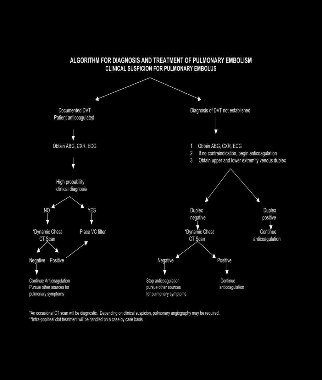
168 168
169 Revised 10/08 University of Kentucky Medical Center Trauma Service Stress Ulcer Prophylaxis Guidelines Incidence of Gastrointestinal Stress Ulceration Clinically important stress-related mucosal bleeding affects up to 4% of critically ill patients, with a mortality rate that approaches 50%. Pathophysiology The splanchnic hypoperfusion that causes stress-related mucosal damage in critically ill patients is multifactorial and results from sympathetic nervous system activation, increased catecholamine release and vasoconstriction, hypovolemia, decreased cardiac output, and the release of proinflammatory cytokines. Stress Ulcer Prophylaxis (SUP) 1) Histamine H2-receptor antagonists (H2RA) Preferred Agents a. UK formulary i. PO - Famotidine 20mg tablets ii. IV - Famotidine 20mg injection 2) Proton Pump Inhibitors (PPI) a. UK formulary i. PO 1. Pantoprazole 40mg tablets 2. Omeprazole/Sodium Bicarbonate 40mg packets ii. IV 1. Pantoprazole 40mg injection Recommended Prophylaxis at the University of Kentucky Who Requires Prophylaxis Prophylaxis for stress ulceration is not necessary in all patients. Only patients with one or more of the following risk factors require prophylactic therapy: 1. Respiratory failure requiring mechanical ventilation > 48 hours. 2. Coagulopathy (platelet count < 5,000, INR > 1.5, aptt>2x control) 3. Major Trauma (ISS > 16) 4. Major Burns (TBSA > 30%) 5. Major head and spinal cord injury 6. Stroke 7. Known history of peptic ulcer disease 8. Organ transplant recipients 9. Patients receiving NSAID s or steroids Other Considerations Early enteral nutrition may be an important factor for preventing gastrointestinal mucosal ischemia. Enteral feedings produce splanchnic vasodilation and increased mucosal blood flow thereby preventing mucosal ischemia and its untoward consequences. 169
170 UK Trauma Service Stress Ulcer Prevention Guidelines Hospital Admission Screen for Risk Factors Respiratory failure requiring mechanical ventilation > 48 hours. Coagulopathy (platelet count < 50,000, INR > 1.5, aptt >2x control) Major Trauma (ISS > 16) Major Burns (TBSA > 30%) Major head and spinal cord injury Stroke Known history of peptic ulcer disease Organ transplant recipients Patients receiving scheduled NSAID s or steroids 1 Risk Factor? Yes No Famotidine 20mg BID* Enteral Access Available? Yes No Give PO Give IV No SUP necessary Consider switch to a PPI ONLY WHEN: Current or recent upper GI bleed Famotidine allergy Ongoing treatment for H. pylori Unexplained mental status changes Receiving PPI at home *For renal insufficiency/failure, adjust dosing according to pharmacy recommendations. 170
171 Diagnosis and Treatment of Stress Related Mucosal Bleeding Diagnosis Endoscopy is the diagnostic modality of choice. Overt Bleeding Overt bleeding is defined as hematemesis, melena, hematochezia, gross blood > (100cc) in the NG tube, or coffee ground emesis or nasogastric drainage. Clinically Important Bleeding Clinically important bleeding is defined as overt bleeding complicated by one or more of the following within 24 hours of the onset of bleeding (in the absence of other causes): 1. spontaneous decrease of > 20 mmhg in the systolic blood pressure 2. an increase in HR > 20 beats/min 3. a decrease in [Hgb} > 2 gm/dl (Hct of > 8%) 4. subsequent transfusion where the Hgb & Hct do not rise appropriately for the number of units transfused Only clinically important bleeding warrants further investigation and if necessary treatment. Endoscopy is the diagnostic modality of choice. Treatment Initial bolus of pantoprazole 80mg, followed by pantoprozole 8mg/hr continuous infusion for 72 hours. Follow with once daily PPI for months. References Cook DJ, Fuller HD, Guyatt GH, et al. Risk factors for gastrointestinal bleeding in critically ill patients. The New England Journal of Medicine. 1994; 330(6): Tryba M. Sucralfate versus antacids or H2-antagonists for stress ulcer prophylaxis: a meta-analysis on efficacy and pneumonia rate. Critical Care Medicine. 1991; 19(7): Schepp W. Stress ulcer prophylaxis: Still a valid option in the 1990s? Digestion. 1991; 54: Pickworth KK, Falcone RE, Hoogeboom JE, Santanello SA. Occurrence of nosocomial pneumonia in mechanically ventilated trauma patients: a comparison of sucralfate and ranitidine. Critical Care Medicine. 1992; 21(12): Pemberton LB, Schafer N, Goehring, et al. Oral ranitidine as prophylaxis for gastric stress ulcers in intensive care unit patients: Serum concentrations and cost comparisons. Critical care Medicine. 1993; 21(12): Loperfido S, Monica F, Maifreni L, et al. Bleeding peptic ulcer occurring in hospitalized patients: Analysis of predictive and risk factors and comparison with out-ofhospital onset of hemorrhage. Digestive Diseases and Sciences. 1994; 39(4): Cook D, Gordon G, Marshall J, et al. A Comparison Of Sucralfate And Ranitidine For The Prevention Of Upper Gastrointestinal Bleeding In Patients Requiring Mechanical Ventilation. The New England Journal of Medicine. 1998; 338(12):
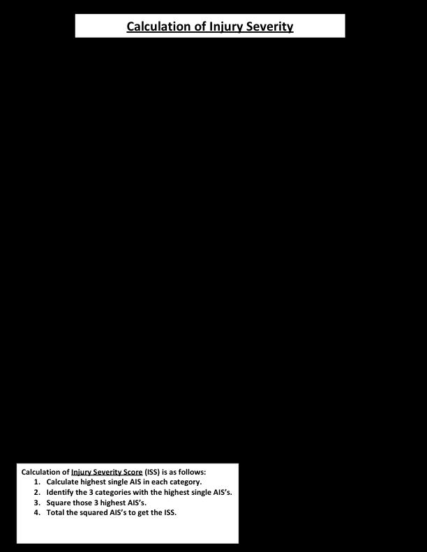
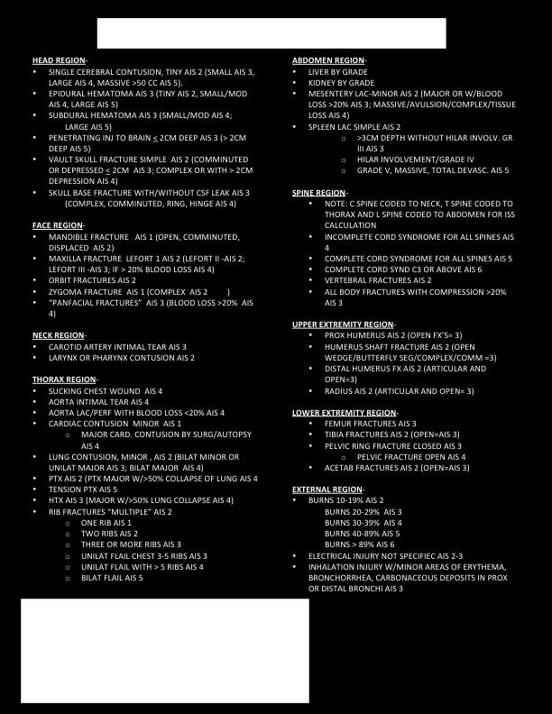
172 172
173 Revised: 09/08 University of Kentucky Medical Center Trauma Service Guideline for Maintenance of Endoscopic Feeding Tubes Early enteral nutrition is very important in the management of critically ill patients. Obtaining and maintaining access to the GI tract is an essential component of patient care. The gastric dysfunction that accompanies critical illness necessitates post pyloric placement of small-bore feeding tubes to insure tolerance of enteral formulas and the administration of medication. Reliable post-pyloric placement of feeding tubes using a blind technique is difficult. Surgical and endoscopic methods are both difficult and expensive. Maintaining patency of these important enteral access devices to insure continuous nutrient flow and to prevent costly tube placement is essential for proper patient care. These guidelines were developed to assist the clinician with medication administration, maintenance of the feeding tube, and trouble shooting guideline when the feeding tube becomes clogged. Maintenance Routine flushing with ml of water before and after medications is essential for maintaining patency, especially with medications containing sorbitol. If enteral nutrition is held for any length of time the feeding tube should be flushed with 30 ml of water. Adequate flushing of the feeding tube is the key to maintain patency and cannot be over emphasized. Common Causes of Obstruction 1. Infrequent or inadequate water flushes pre/post medication delivery. 2. Viscous medications and tablet fragments may adhere to lumen wall causing obstruction within 15 minutes to one hour. 3. Small caliber feeding tube collapse when aspirated. Do not aspirate small bore feeding tubes! 4. Aspirated tube feeding formula mixes with gastric acid and coagulates, producing tube obstruction. 5. Feeding flow rates less than 50cc/hr. may cause residue buildup and obstruction. 6. Viscous feeding formulas such as those containing additives like Promod. 7. Tube type effects patency. Silicone tubes have a higher obstruction rate than polyurethane tubes. 8. Tube kinking from dislodgment. 173
174 Assessment of Small Caliber Feeding Tubes and Feeding Tube Regimen 1. Auscultation is not adequate to verify feeding tube location initially or during ongoing assessment. An x-ray must be done to confirm position. 2. Monitor for potential regurgitation of tube feeding into the stomach by aspiration from the nasogastric tube every 4 hours. 3. Change tube feeding canister every 24 hours, formula every 12 hours for canned preparations, and every 6 hours if pre mixed by dietary. 4. Document cessation of tube feeding, amount given, versus the amount prescribed. Administration of Medications 1. Consider whether medications should be taken on an empty/full stomach. If a patient is receiving intermittent feedings schedule medications accordingly. 2. Check nasogastric or PEG residual prior to medication delivery. If residual is equal/greater than 75% of hourly rate, wait a short period and check again before giving medication. 3. Flush feeding tube with ml of water pre/post medication. 4. Utilize liquid form of medications instead of tablets when possible. 5. Drugs designated SA, E.E.S., SL, XL, EC, LA, MT should never be crushed or dissolved unless formulated for that purpose. 6. Never add medications to enteral formulas in tube feeding container. This practice may cause coagulation and feeding tube obstruction. 7. Administer multiple medications separately to avoid drug interactions; Flush with 5ml of water between medications, when possible. 8. Determine whether medication should be given in the stomach or small bowel. Drugs such as sucralfate, antacids, iron salts, ketoconazole, should be administered via the nasogastric tube to insure efficacy. 9. Phenytoin administrations; stop tube feeding one hour pre/post administration. 10. Some medications require special preparation to insure proper efficacy. If unsure of how to administer the drug, please check with the pharmacist. Nursing Guidelines 1. Ask pharmacy if medication is available in solution. 2. Always flush feeding tube with ml of water pre/post medication administration. 3. Elevation of head of bed 30 is necessary when feeding tube is in the stomach. Elevation is not necessary but is recommended when the feeding tube is in the postpyloric position. 4. Monitor the patient for diarrhea or constipation (refer to appropriate algorithm). 5. Send the patient to the operating room with enough formula for anticipated length of case (May send 12 hours of formula in the container, if case is expected to go beyond 12 hours, please send cans of formula with patient). 6. The nurse should notify the patient s primary service in the event the anesthesiologist or consulting service requests that the tube feeding be stopped. 174
175 Troubleshooting Obstructions for Small Caliber Feeding Tubes 1. If a small caliber feeding tube becomes clogged, attempt unclogging with warm (not hot) water or carbonated soda. 2. Pancreatic enzymes (Viokase) can be used to unclog tubes. Prior to administration of above agent, aspirate tube-feeding formula using a ml syringe. This will clear tube up to the obstruction site. Clamp tube post administration for 15 minutes, and then flush with water. 3. Due to rare instances of tube perforation, reinsertion of the stylet is not recommended without the physician s approval or supervision See Clogged Feeding Tube Algorithm 175
176 Clogged Feeding Tube Slowly aspirate tube feeding formula using syringe Flush tube with 30 ml of warm water. Clamp tube for 15 minutes Clogged Unclogged Administer the following: Continue tube feeding Viokase + sodium bicarbonate (obtained from pharmacy) Clamp feeding tube for 15 minutes Clogged Unclogged Raise HOB 30 degrees Turn patient on side Continue tube feedings Clogged Is tube kinked? Obtain KUB, reposition tube with MD order. Flush with 30 ml warm water Clogged Unclogged Consider tube replacement Continue tube feeding 176
177 References Byerly WG. Drug information questions and answers. Hospital Pharmacy Hotline. 1992; 5: 2. Drug Information Center, University of Kentucky Medical Center. Pharmacy News for Nurses. 1992; 13(4): Kohn CL, Keithley JK. Enteral nutrition-potential complications and patient monitoring. Nursing Clinics of North America. 1989; 24: Lehmann S, Barber J. Giving medications by feeding tube. How to avoid problems. Nursing ; 21: Metheny N. Minimizing respiratory complications of nasoenteric tube feedings; State of science. Heart and Lung. 1993; 22: Paauw JD, Fagerman KE, McCamish, et al. Enteral nutrition solutions. The American Surgeon. 1984; 50(6): Potts RG, Zaroukian MH, Guerrera PA, et al. Comparison of blue dye visualization and glucose oxidase test strip methods for detecting pulmonary aspiration of enteral feedings in intubated patients. Chest. 1993;103: Powell KS, Marward SP, Farrior ES, et al. Aspirating gastric residuals causes occlusion of small-bore feeding tubes. Journal of Parenteral and Enteral Nutrition. 1993; 17: Thurlow PM. Bedside enteral feeding tube placement into duodenum and jejunum. Journal of Parenteral Enteral Nutrition. 1986; 10: White WT, Acuff TE, Skyes TR, et al. Bacterial contamination of enteral nutrient solution: A preliminary report. Journal of Parenteral and Enteral Nutrition. 1979; 3(6): Zaloga GP. Bedside method for placing small bowel feeding tubes in critically ill patients. A prospective study. Chest. 1991; 100(6):
178 UNIVERSITY OF KENTUCKY HOSPITAL POLICY NUMBER: HP08-39 CHANDLER MEDICAL CENTER FIRST ISSUED: 7/04 HOSPITAL POLICY CURRENT AS OF: 9/06 SUBJECT: Enteral Feeding: Verification and Maintenance of Small-Bore Feed Tubes in Adults SEE ALSO: NN08-03, Placement and Maintenance of Small-Bore Tubes (Adults) INFORMATION To facilitate delivery of enteral nutrition in adults, the Hospital has established guidelines for the verification and maintenance of small-bore feed tubes. Clinical Nutrition s consultation service should be involved with all patients receiving enteral nutrition, and will be responsible for ordering the feeds. Confirmation of Tube Placement Verification of small-bore feeding tube position requires initial radiographic confirmation with KUB or lower chest x-ray prior to initiation of feeding. Exception: Feeding tubes placed during laparotomy do not require radiographic confirmation. In patients with surgical disruption between the GI tract and larynx, the physician may document that radiologic confirmation of the feeding tube is not necessary. 1. The physician will notify the nurse of the location of the feeding tube and give an order to begin enteral feeding. 2. The nurse should document the location initially on the nurses note and on the patient s care plan or the electronic Assessment and Intervention record, and should document that tube feeding is started per physician s order. This documentation is required each time a feeding tube is inserted and with each time feeding started. 3. If tube is not in the small bowel as confirmed by x-ray, then nurse may reattempt insertion twice. If tube continues to be in gastric position after both subsequent attempts, the nurse will document and notify physician for endoscopic tube placement. Administration of Formula (Open System) Formula administration should be continuous via a feeding pump, unless otherwise ordered by the physician. Bolus feedings should be avoided in the adult ICU patient. Cyclic administration that delivers feeding over a few hours (either daily or intermittently during a 24-hour period) using a feeding pump may be appropriate in some patients and should be ordered by the physician. Food coloring or dye on an on-going basis is contraindicated and should not be added to the formula or container. Physicians may order food coloring added to tube feeding in specific situations in which aspiration of tube feeding is suspected. Nurses should follow the guidelines for use of food coloring in these instances. Prevention/Assessment of Aspiration For All Patients on Tube Feeding 1. Unless contraindicated (i.e., patient with cervical spine injury or subdural drain), elevate the head of the bed to 30 to 45 degrees to reduce the risk of aspiration. Patients with spinal injuries may be able to be placed in reverse Trendelenburg position at 30 to 45 degrees to reduce the risk of aspiration. Note: The nurse should verify head of bed elevation with the physician HP08-39, Verification and Maintenance of Small-Bore Feed Tubes in Adults 178
179 For Critically Ill Patients with One Major Risk Factor (refer to following list of risk factors), it is recommended: to provide all care listed above, maintain tight glycemic control (blood glucose between mg/dl), and use continuous infusions rather than intermittent boluses. For Critically Ill Patients with Two or More Major Risk Factors (refer to following list of risk factors), it is recommended: to provide all care listed above, that all feeding tubes be in the small bowel, and to use prokinetic agents (Metoclopramide). Major Risk Factors previous episode of aspiration decreased LOC (sedation or elevation in intracranial pressure) neuromuscular disease neuromuscular blocking agents endotracheal tube or intubation vomiting persistent high residuals For Patients Receiving Gastric Tube Feeding 1. If the patient has any of the risk factors for aspiration listed above, tube feeding tolerance should be assessed by checking residuals on gastric tube feedings every four hours. 2. For gastric residuals of > 300 ml, hold tube feeding for 1 hour, then recheck residual. If residual remains > 300 ml after 1 hour, then hold tube feeding, discard residual, and notify service. 3. If second gastric residual is < 300 ml, resume previous tube feeding orders and return residual to patient. If second gastric residual is > 300 ml, hold tube feeding and notify service for specific orders (see Enteral Nutrition Physician/Patient Care Orders, form J703). Glucose Monitoring in Patients Who are Diabetic or Receiving Insulin 1. Fingerstick glucose should be monitored at least every six hours in diabetic patients or in those receiving insulin. 2. Patients receiving insulin infusions require more frequent glucose monitoring, which should be specified by the physician. Approved by Karen Stefaniak, Chief Nursing Officer Approved by Joseph Conigliaro, M.D., Associate CMO, Quality & Patient Safety Authorized by Murray B. Clark, Jr., Associate VP for MC Operations 179
180 Revised 9/08 University of Kentucky Medical Center Trauma Intensive Care Unit Bowel Regimen Program The injured/critically ill patient undergoes physiologic alterations in gut function. Gastric motility slows; use of narcotics slows intestinal motility, warranting bowel function surveillance to ensure timely identification and intervention of alterations. All trauma service ICU patients will be started on a bowel protocol upon admission consisting of Docusate Sodium 250 mg twice daily and Biscodyl suppository twice daily prn. Assessment of the patient should include bowel patterns pre-injury, other medical conditions that predispose patient to alterations in bowel function, medications, High risk indicators: use of narcotic analgesia, lack of BM x 24 hours. Diarrhea and constipation in critically ill patients may either be serious or benign conditions. There are many causes for constipation such as improper diet, intestinal obstruction, tumors, excessive use of laxatives and weakness of the intestinal musculature. What constitutes constipation is very subjective and individualized. However, absence of stool > 3 days in patients receiving enteral nutrition should be investigated and appropriate therapy initiated when indicated. Diarrhea is common in critically ill patients, occurring in 24% of patients admitted to intensive care units. Many factors contribute to diarrhea, such as infection, antibiotics, and drugs containing sorbitol, bowel edema, inflammation, enteral feeding rate, and hyperosmolar enteral feedings. Incomplete digestion may occur in patients who have had feedings withheld for several days, therefore low rates with slow advance are recommended when beginning enteral feedings. However, diarrhea associated with enteral feedings is usually seen in patients who have gut atrophy from prolonged fast or patients on high hourly feeding rates (> 75 ml/hour). Hypoalbuminemia (<2.5g/dl) and attendent bowel edema have been implicated as a cause of diarrhea, but this association has never been firmly established. Diarrhea associated with antibiotic therapy is usually benign, but can be serious. Antibiotic therapy alters colonic flora which may allow overgrowth of pathogenic and or diarrheagenic bacteria. Clostridium difficile is present in the normal colonic flora of some patients. Overgrowth by toxigenic C. difficile can occur in patients receiving antibiotic therapy. C. difficile infection can present as diarrhea that is mild to moderate or can cause severe colitis with pseudomembrane formation. Pseudomembraneous colitis can lead to severe diarrhea, hypovolemic shock, toxic megacolon, perforation and death. Specific findings of pseudomembraneous colitis include watery, green, foul-smelling, non-bloody or bloody diarrhea, cramping abdominal pain, fever (> 39.5 C) and leukocytosis. Stool specimen should be sent for C. difficile toxin. C. difficile culture is a less efficient method and does not differentiate between nontoxiocogenic colonic flora and toxic strains of C. difficile. Turnover time for the C. difficile toxin assay is usually 24 to 48 hours. Severe diarrhea, abdominal distention, tenderness, unexplained SIRS, megacolon on plain film and a high index of suspicion should prompt an endoscopic evaluation to exclude pseudomembranes. Treatment varies according to the severity of the illness. Enteral metronidazole is the first line drug of choice. If a patient cannot tolerate oral medication intravenous metronidazole should be given. Intravenous vancomycin is not effective in the treatment of C. difficile enterocolitis. Enteral vancomycin is the drug of choice for severe colitis and treatment failures on metronidazole. 180
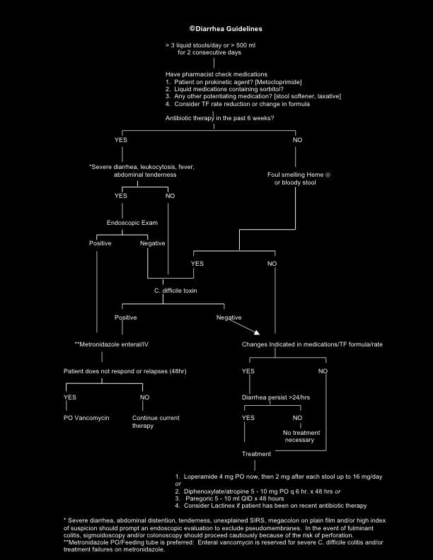
181 181
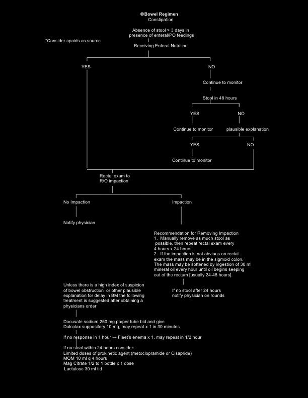
182 182
183 Nursing Guidelines: Diarrhea: 1. Document frequency and amount of stool 2. Inform physician of greater than 3 liquid stools day or greater than 500 ml for 2 consecutive days 3. Use skin barrier or fecal bag to prevent skin breakdown from diarrhea stools 4. If oral medication is administered in the nasogastric tube clamp for at least 30 minutes 5. Flush feeding tube with 20 ml - 30 ml of water pre and post medication 6. Based on algorithm send stool specimen for C. difficile toxin per physicians order [turn around time is usually 24 to 48 hours] 7. Notify H.O. if patient does not respond to antibiotic therapy within 48 hours or if diarrhea returns < 72 hours after antibiotic stopped Constipation: 1. Notify physician when patient who is receiving PO/enteral feeding has not had a stool for > 3 days 2. Ensure patients are receiving adequate hydration 3. Assess for medications or dietary intake (low residue, low fiber) that may contribute to constipation 4. Lubricant should be used when manually removing impaction 5. Abdominal pain and/or distention may be a clinical manifestation of a serious problem References Thomspon, et al. (1992) cited in Powell, M and Rigby D. Management of bowel dysfunction: evacuation difficulties. Nursing Standard. 2000; 14(47): Winney, J. Constipation. Nursing Standard. 1998; 13(11): Sykes N. The treatment of morphine induced constipation. European Journal of Palliative Care. 1998; 15(1): Fekety R, Shah AB. Diagnosis and treatment of clostridium difficile colitis. JAMA. 1993; 269: Kelly CP, Pothoulakis C, LaMont JT. Clostridium difficile colitis. The New England Journal of Medicine. 1994; 33(94): Hwang TL, Lue MC, Nee YJ, et al. The incidence of diarrhea in patients with hypoalbuminemia due to acute or chronic malnutrition during enteral feeding. American Journal of Gastroenterology. 1994; 89(3): Johnson, DA. Diarrhea in critically ill Patients. Problems in Critical Care. 1989; 3(3):
184 Blue Surgery Bladder Management Protocol for the Urologically Non-complicated Patient UTIs account for 31% of nosocomial infections in US medical ICUs, thus it is essential to understand the logistics of Foley monitoring and maintenance. The risk of bacteriuria from an indwelling catheter is 3-10% per day. Daily assessment for the continued necessity of an indwelling catheter and removal when they are no longer indicated are simple but important steps to reduce infection, encourage a return to normal physiologic function, and improve patient care. Intermittent catheterization deals with draining the bladder at specific time intervals to simulate normal physiologic function. An adult bladder holds approximately 400 ml of urine and voids 4-5 times/day. Intermittent catheterization should likewise be performed a 4-5 times/day with adjustments based on volume drained. The total volume drained should not exceed 400 ml. If volume drained is high, either the frequency of intermittent catheterizations should be increased or fluid intake should be decreased. Spinal Cord Injuries: According to several studies, intermittent catheterization is a better option than indwelling catheterization for both male and female patients. Some studies show that for patients who are undergoing catheterizations approximately 4 times/day, bacteriuria occurs at an incidence of 1-3% per catheterization. Antibiotics are usually not required during intermittent catheterizations unless the patient is in a high-risk population (immunosuppressed, internal prosthesis, etc.). Raymond et al. Neurogenic Bladder. emedicine May 22, Richards et al. Crit Care Med 1999; 27:887. Saint et al. Arch Intern Med 1999; 159:
185 185
186 University of Kentucky Trauma and Critical Care Recommendations for Central Venous Catheter Insertion Adapted from the American College of Surgeons Guidelines for Central Venous Access* Indwelling vascular catheters are essential for patient care. All indwelling vascular catheters have associated mechanical and infection risks. Catheters should be place when needed and the insertion should be performed properly using strict aseptic technique. Catheter maintenance should follow accepted guidelines including sterile technique for infusions, lines, and hubs. Site inspection and care are essential for preventing infection. Catheters should never remain in place for caregiver convenience and should be removed when no longer needed. Placement 1. Appropriately trained and experienced personnel identified to place CVC 2. Time out performed prior to procedure (patient ID, consent, site selection, diagnosis) 3. Choose the most appropriate site for CVC insertion based on patient s needs 4. Full sterile precautions must performed on ALL non-emergent CVC insertions. This includes sterile hat, mask w/shield, gown and gloves. A fully body sterile sheet is applied after appropriate antisepsis. 5. Appropriate antisepsis is achieved with 2% chlorhexidine gluconate for 30 seconds and then allowed to air dry. If this is not available, it is appropriate to use iodine, iodophore or 70% ethanol, no organic solvents. 6. Choose a central venous catheter with the minimum number of lumens for your patient s needs. 7. All internal jugular and femoral lines should be placed under ultrasound guidance unless emergent line placement is required. 8. If a CVC is malfunctioning then a new one may be placed by exchange over a guidewire ONLY if there are no signs of bacteremia and/or infection 9. >3 needle sticks for access increases risk of insertion complications and consideration should be given to more experienced personnel insertion and/or new stick site 10. After successful insertion a chlorhexidine impregnated sponge should be placed around the catheter at the insertion site. 11. Position of CVC must be evaluated with a STAT chest x-ray. Correct CVC should have the tip near the SVC and right atrial junction. All CXR should be evaluated for evidence of pneumothorax. 12. If line is in satisfactory position the line needs to be cleared for use in SCM. Alert the nursing staff that the line is ok to use and that the order for clearance is in SCM. 13. A procedure note is to be performed on all CVC insertion attempts, successful or not. SCM has a procedure note dedicated to CVC insertion. This is the default and expected method of procedure note completion. A brief note in the chart should indicate the patient name, date and time with reference to the complete note in SCM. Maintenance 1. The routine replacement of central lines does not prevent CRBSI and is not recommended in the absence of CRBSI 2. The CVC site should be inspected daily and PRN 3. Before manipulation of any CVC proper hand aspesis should be performed by washing with soap and water or alcohol scrub (even when gloves are worn) 4. Clean gloves should always be donned before CVC manipulation 186
187 5. If CVC lumen access is attempted via the injection ports or caps then asepsis should be applied to the ports/caps with 2% chlorhexidine gluconate or another appropriate antisepsis 6. 2% chlorhexidine gluconate, iodine, iodophore or 70% ethanol (no organic solvents) should be used for all CVC dressing care and dressing changes. All antisepsis should be allowed to air dry before manipulation/dressing changes. 7. Use a sterile, transparent dressing over catheter site with clean gloves and a no-touch technique. If the site is not dry, then apply a sterile dry gauze and change dressing when it becomes saturated. Change to a transparent sterile dressing as soon as possible. 8. Sterile dressing should be replaced q7 days unless it becomes loose. 9. There is no indication for antimicrobial prophylaxis (systemic or local) with an indwelling CVC. 10. Avoid anticoagulants for clot or CRBSI infection unless certain patient conditions mandate their use 11. Use sterile NaCl (heparin if indicated) to flush and lock to maintain patency 12. The use of needle free connectors is encouraged to prevent needle stick injuries, and aseptic technique should always be followed. Catheter Related Infection and/or Complication Evaluation 1. The occurrence of fever should not prompt CVC removal. It is expected that clinical judgment be applied. If patient demonstrates a strong suspicion of CRBSI then CVC removal is considered. 2. If CRBSI is suspected or confirmed, the CVC or introducer should be removed. The line should be cultured only if CRBSI is suspected. 3. The tip or intracutaneous segment only should be cultured. 4. If the patient demonstrates erythema or purulence at the catheter site, sepsis, + blood culture results the CVC should be removed. 5. If the CVC that was exchanged demonstrates quantitative cultures suggestive of CRBSI (>15 Colony Forming Units, CFUs) then the catheter should be removed and placed at a new site sets of blood cultures, one from a peripheral stick, in all patients with suspected CRBSI 7. After removal of CVC in patients with CRBSI, a non-tunneled CVC may be placed at a new site after systemic antimicrobial therapy is begun 8. If after removal of CVC there is persistence of bacteremia and/or fungemia, lack of clinical improvement (after 3 days of CVC removal and appropriate antimicrobial therapy) then the clinician should seek septic foci (i.e. endocarditis, septic thrombi, and mestastatic infections. 9. All institutions should have monitoring systems for outcomes, infections and complication rates. They should also have systems in place if complications exceed the standard of care to identify and correct these occurrences 10. Venous thrombosis, documented at insertion site, the CVC should be removed and placed a new site if CVC is still needed. Central venous, pulmonary artery, and peripheral venous lines placed outside the Intensive Care Unit All deep lines place outside of the intensive care areas must be changed to a new sight within 24 hours of admission to the unit. Exceptions to this policy are as follows: 1. Lines placed under aseptic conditions in the operating room, on the floor, or in another ICU where sterility of the procedure can be documented. 187
188 2. All emergent lines, placed under non-sterile conditions, placed in the ED for emergency resuscitation must be removed within 24 hours of admission to the ICU without exception. Pulmonary Artery Catheter Important Notes 1. PAC should not be left in for > 10 days due to the significant risk of CRBSI 2. If the PAC is removed, under no circumstance should the introducer sheath be left in place. 3. Guide wire PAC exchanges are only indicated for PAC malfunction only. If there are extenuating circumstances then chief, fellow or attending approval is needed. Insertion, maintenance, removal, and replacement of Arterial lines Route of Insertion 1. The percutaneous route of arterial line placement is preferred to surgical cutdown. a. Surgical cutdown for arterial cannulation should only be performed after approval by the critical care attending. Site of Insertion 1. The preferred site of arterial cannulation is the radial artery. Alternate sites of arterial cannulation are for most patients listed in descending order preference. a. Femoral artery* b. Dorsalis Pedis artery* *In patients with peripheral arterial disease, percutaneous femoral arterial cannulation has a much higher complication rate and should be avoided. Dorsalis Pedis cannulation may lead to inaccurate blood pressure measurements. Caution must be exercised in choosing the site of arterial cannulation in this group of patients. 2. The complications associated with brachial artery and axillary artery cannulation are higher than other routes. a. Brachial artery: Used uncommonly because of the high complication rates. The major complication is thromboembolic occlusion usually without ischemia. b. Axillary artery: Limb threatening ischemia negligible. However, catheter infection rate is much higher than other sites and line care is difficult. Insertion Procedure 1. The site chosen for arterial cannulation should be prepped and draped to create a sterile field. 2. Sterile gloves and mask should be worn during insertion of the line. 3. The line should be sutured in place using aseptic technique. 4. At the completion of the procedure, a chlorhexidine impregnated sponge is applied to the insertion site and covered with a sterile MVP (Tegaderm or OP-Site ) dressing. Line Changes 1. The infection rate in percutaneously placed arterial cannulas are very low. Routine catheter removal and change to a new site is unnecessary. Arterial cannulas can remain at the original site of insertion until no longer needed. Exception: Catheters should be changed to a new site when: a. There is evidence of infection at the insertion site manifested as pain, redness, swelling, or purulence. Catheters should always be cultured in this situation. 188
189 b. There is evidence of ischemia distal to the site of insertion. c. The catheter is implicated as a potential source of unexplained systemic sepsis. 2. The decision to change the arterial line to a new site or to culture the catheter tip is left to the discretion of the treating physician. It is recommended that catheter tips be submitted for culture when there is evidence of infection at the site of insertion or when the patient exhibits unexplained sepsis. Reference Freel AC, et al. American College of Surgeons Guidelines Program: A Process for Using Existing Guidelines to Generate Best Practice Recommendations for Central Venous Access June
190 Revised: 11/08 University of Kentucky Hospital Trauma Intensive Care Unit Hypnosedative/Alcohol Withdrawal Prophylaxis Introduction: Alcohol abuse and/or addiction are frequent problems in the trauma patient population. Acute alcohol withdrawal increases mortality and morbidity for the critically ill and/or injured patient. It is essential that physicians and nurses obtain a thorough history to ascertain if the patient has a history of alcohol abuse or dependency. Symptoms of alcohol withdrawal begin 6-12 hours after cessation of alcohol and can be difficult to differentiate from injury/illness related symptoms. Tremor, nausea, vomiting, tachycardia, hypertension, diaphoresis, irritability and profound anxiety are frequently seen. Seizures are the most life threatening symptom of alcohol withdrawal. Seizures cause additional stress on body reserves which will further compromise the critically ill patient. Delirium tremens is classified as the late phase or major withdrawal. Patients will often have a combined alcohol and hypnosedative abuse which requires a different treatment approach to prevent withdrawal symptoms. The goal of medical management is to recognize symptomatology of hypnosedative/alcohol withdrawal and prevent progression into delirium tremens for patients at high risk. Early pharmacological intervention is designed to prevent and/or mitigate withdrawal symptoms thereby minimizing risk to the critically ill patient. Criteria for Implementing Alcohol Protocol: 1. Alcohol level > 200 with GCS of Alcohol level > 200 with prior history of Delirium Tremens and/or prior enrollment in a detoxification program 3. History of Delirium Tremens or Detoxification program with clinical manifestations of withdrawal Criteria for Implementing Alcohol/Hypnosedative Protocol: 1. Above Criteria plus 2. History of daily use of benzodiazepines or barbiturates or 3. Positive drug screen with history of daily use. 4. Enter Chemical Dependency Consult. Nursing Care Guidelines Signs and Symptoms of Withdrawal: agitation, anxiety, tremors, nausea, vomiting, hypertension, tachycardia, diaphoresis 190
191 [Differentiation from behaviors associated with closed head injuries, and agitation/anxiety related to uncontrolled pain is essential] 1. Monitor vital signs q 1 hour x 24 hours 2. Monitor agitation/sedation level q 1 hour x 72 hours then q 4 hours and PRN A. Diazepam Load and PRN Diazepam Use PRN diazepam only if patient is having tremors Use lower dose of haloperidol and diazepam for non-intubated patients Assess and record the following vital signs parameters prior to each dose: Heart Rate MAP Respiratory Rate SpO 2 Level of sedation Hold next dose and/or notify physician if : Heart rate < 60 [if a change from baseline] Respiratory rate < 16 MAP < 60 Sedation level > 4 SpO 2 < 92% B. Carbamazepine: Carbamazepine level prior to starting maintenance dose if patient is having signs and symptoms of withdrawal Goal is to have level > 12. Assess and record the following vital sign parameters prior to each dose: Heart Rate MAP Respiratory Rate SpO 2 Level of sedation Hold next dose and/or notify physician if: Heart rate < 60 [if a change from baseline] Respiratory rate < 16 MAP < 60 Sedation level > 4 SpO 2 <
192 C. Haloperidol: 1. Administer 10 mg IV over 1-2 minutes [administer over 5 minutes if SBP< 100] 2. Not to exceed 240 mg/24 hours. 3. Monitor for extrapyramidal symptoms and notify physician if symptoms observed. 4. Prolonged QT interval & incidence of Torsades des pointes is rare. 5. Monitor for signs and symptoms of rigidity, consult physical therapy if present Sedation Status: Anxiety Scale: Tremors 1 - Wide Awake 1 - No Anxiety 0 - Non Existent 2 - Drowsy 2 - Mild Anxiety 1 - Moderate 3 - Dozing Intermittently 3 - Anxiety 2 - Profound 4 - Mostly Sleeping 4 - Severe Anxiety 5 - Only Awakens When Aroused 5 - Extreme anxiety References: Ries, RK, Roy-Byrne, PP, Ward, NG, et al. Carbamazepine treatment for benzodiazepine withdrawal. American Journal of Psychiatry. 1989; 146: Klein, EK, Uhde TW, Post RM. Preliminary Evidence for the utility of carbamazepine an alprazolam withdrawal. American Journal of Psychiatry. 1986; 143: Butler D, Messsiha FS. Alcohol withdrawal and carbamazepine. Alcohol; 1986; 3: Denicoff KD, Meglathery SB, Post RM, et al. Efficacy of carbamazepine compared with other agents; a clinical practice survey. Journal of Clinical Psychiatry. 1994; 55(2): Miller, R. Protracted alcohol withdrawal delirium. Journal of substance abuse and treatment.1994; ll(3): Strasen, L. Acute alcohol withdrawal syndrome in the critical care unit. Critical Care Nurse. 1982: Menza MA, Murray GB, Holmes VF, et al. Decreased extrapyramidal symptoms with intravenous haloperidol. Journal of Clinical Psychiatry. 1987; 48(7): Fraser GL, Riker RR. Controlling severe agitation in the critically ill with intravenous haloperidol. Hospital Pharmacy. 1994; 29(7): Skinner MH, Thompson DA. Pharmacologic considerations in the treatment of substance abuse. Southern Medical Journal. 1992; 85(12):
193 Hyatt M. Anxiolytic Drug Abuse, Addiction and Withdrawal Use of Neuraxial Pain Control Methods For Trauma and Acute Care Surgery Julie McWhorter, MD, Department of Anesthesia and Jeffrey Coughenour, MD, Department of Surgery, Section of Trauma and Critical Care The use of Anti-Xa levels is not predictive of the risk of bleeding. Antiplatelet or oral anticoagulant medications used in combination with LMWH may increase the risk of spinal hematoma. Patients who present with chronic use of Coumadin should be off therapy for 4-5 days prior to neuraxial procedures. An INR should be checked and should be <1.5. If therapy is reinstituted with an indwelling epidural catheter, INR should be monitored closely. The catheter can be removed at any time assuming the INR is <1.5. The site should be monitored for 24 hours after removal for complications. NSAIDs pose no significant added risk of bleeding or spinal hematoma after neuraxial procedures. GP IIb/IIIa antagonists (Ticlid, Plavix) should be discontinued for 14 days (Ticlid) and 10 days (Plavix) respectively prior to procedures. Administration of platelets may be of assistance in urgent situations, but no clear risk stratification has been established. Patients taking herbal medications pose no increased risk from neuraxial procedures, unless they are taken in conjunction with other conventional anticoagulants. No specific consensus statement exists regarding risk and the use of direct thrombin inhibitors and fondaparinux. Spines should be evaluated and cleared. Method of evaluation and a negative tertiary exam should be documented by the Trauma, NS, or Ortho spine service. Prophylactic Lovenox should be held at least 12 hours prior to epidural placement. Epidural catheters may be placed in intubated patients. Some attending anesthesiologists are less comfortable with this as the ability to determine development of a neurologic deficit is difficult. Therefore placement may be on a case-by-case basis. Literature clearly supports placement of epidural catheters for pain control and improvement of pulmonary mechanics in the setting of rib fractures. Pre-operative placement of epidurals for thoracic operations and many abdominal operations is associated with lower incidence of DVT, quicker return of bowel function, improved pulmonary toilet, improved pain scores and increased ambulation postop. Contrary to popular belief, Foley catheters are unusually required for thoracic epidurals. 193
194 Minimum time from last dose to catheter placement/spinal injection Minimum time from last dose to catheter removal Standard Anticoagulants Warfarin INR < 1.5 Avoid use while Heparin, full dose IV/SQ INR < 1.5 catheter in place Heparin prophylaxis (BID, TID) No restrictions Newer Anticoagulants Xigris (Drotrecogin Alfa) 24 hours Arixtra (Fondaparinux) hours Avoid use while Lovenox (1 mg/kg BID) 24 hours catheter in place Lovenox (30 mg SQ BID) hours Lovenox (40 mg SQ QD) hours hours Direct Thrombin Inhibitors Argatroban aptt < 40 Avoid use while Lepirudin aptt < 40 Antiplatelet Agents ASA/NSAIDs catheter in place No restrictions Plavix 7 days Avoid use while Ticlid 14 days catheter in place Thrombolytic Agents TPA (full dose for stroke, MI, etc.) 10 days Avoid use while catheter in place TPA (2 mg for catheter clearance) No restrictions References: University of Washington Medical Center, Nursing Epidural Policy and Procedure Accessed May 14, 2008 Second ARSA Consensus Conference on Neuraxial Anesthesia and Anticoagulation, Reg Anesth Pain Med 2003;28: /08 194
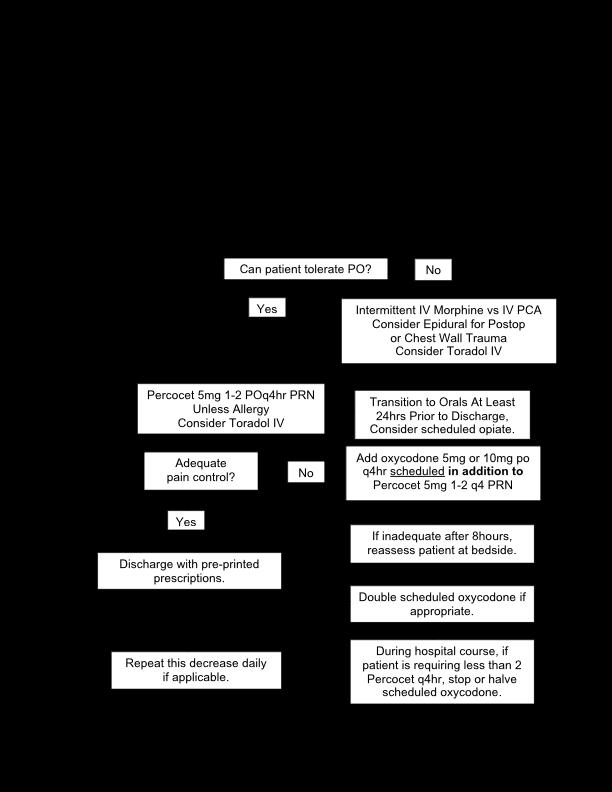
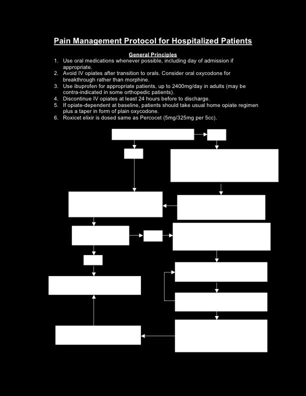
195 195
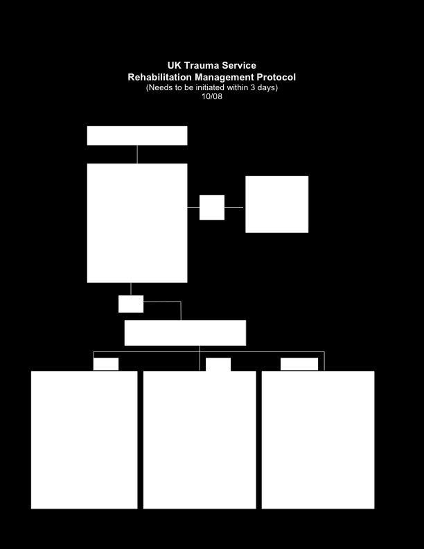
196 196

197 SECTION 6: DISCHARGE INSTRUCTIONS 197
198 Discharge Instructions D/C Home: Please be thinking ahead of time on patient s D/C home plans. Please inform the bedside RN of D/C plans so home care instructions can be completed. If the pt. needs home health or home equipment, please contact patient care facilitator assigned to Blue Surgery for this before the day of D/C. The Care Facilitator will contact you if they need written prescriptions for any of the Home Health/DME orders. On the weekends, the weekend social worker can set up Home Health; the patients do not need to be kept over the weekend for home health needs only. Next, complete the following: 1. Complete the UK Discharge Orders form (H341) fully 2. Controlled substance prescriptions must be written on a separate (green) prescription pad. Record type and amount of any controlled substance prescription on the white medical record copy, of form H341, in order to have documentation of all meds given to pt. at D/C 3. Make sure the need for all f/u appointments have been documented on form H341 (may need to call consulting services to find out if /when they need to see pt. back) 4. Write D/C to home order in routine MD orders 5. Complete an automated D/C summary* *For patient s who live out-of-state, you will need to do a STAT D/C summary to send with the patient in case they can find a local MD to provide their follow-up care. Inter-Facility Transfers: Such as to: Acute Rehab, Subacute Rehab, ECF, or Hospital to Hospital The Social Worker for our service can assist you with inter-facility transfers. If you suspect the pt. will need some type of placement, contact the S.W. on pager number found on Blue Surgery Census. Inform the pt./family that they may have some rehabilitation needs and that our S.W. will come by to talk with them. Please never tell a pt./family we will send them to a specific place (such as Cardinal Hill). Many factors need to be looked at before a pt. can go to a facility and not all pts. are candidates for CHH. Once a facility has accepted the pt. please complete the following: 1. Complete a STAT D/C summary. This will have to go with the pt. It takes several hours to get this done so please plan accordingly. 2. Complete the UK Discharge Orders Form (H341). This form still needs to be completed except for the prescriptions, since the list of meds are in the D/C summary. Please make sure you write for all needed F/U appointments on the form so the clerk can schedule these before the pt. is transferred. 198
199 3. There is an Inter-Facility Form (J076) that has to be completed prior to transfer. This form has 2 pages. You are responsible for completing the M.D. section on page 1 and the Risk & Benefit section on page 2 4. Write an order Transfer to in routine M.D. Orders Subject: Physician responsibility in completion of Inter-Facility Transfer Form (J076) that must be completed by the physician prior to transfer of patient to another facility that will assume care of the patient. Patient Condition (page 1, gray section) Includes: Diagnosis/initial impressions Current condition/observations Treatment rendered Current medications ATTENDING PHYSICIAN SIGNATURE AND DATE Transfer Information (last section of page 1) The physician completing the Inter-Facility transfer form MUST WRITE THE NAME OF THE RECEIVING PHYSICIAN on the appropriate line. Authorization of Transfer (page 2, gray section) Includes: Risks of transfer Benefits of transfer Condition of patient at time of transfer Attending physician s signature and date NOTE: See H & P cannot substitute completion of the form by a physician. 199
200 Discharge Pain Medication Algorithm Outpatient Surgery, Including Laparoscopy-8 Day Taper Lortab 5/500mg or Percocet 5/325mg 1 PO q4hr PRN x 2 days then 1 PO QID PRN x 2 days then 1 PO TID PRN x 2 days then 1 PO BID PRN x 2 days and stop Disp #30 Tylenol and/or Motrin thereafter unless contraindicated Long-Bone & Pelvic Fractures and Ribs-20 Day Taper (until ortho follow-up) Percocet 5/325mg 1-2 PO q4hr PRN x 5 days then 1-2 PO QID PRN x 5 days then 1-2 PO TID PRN x 5 days then 1-2 PO BID PRN x 5 days and stop Disp #150 Tylenol or Motrin thereafter unless contraindicated Soft-Tissue Trauma, Laparotomy & Non-Long Bone Fractures-16 Day Taper Percocet 5/325mg 1-2 PO q4hr PRN x 4 days then 1-2 PO QID PRN x 4 days then 1-2 PO TID PRN x 4 days then 1-2 PO BID PRN x 4 days and stop Disp #100 Tylenol or Motrin thereafter unless contraindicated Lortab is available as 5/500mg. Therefore, tapers with Lortab must not exceed 2 tabs q6hr so that total Tylenol dosage does not exceed 4g/day. Instruct the patient that they may take ibuprofen 600mg QID with Percocet or Lortab but that Tylenol/acetaminophen should not be taken with Percocet/Lortab. Discharge Pain Medication Protocol 200

201 201
The Ohio State University Department of Orthopaedics. Residency Curriculum. PGY1 Rotations
 The Ohio State University Department of Orthopaedics Residency Curriculum PGY1 Rotations Goals and Objectives Anesthesiology Rotation PGY1 Level I. Core Competency Areas By the end of the PGY1 rotation
Title: ED Management of Trauma Patient Protocol
 Title: ED Management of Trauma Patient Protocol Document Category: Clinical Document Type: Protocol Department/Committee Owner: Emergency Department Original Date: August 2009 Approver(s) last review:
AMERICAN COLLEGE OF SURGEONS 1999 TRAUMA FACILITIES CRITERIA (minus the Level IV criteria)
") AMERICAN COLLEGE OF SURGEONS 1999 TRAUMA FACILITIES CRITERIA (minus the Level IV criteria) Note: In the table below, (E) represents essential while (D) represents desirable criteria. INSTITUTIONAL ORGANIZATION
AMERICAN COLLEGE OF SURGEONS 1999 TRAUMA FACILITIES CRITERIA (minus the Level IV criteria) Note: In the table below, (E) represents essential while (D) represents desirable criteria. INSTITUTIONAL ORGANIZATION
ADC ED/TRAUMA POLICY AND PROCEDURE Policy 221. I. Title Trauma team Activation Protocol/Roles & Responsibilities of the Trauma Team
 Section: ADC Trauma ADC ED/TRAUMA POLICY AND PROCEDURE Policy 221 Subject: Trauma Team Activation Protocol/Roles & Responsibilities of the Trauma Team Trauma Coordinator UTMB respects the diverse culture
Emergency Medical Services Program
 County of Santa Cruz HEALTH SERVICES AGENCY 1080 EMELINE AVENUE, SANTA CRUZ, CA 95060 (831) 454-4120 FAX: (831) 454-4272 TDD: (831) 454-4123 EMERGENCY MEDICAL SERVICES PROGRAM Policy No. 7000 Reviewed
Alabama Trauma Center Designation Criteria
 2 Alabama Trauma Center Designation Criteria Office of Emergency Medical Services Master Checklist Alabama Trauma Center Designation Trauma Center Criteria: APPENDIX A Trauma Rules The following table
Penn State Milton S. Hershey Medical Center. Division of Trauma, Acute Care & Critical Care Surgery
 Penn State Milton S. Hershey Medical Center Division of Trauma, Acute Care & Critical Care Surgery Residency-Trauma Curriculum The Medical Director for the Penn State Shock Trauma Center is Dr. Heidi Frankel.
TRAUMA CENTER REQUIREMENTS
 California Trauma Center Level III Criteria California Code of Regulations,, Chapter 7 - Trauma Care System with American College of Surgeons (Green Book) references; includes FAQ clarifications TRAUMA
Trauma Rotation UMASS Memorial University Campus
 Trauma Rotation UMASS Memorial University Campus * The following objectives include goals and achievements set forth for successful completion in the acute surgery & trauma rotation such that residents
ROTATION: TRAUMA AND CRITICAL CARE (L AND A SURGERY)
") July 2011 ROTATION: TRAUMA AND CRITICAL CARE (L AND A SURGERY) ROTATION DIRECTOR: Areti Tillou, M.D. CHIEF OF TRAUMA SURGERY: Henry G. Cryer, M.D. SITE: RRUMC GOALS AND OBJECTIVES: To provide trainees
July 2011 ROTATION: TRAUMA AND CRITICAL CARE (L AND A SURGERY) ROTATION DIRECTOR: Areti Tillou, M.D. CHIEF OF TRAUMA SURGERY: Henry G. Cryer, M.D. SITE: RRUMC GOALS AND OBJECTIVES: To provide trainees
Comer Emergency Department (ED) Clinical Guidelines: Pediatric Trauma Service Manual
 Clinical Guidelines: Pediatric Trauma Service Manual") INTRODUCTION: This manual is provided to all personnel caring for the injured pediatric patient. Since the Trauma Service at the University of Chicago Children s Hospital is an interdisciplinary organization
INTRODUCTION: This manual is provided to all personnel caring for the injured pediatric patient. Since the Trauma Service at the University of Chicago Children s Hospital is an interdisciplinary organization
Optimizing the clinical role of the ACP in Trauma Gena Brawley, ACNP Carolinas Healthcare Systems NPSS Asheville, NC
 Optimizing the clinical role of the ACP in Trauma Gena Brawley, ACNP Carolinas Healthcare Systems 2017 NPSS Asheville, NC Objectives Discuss the role of the Critical Care Nurse Practitioner in Trauma Identify
Supervision of Residents/Chain of Command
 Supervision of Residents/Chain of Command Creighton University Department of Surgery Residency Training Program Chain of command for Surgery residents at CUMC PGY1: The intern on call covers the two general
1. Receives report from EMS and/or outlying facility. 5. Adheres to safety and universal precaution guidelines.
 Trauma Nurse Specialist 1. Receives report from EMS and/or outlying facility. 2. Reports to trauma room and signs in. 3. Relays reports to trauma team members. 4. Assists with resuscitation readiness:
Trauma Logistics: The things to know ED Charge RN
 The University East Bank Campus is verified by the American College of Surgeons as a Level II Trauma Center. We serve the metro and referring areas as a definitive care trauma center for our patients.
RESOURCES FOR OPTIMAL CARE OF THE INJURED PATIENT
 CALIFORNIA TRAUMA REGULATIONS (Title 22) versus ACS RESOURCES FOR OPTIMAL CARE OF THE INJURED PATIENT 2006 (Green Book) (Level I/II Trauma Centers Only) Requirement TITLE 22 ACS GREEN BOOK Trauma Medical
Subject: Trauma Team Roles and Responsibilities for TRAUMA ACTIVATION patients
 UNM Trauma & EM Operational Policies Subject: Trauma Team Roles and Responsibilities for TRAUMA ACTIVATION patients Purpose: To define the roles and responsibilities of personnel responding to trauma activations,
POLICIES AND PROCEDURES
 POLICIES AND PROCEDURES POLICY: 553.25 TITLE: EFFECTIVE: 4/13/17 REVIEW: 4/2022 SUPERCEDES: APPROVAL SIGNATURES ON FILE IN EMS OFFICE PAGE: 1 of 5 I. AUTHORITY Division 2.5, California Health and Safety
SURGICAL RESIDENT CURRICULUM FOR NORTH CAROLINA JAYCEE BURN CENTER. Residency years included: PGY1 _X PGY2 PGY3 _X PGY4 PGY5 Fellow
 SURGICAL RESIDENT CURRICULUM FOR NORTH CAROLINA JAYCEE BURN CENTER Residency years included: PGY1 _X PGY2 PGY3 _X PGY4 PGY5 Fellow I. Clinical Mission of the North Carolina Jaycee Burn Center The clinical
Level 3 Trauma Hospital Criteria
 Level 3 Trauma Hospital Criteria Hospital Commitment The board of directors, administration, and medical, nursing and ancillary staff shall make a commitment to providing trauma care commensurate to the
The following policy was adopted by the San Luis Obispo County EMS Agency and will become effective March 1, 2012 at 0800 hours.
 SLO County Emergency Medical Services Agency Bulletin 2012-02 PLEASE POST New Trauma System Policies and Procedures February 9, 2012 To All SLO County EMS Providers and Training Institutions: The following
NORTH CAROLINA S TRAUMA CENTER CRITERIA October 1, 2000
 NORTH CAROLINA S TRAUMA CENTER CRITERIA October 1, 2000 Levels INSTITUTIONAL ORGANIZATION Trauma program E E E Trauma service 1 E E E Trauma team 2 E E E Trauma registry 3 E E E Trauma program medical
GENERAL PROGRAM GOALS AND OBJECTIVES
 BENJAMIN ATWATER RESIDENCY TRAINING PROGRAM DIRECTOR UCSD MEDICAL CENTER DEPARTMENT OF ANESTHESIOLOGY 200 WEST ARBOR DRIVE SAN DIEGO, CA 92103-8770 PHONE: (619) 543-5297 FAX: (619) 543-6476 Resident Orientation
Course: Acute Trauma Care Course Number SUR 1905 (1615)
") Course: Acute Trauma Care Course Number SUR 1905 (1615) Department: Faculty Coordinator: Surgery Dr. Joseph P. Minei Hospital: Periods Offered: Length: Parkland Health & Hospital System All year 4 weeks
Course: Acute Trauma Care Course Number SUR 1905 (1615) Department: Faculty Coordinator: Surgery Dr. Joseph P. Minei Hospital: Periods Offered: Length: Parkland Health & Hospital System All year 4 weeks
Appendix 1 - Licensing and Audit Requirements for Emergency Department Services
 Appendix 1 - Licensing and Audit Requirements for Emergency Department Services Number Urgent Care Centres Emergency Department Emergency Department with Major Trauma Centre 1. Access 24/7 (This requirement
POLICIES AND PROCEDURES
 POLICIES AND PROCEDURES POLICY: 535.10 TITLE: EFFECTIVE: 4/13/17 REVIEW: 4/2022 SUPERCEDES: APPROVAL SIGNATURES ON FILE IN EMS OFFICE PAGE: 1 of 14 I. AUTHORITY Division 2.5, California Health and Safety
Emergency Department Student Elective Goals and Objectives
 Emergency Department Student Elective Goals and Objectives Goals: During the Emergency Department (ED) rotation, the student will develop his/her knowledge and skills associated with the evaluation, treatment
Level 4 Trauma Hospital Criteria
 Level 4 Trauma Hospital Criteria Hospital Commitment The board of directors, administration, and medical, nursing and ancillary staff shall make a commitment to providing trauma care commensurate to the
The curriculum is based on achievement of the clinical competencies outlined below:
 ANESTHESIOLOGY CRITICAL CARE MEDICINE FELLOWSHIP Program Goals and Objectives The curriculum is based on achievement of the clinical competencies outlined below: Patient Care Fellows will provide clinical
SURGICAL RESIDENT CURRICULUM FOR THE DIVISION OF GENERAL and PEDIATRIC SURGERY
 SURGICAL RESIDENT CURRICULUM FOR THE DIVISION OF GENERAL and PEDIATRIC SURGERY I. The Clinical Mission of the Division of Pediatric Surgery The clinical mission of the Division of Pediatric Surgery at
Developing a Trauma Center
 Developing a Trauma Center Amy Koestner, RN, BSN, MSN Trauma Program Manager Spectrum Health Medical Center Carol Spinweber, MS, RN Trauma Program Manager St. Joseph Mercy Oakland Objectives: Describe
SURGICAL RESIDENT CURRICULUM FOR THE DIVISION OF CARDIOTHORACIC SURGERY
 SURGICAL RESIDENT CURRICULUM FOR THE DIVISION OF CARDIOTHORACIC SURGERY Residency Years Included: PGY1_X_ PGY2_X_ PGY3 PGY4 PGY5 Fellow I. The Clinical Mission of the Division of Cardiothoracic Surgery
Wadsworth-Rittman Hospital EMS Protocol
 Wadsworth-Rittman Hospital EMS Protocol Prehospital Advanced Life Support Protocol Revised: May 2004 Version 04.1 DISCLAIMER Every attempt has been made to reflect sound medical guidelines and protocols
WESTCHESTER REGIONAL
 WESTCHESTER REGIONAL EMERGENCY MEDICAL SERVICES COUNCIL POLICY STATEMENT Supersedes/Updates: New Policy No. 11-02 Date: February 8, 2011 Re: EMS System Resource Utilization Pg(s): 5 INTRODUCTION The Westchester
Guidelines for Supervising Residents Updated July 2017
 NORTHWESTERN UNIVERSITY FEINBERG SCHOOL OF MEDICINE DEPARTMENT OF SURGERY POLICIES & PROCEDURES Guidelines for Supervising Residents Updated July 2017 PURPOSE To clearly define the level of patient care
Oakland County Medical Control Authority System Protocols Transportation Protocol Section Transportation Protocol.
 Purpose: To define the decision-making process to be followed by EMS personnel in order to ensure patients are transported to a facility appropriate for their condition. I. Transportation Procedure A.
Trauma Center Pre-Review Questionnaire Notes Title 22
 This Pre-Review Questionnaire is designed to accompany the spread sheet appropriate for the Trauma Center being reviewed For use with review of Level III Trauma Center with American College of Surgeons'
HOSPITALS TO ENTER PATIENTS INTO THE
 PATIENT CRITERIA FOR HOSPITALS TO ENTER PATIENTS INTO THE TRAUMA SYSTEM 1 THE ALABAMA TRAUMA SYSTEM IS UNIQUE NOT ONLY ARE THE TRAUMA HOSPITALS INSPECTED AND CERTIFIED BUT ALSO THEIR CRITICAL RESOURCES
REVIEW AGENDA AND LOGISTICS
 REVIEW AGENDA AND LOGISTICS The purpose of the American College of Surgeons Verification, Review, & Consultation (VRC) Program is to verify a hospital s compliance with the ACS standards for a trauma center.
PLASTIC AND HAND SURGERY CORE OBJECTIVES
 PLASTIC AND HAND SURGERY CORE OBJECTIVES Through rotation on the plastic and hand surgery service, residents shall attain the following goals: I. Patient Care A. Preoperative Care: Residents will evaluate
To teach residents the fundamentals of patient triage and prioritization of medical care.
 EMERGENCY MEDICINE Overview Most of the Emergency Medicine Experience occurs predominantly during PGY-1 or PGY-2 Emergency Blocks. In addition, all inpatient rotations provide residents varying degrees
Teaching Methods. Responsibilities
 Avera McKennan Critical Care Medicine Rotation Goals and Objectives Pulmonary/Critical Care Medicine Fellowship Program University of Nebraska Medical Center Written: May 2011 I) Rotation Goals A) To manage
Basic Standards for Residency Training in Anesthesiology
 Basic Standards for Residency Training in Anesthesiology American Osteopathic Association and American Osteopathic College of Anesthesiologists Adopted BOT 7/2011, Effective 7/2012 Revised, BOT 6/2012,
BAYHEALTH MEDICAL STAFF RULES & REGULATIONS
 BAYHEALTH MEDICAL STAFF RULES & REGULATIONS Rules and Regulations initial approval by the Board of Directors: Amendments approved by the Board of Directors: Revised 1/21/13 Revised 4/17/13 Revised 9/16/13
Beth Israel Deaconess Medical Center Perioperative Services Manual. Guidelines for Perioperative Handoffs from OR to receiving units.
 Beth Israel Deaconess Medical Center Perioperative Services Manual Title: Guidelines for Perioperative Handoffs from OR to receiving units. Policy #: PSM 100-102A Purpose: This guideline provides a standard
St. Vincent s Health System Page 1 of 11. TITLE: Mass Casualty Plan Code Yellow 12/11/07 12/11/07
 St. Vincent s Health System Page 1 of 11 TITLE: Mass Casualty Plan Code Yellow FACILITY: St. Vincent s East FUNCTION: ORIGINATING DEPT: Safety HOSPITAL SHARED POLICY? Yes No DOCUMENT NUMBER: 802 ORIGINATION
Effective Date: 7/2004
 MEDICAL STAFF POLICY & PROCEDURE Page 1 of 6 Effective Date: 7/2004 Review/Revised: 9/1/2011 Policy No. MSP 003 Purpose: To assure that physicians at all levels are familiar with their roles during the
Neurocritical Care Fellowship Program Requirements
 Neurocritical Care Fellowship Program Requirements I. Introduction A. Definition The medical subspecialty of Neurocritical Care is devoted to the comprehensive, multisystem care of the critically-ill neurological
Institutional Handbook of Operating Procedures Policy
 Section: Admission, Discharge, and Transfer Institutional Handbook of Operating Procedures Policy 9.1.29 Responsible Vice President: EVP & CEO Health System Subject: Admission, Discharge, and Transfer
POLICY - RESIDENT SUPERVISION DEPARTMENT OF UROLOGY (2008) - Approved UTHSCSA GME 2009
 - Approved UTHSCSA GME 2009") POLICY - RESIDENT SUPERVISION DEPARTMENT OF UROLOGY (2008) - Approved UTHSCSA GME 2009 Section I. Introduction The Urology Department has adopted the general supervision policy as provided by the UTHSCSA-GMEC.
POLICY - RESIDENT SUPERVISION DEPARTMENT OF UROLOGY (2008) - Approved UTHSCSA GME 2009 Section I. Introduction The Urology Department has adopted the general supervision policy as provided by the UTHSCSA-GMEC.
PGY-7 (2 nd Year) GOALS AND OBJECTIVES VANDERBILT UNIVERSITY MEDICAL CENTER VASCULAR SURGERY PROGRAM ROTATION-BASED GOALS AND OBJECTIVES
 GOALS AND OBJECTIVES VANDERBILT UNIVERSITY MEDICAL CENTER VASCULAR SURGERY PROGRAM ROTATION-BASED GOALS AND OBJECTIVES") PGY-7 (2 nd Year) GOALS AND OBJECTIVES VANDERBILT UNIVERSITY MEDICAL CENTER VASCULAR SURGERY PROGRAM ROTATION-BASED GOALS AND OBJECTIVES A. VANDERBILT HOSPITAL VASCULAR SURGERY SERVICE COMPETENCY BASED
PGY-7 (2 nd Year) GOALS AND OBJECTIVES VANDERBILT UNIVERSITY MEDICAL CENTER VASCULAR SURGERY PROGRAM ROTATION-BASED GOALS AND OBJECTIVES A. VANDERBILT HOSPITAL VASCULAR SURGERY SERVICE COMPETENCY BASED
WELCOME TO THE PEDIATRIC SURGERY SERVICE
 We re happy to welcome you to the Pediatric Surgery team. If you haven t done much pediatrics, sick children can be a bit intimidating but you will quickly discover a few things: it s easy to recognize
TITLE: Trauma Triage and Patient Destination EMS Policy No. 5210
 PURPOSE: The purpose of this policy is to establish triage criteria for trauma patients, identify appropriate receiving hospital destinations for trauma patients, and direct the actions of the prehospital,
The 2013 Boston Marathon Bombings
 The 2013 Boston Marathon Bombings Lessons Learned from a Resource-Rich Urban Battlefield Presented at the 41 st Convention of the American Society of Plastic Surgical Nurses Boston, Massachusetts October
Beth Israel Deaconess Medical Center Department of Anesthesia, Critical Care, and Pain Medicine Rotation: Post Anesthesia Care Unit (CA-1, CA-2, CA-3)
") Beth Israel Deaconess Medical Center Department of Anesthesia, Critical Care, and Pain Medicine Rotation: Post Anesthesia Care Unit (CA-1, CA-2, CA-3) Goals GOALS AND OBJECTIVES To analyze and interpret
Beth Israel Deaconess Medical Center Department of Anesthesia, Critical Care, and Pain Medicine Rotation: Post Anesthesia Care Unit (CA-1, CA-2, CA-3) Goals GOALS AND OBJECTIVES To analyze and interpret
UNIVERSITY OF COLORADO HEALTH SCIENCES CENTER PULMONARY ELECTIVE HOUSESTAFF ROTATION CURRICULUM AND OBJECTIVES
 January 2007 UNIVERSITY OF COLORADO HEALTH SCIENCES CENTER PULMONARY ELECTIVE HOUSESTAFF ROTATION CURRICULUM AND OBJECTIVES This paragraph only applies if you are rotating at the University of Colorado
Policy on Resident Supervision. University of South Florida College of Medicine General Surgery Residency Rev. July 2013
 Policy on Resident Supervision University of South Florida College of Medicine General Surgery Residency Rev. July 2013 Policy Definitions: 1. Resident: A medical school graduate who is enrolled in the
South Central Region EMS & Trauma Care Council Patient Care Procedures
 South Central Region EMS & Trauma Care Council Patient Care s Table of Contents PCP #1 Dispatch PCP #2 Response Times PCP #3 Triage and Transport PCP #4 Inter-Facility Transfer PCP #5 Medical Command at
Pediatric Surgery Curriculum Clinical Base Year
 Pediatric Surgery Curriculum Clinical Base Year Collaborating Faculty: Cindi Graves, MD Residency Program Director Department of Surgery Purpose and Educational Value The purpose of the Pediatric Surgery
COURSE DESCRIPTIONS. Emergency Health Sciences (EMSP)
") ` COURSE DESCRIPTIONS Emergency Health Sciences (EMSP) EMSP 4010. Emer Med Serv-Ambulance. 4 Credit Hours. Orientation to the San Antonio Fire Department Standard Medical Operating Procedures (SMOPs) and
` COURSE DESCRIPTIONS Emergency Health Sciences (EMSP) EMSP 4010. Emer Med Serv-Ambulance. 4 Credit Hours. Orientation to the San Antonio Fire Department Standard Medical Operating Procedures (SMOPs) and
RESIDENT SUPERVISION DEPARTMENT OF UROLOGY (Revised )
") RESIDENT SUPERVISION DEPARTMENT OF UROLOGY (Revised 12-31-2011) Section I. Introduction The Urology Department has adopted the general supervision policy as provided by the UTHSCSA-GMEC. A link to the
RESIDENT SUPERVISION DEPARTMENT OF UROLOGY (Revised 12-31-2011) Section I. Introduction The Urology Department has adopted the general supervision policy as provided by the UTHSCSA-GMEC. A link to the
General OR-Stanford-CA-1 revised: Tuesday, February 02, 2016
 Stanford University Anesthesiology Residency Program Rotation specific goals and objectives for residents Core Curriculum for PGY 1 Surgery Residents on the Anesthesia Rotation Description: The General
SAN FRANCISCO EMERGENCY MEDICAL SERVICES AGENCY DESTINATION POLICY
 SAN FRANCISCO EMERGENCY MEDICAL SERVICES AGENCY I. PURPOSE DESTINATION POLICY Policy Reference No.: 5000 Supersedes: February 1, 2015 A. To identify the approved ambulance-transport destinations for the
The Royal College of Surgeons of England
 The Royal College of Surgeons of England Provision of Trauma Care Policy Briefing This policy briefing outlines the view of the Royal College of Surgeons of England in relation to the planning and provision
Anesthesia Elective Curriculum Outline
 Department of Internal Medicine Texas Tech University Health Sciences Center Odessa, Texas Anesthesia Elective Curriculum Outline Revision Date: July 10, 2006 Approved by Curriculum Meeting September 19,
Base Hospital Advanced Life Support Program for Durham Region
 Title: Purpose and Goals of the Base Hospital Program Number: 2.1 Category: 2.0 Base Hospital Roles and Responsibilities Written By: M. Epp Approved By: Dr. R. Vandersluis Issue Date: October 2002 Review
Critical Care Curriculum for Two-Month Rotation as Part of an Anesthesiology Residency
 DEPARTMENT OF ANESTHESIA Critical Care Curriculum for Two-Month Rotation as Part of an Anesthesiology Residency 1. An anesthesiology resident, during a two month rotation should gain exposure to the scope
Comparison: ITLS Provider and Trauma Nursing Core Course (TNCC)
") Overview International Trauma Life Support (ITLS) is a global organization dedicated to preventing death and disability from trauma through education and emergency care. ITLS educates emergency personnel
Overview International Trauma Life Support (ITLS) is a global organization dedicated to preventing death and disability from trauma through education and emergency care. ITLS educates emergency personnel
MEDICAL STAFF ORGANIZATION MANUAL OF THE BYLAWS OF THE MEDICAL STAFF UNIVERSITY OF NORTH CAROLINA HOSPITALS
 MEDICAL STAFF ORGANIZATION MANUAL OF THE BYLAWS OF THE MEDICAL STAFF UNIVERSITY OF NORTH CAROLINA HOSPITALS Approved by the Executive Committee of the Medical Staff, November 5, 2001. Approved and adopted
Central Zone Trauma Program Annual Report
 2011-2012 Central Zone Trauma Program Annual Report Central Zone Trauma Program Red Deer Regional Hospital 2011-2012 Table of Contents Alberta Health Services Mission and Strategy... Page 4 Central Zone
Standard of Care for MTC inpatients
 Standard of Care for MTC inpatients The following document is intended to summarise the model of care for patients admitted under the care of the Leeds Major Trauma System. It will outline expected duties
Guidelines for Kuakini Medical Center General Surgery Rotation (Formulated by a previous Chief Surgical Resident)
") Guidelines for Kuakini Medical Center General Surgery Rotation (Formulated by a previous Chief Surgical Resident) Welcome to Kuakini Medical Center! The typical patient is in the Geriatric age group. As
Guidelines for Kuakini Medical Center General Surgery Rotation (Formulated by a previous Chief Surgical Resident) Welcome to Kuakini Medical Center! The typical patient is in the Geriatric age group. As
Department of Health and Wellness Emergency Care Standards April 2014
 Background In September 2009, the Nova Scotia government appointed Dr. John Ross as its provincial advisor on emergency care. Dr Ross s report, The Patient Journey Through Emergency Care in Nova Scotia
Minor/technical revision of existing policy X Major revision of existing policy Reaffirmation of existing policy
 Name of Policy: Policy Number: 3364-100-45-06 Department: Approving Officer: Responsible Agent: Scope: Heart and Vascular Center, Hospital Clinics, the George Isaac Outpatient Surgical Center, the First
Trauma. Level 2. This resident can lead a to recognize common. This resident can. accurately diagnose. team that cares for traumatic conditions and
 Page 1 of 7 Trauma Subject Name Status Employer Program Rotation Evaluation Dates Evaluated by: Evaluator Name Status Employer Program 1 (Trauma) Patient Care: Ward Care This resident is not able lead
TRAUMA AND EMERGENCY SURGERY CORE OBJECTIVES: PGY 4
 TRAUMA AND EMERGENCY SURGERY CORE OBJECTIVES: PGY 4 GOALS Through rotation on the trauma and emergency surgery service, residents shall attain the following goals: I. Patient Care A. Trauma Resuscitations
Standards for Trauma Center Accreditation Pediatric Levels I & II. Effective Date: October 1, 2014
 2014-2015 Standards for Trauma Center Accreditation Pediatric Levels I & II ffective Date: October 1, 2014 PRFAC In 1985 Pennsylvania became the eighth state in the country to develop a trauma system through
Medical Staff Rules & Regulations Last Updated: October University Hospital Medical Staff. Rules & Regulations
 University Hospital Medical Staff Rules & Regulations 1 UNIVERSITY HOSPITAL MEDICAL STAFF RULES AND REGULATIONS The Medical Staff shall adopt Rules and Regulations as may be necessary to implement the
Activation of the Rapid Response Team
 Approved by: Activation of the Rapid Response Team Senior Operating Officer, Acute Services, GNCH; and Senior Operating Officer, Acute Services, MCH Edmonton Acute Care Patient Care Policy & Procedures
Standard Policies Policy 4002
 I. PURPOSE This policy identifies the procedure for determining the appropriate receiving facility for patients transported by ground ambulance to the Emergency Department (ED) of an acute care hospital.
Delineation of Privileges and Credentialing for Critical Care Procedures
 Delineation of Privileges and Credentialing for Critical Care Procedures Marialice Gulledge, DNP, ANP-BC Chief, Nurse Practitioner Trauma and Acute Care Surgery Disclosure Faculty/presenters/authors/content
Family Medicine Residency Surgery Rotation
 Family Medicine Residency Surgery Rotation Rotation Goal The overall goal for the educational experience provided in the areas of general surgery, trauma surgery, office orthopedic surgery and sports medicine,
Modesto Junior College Course Outline of Record EMS 350
 Modesto Junior College Course Outline of Record EMS 350 I. OVERVIEW The following information will appear in the 2011-2012 catalog EMS 350 First Responder with Healthcare Provider CPR 3 Units Formerly
Inpatient Rehabilitation. Scope of Services
 Inpatient Rehabilitation Scope of Services Inpatient Rehabilitation is a 12-bed inpatient unit located within Nationwide Children s Hospital. Nationwide Children s is a 451-bed, Level I Trauma Center.
interventional cardiac facility (see Appendix 2). Notify receiving hospital, as soon as possible of impending arrival of the patient and give ETA.
. Notify receiving hospital, as soon as possible of impending arrival of the patient and give ETA.") Page 1 of 9 Purpose: To define the decision-making process to be followed by EMS personnel in order to ensure patients are transported to a facility appropriate for their condition. I. Transportation Procedure
Page 1 of 9 Purpose: To define the decision-making process to be followed by EMS personnel in order to ensure patients are transported to a facility appropriate for their condition. I. Transportation Procedure
UNIVERSITY OF MASSACHUSETTS MEDICAL SCHOOL ANESTHESIOLOGY RESIDENCY PROGRAM GOALS AND OBJECTIVES
 UNIVERSITY OF MASSACHUSETTS MEDICAL SCHOOL ANESTHESIOLOGY RESIDENCY PROGRAM GOALS AND OBJECTIVES CA-2/CA-3 REQUIRED ROTATIONS IN PEDIATRIC ANESTHESIOLOGY The Department of Anesthesiology has established
Iowa Methodist Medical Center Department of Surgery Education Resident Rotation Description
 Iowa Methodist Medical Center Department of Surgery Education Resident Rotation Description Rotation: Trauma Surgery Service, PGY-1 General Information: 1. Postgraduate year: PGY-1 2. Rotation Length:
Pediatric ICU Rotation
 Pediatric Anesthesia Fellowship Program Department of Anesthesiology 800 Washington Street, Box 298 Boston, MA 02111 Tel: 617 636 6044 Fax: 617 636 8384 Pediatric ICU Rotation ROTATION DIRECTOR: RASHED
Caldwell Medical Center Departments
 Caldwell Medical Center Departments Surgery Medical / Surgery Same Day Surgery Lab Education Administration Special Care Unit Women s Center Admission Emergency Services Radiology Cardiac Rehab Admission
Busy Lots of variety Chance to do Procedures Mix of didactics and practical experience Amount of practical experience is up to you Trauma and General
 Busy Lots of variety Chance to do Procedures Mix of didactics and practical experience Amount of practical experience is up to you Trauma and General Surgery Trauma bay, ICU, OR, floor, clinic In your
Neurocritical Care Program Requirements
 Neurocritical Care Program Requirements Approved October 17, 2014 Page 1 Table of Contents I. Introduction 3 II. Institutional Support 3 A. Sponsoring Institution 4 B. Primary Institution 4 C. Participating
Course: Sub Internship Emergency Medicine Course Number: EMED 1902
 Course: Sub Internship Emergency Medicine Course Number: EMED 1902 Department: Course: Faculty Coordinator: Assoc Faculty Hospital: Periods Offered: Length: Max students: First Day Administrative Contact
OVERALL GOALS AND OBJECTIVES FOR EACH RESIDENT LEVEL 3 rd YEAR GENERAL SURGERY RESIDENT PATIENT CARE
 OVERALL GOALS AND OBJECTIVES FOR EACH RESIDENT LEVEL CRITERIA FOR ADVANCEMENT TO PGY-4 YEAR: Satisfactory completion of all rotations and fulfillment of all performance objectives listed above as judges
TRAUMA SERVICE - ROUTINES AND PROTOCOL REVIEW* [* From QA ISSUES]
![TRAUMA SERVICE - ROUTINES AND PROTOCOL REVIEW* [* From QA ISSUES]](/thumbs/78/77483194.jpg "TRAUMA SERVICE - ROUTINES AND PROTOCOL REVIEW* [* From QA ISSUES]") TRAUMA SERVICE - ROUTINES AND PROTOCOL REVIEW* [* From 1989-2003 QA ISSUES] File: Routine.outRevised 1/03 I. NO PATIENT IS TRANSFERRED TO ANOTHER SERVICE OR FACILITY DURING THE FIRST 24 HOURS OF ADMISSION.
TRAUMA SERVICE - ROUTINES AND PROTOCOL REVIEW* [* From 1989-2003 QA ISSUES] File: Routine.outRevised 1/03 I. NO PATIENT IS TRANSFERRED TO ANOTHER SERVICE OR FACILITY DURING THE FIRST 24 HOURS OF ADMISSION.
BASIC Designated Level
 County Date of Survey BASIC Designated Level Type of Survey Name of Facility Hospital License # Address Telephone ( ) Manager / Director Fax ( ) License / Certificate # # of Bays Surveyor s Signature Date
For Vanderbilt Medical Center Carolyn Buppert, NP, JD Law Office of Carolyn Buppert
 For Vanderbilt Medical Center Carolyn Buppert, NP, JD Law Office of Carolyn Buppert www.buppert.com Describe the services in critical care that nurse practitioners perform that are billable Discuss what
Northwest Georgia - Region 1 EMS Regional Trauma Plan
 Northwest Georgia - Region 1 EMS Regional Trauma Plan "Building Excellence in a Page 1 of 203 Trauma System of Care through Collaboration" November 29, 2012 TABLE OF CONTENTS Executive Summary 2 Mission,
ENVIRONMENT Preoperative evaluation clinic, Preoperative holding area. Preoperative evaluation clinic, Postoperative care unit, Operating room
 Goals and Objectives, Main Operating Room Anesthesia, VAMC, CA-3 year UCSD DEPARTMENT OF ANESTHESIOLOGY OPERATING ROOM CLINICAL ANESTHESIA AT VAMC GOALS AND OBJECTIVES, CA-3 YEAR PATIENT CARE: To provide
Vanderbilt University Medical Center. Division of Trauma and Surgical Critical Care. Clinical Management Guideline: Standard Trauma Resuscitation
 Introduction Vanderbilt University Medical Center Division of Trauma and Surgical Critical Care Clinical Management Guideline: Standard Trauma Resuscitation Good communication and leadership are the keys
Med/Peds Trainee Milestones and Goals and Objectives for Promotion Protocol for when to Call Faculty Johns Hopkins Hospital
 Med/Peds Trainee Milestones and Goals and Objectives for Promotion Protocol for when to Call Faculty Johns Hopkins Hospital PGY 1 Interns should have close supervision by a resident and/or attending and